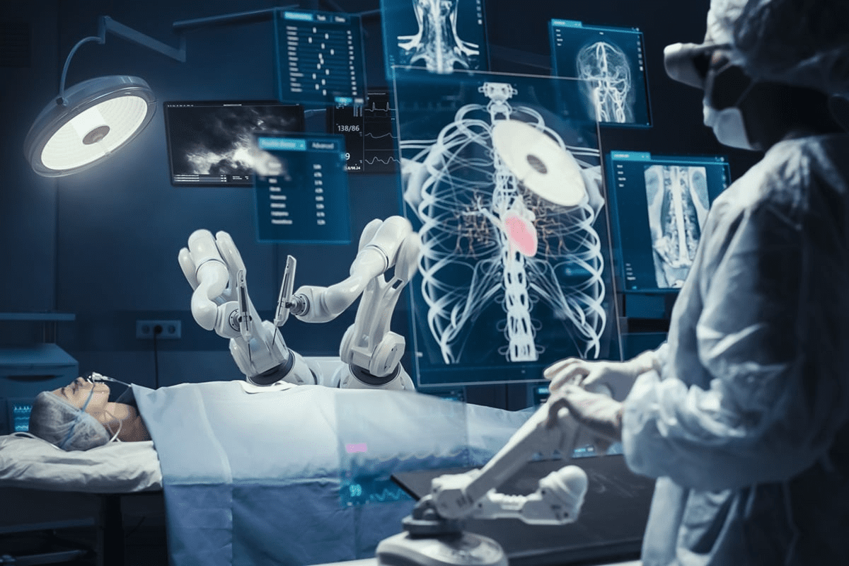
Knowing how well our kidneys work is key to staying healthy. Nuclear renal scans are a top tool for checking kidney health. At Liv Hospitals, we use the latest in nuclear medicine to spot and treat kidney problems.Find kidney imaging options, 7 facts about nuclear scan side effects, and how scans provide essential kidney data.
Nuclear renal scans use tiny amounts of special medicines and cameras to see how kidneys work. This test is great for finding issues that regular tests miss. Knowing how these scans work and their possible side effects helps patients understand their kidney health better.
Key Takeaways
- Nuclear renal scans provide detailed insights into kidney function and structure.
- The procedure involves the use of radiopharmaceuticals and gamma cameras.
- Lasix renal scans are a type of nuclear renal scan used to assess kidney function under stress.
- Understanding the possible side effects of nuclear renal scans is key for patient safety.
- Liv Hospitals offers advanced nuclear medicine techniques for kidney imaging.
The Fundamentals of Nuclear Medicine Kidney Imaging
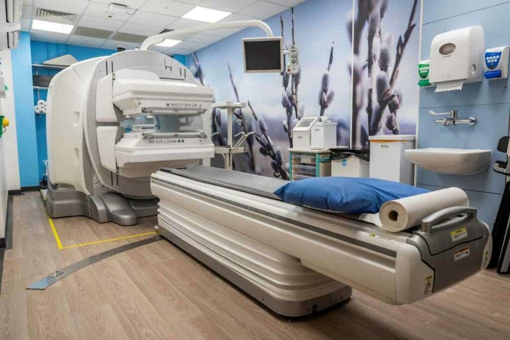
Understanding nuclear medicine kidney imaging is key to seeing its role in diagnosing kidney issues. It includes tests like nuclear renal scans. These tests help check how well the kidneys work and spot problems.
What Is a Nuclear Renal Scan?
A nuclear renal scan is a test that uses tiny amounts of radioactive material. It looks at how well the kidneys work and their structure. It’s important for finding and managing kidney diseases.
The test starts with a radiopharmaceutical, or radiotracer, being injected into the patient. This material is tracked by a gamma camera. The camera takes pictures of the kidneys, showing how they function and drain.
Key Components of Nuclear Kidney Imaging
The main parts of nuclear kidney imaging are the radiotracer injection and the gamma camera. The radiotracer is made to be taken up by the kidneys. This lets doctors check how well the kidneys are working and find any issues.
Key elements of nuclear kidney imaging include:
- Radiopharmaceuticals tailored for kidney function assessment
- Gamma camera technology for image capture
- Advanced image processing software for data analysis
Why Doctors Order This Specialized Test
Doctors use nuclear renal scans to understand kidney function. They look for blockages, check blood flow, and evaluate overall health. This info is vital for making treatment plans.
The diagnostic power of nuclear renal scans lies in their ability to provide both functional and anatomical information about the kidneys.
How Kidney Imaging Works: The Science Behind the Scan
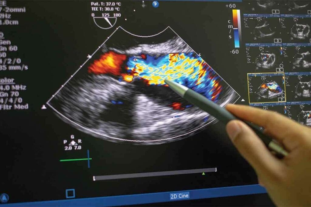
To understand kidney imaging, we need to explore the science behind it. This technology, like nuclear renal scans, uses advanced science to show how kidneys work and what they look like.
Radiopharmaceuticals: The Tracking Agents
Radiopharmaceuticals are key in nuclear kidney imaging. They are special compounds with tiny amounts of radioactive material, like Technetium-99m (Tc-99m). When injected, they go to the kidneys, helping us see how they function. The right radiopharmaceutical is picked based on what we need to know, like blood flow or filtering.
Gamma Cameras and Image Capture Technology
A gamma camera is used to capture images. It detects the gamma rays from the radiopharmaceuticals. As the kidneys absorb the radiopharmaceuticals, the camera takes pictures of their structure and function. This is key for spotting kidney problems like blockages or poor function.
How Images Are Processed and Interpreted
After taking pictures, they are enhanced and analyzed by doctors. This makes the images clearer and helps measure kidney function. For example, how fast the radiopharmaceutical is taken up or cleared tells us a lot about kidney health. This info is vital for diagnosing and treating kidney issues.
To learn more about kidney imaging and how it compares to other tests like ultrasound, check out RadiologyInfo.org. It offers detailed information.
| Aspect of Kidney Imaging | Description | Clinical Significance |
| Radiopharmaceuticals | Compounds containing radioactive material | Enable visualization of kidney function |
| Gamma Cameras | Devices detecting gamma rays emitted by radiopharmaceuticals | Capture detailed images of kidney structure and function |
| Image Processing | Enhancing image quality and quantifying data | Accurate assessment of kidney function and diagnosis |
Key Fact #1: Diagnostic Power of Nuclear Renal Scans
Nuclear renal scans are key in diagnosing kidney issues. They combine functional and anatomical data. This helps doctors understand and treat various kidney problems.
Identifying Kidney Blockages and Obstructions
Nuclear renal scans are great at finding blockages in the kidneys. They track how radiopharmaceuticals move through the kidneys. This helps spot issues like ureteral obstruction or kidney stones.
Seeing the urinary tract in real-time helps doctors understand blockages. They can then choose the best treatment. Often, these scans prevent more invasive tests.
Assessing Renal Blood Flow Problems
Nuclear medicine kidney scans also check blood flow to the kidneys. This is key for those with renovascular disease or kidney transplants. They look at how radiopharmaceuticals spread to the kidneys.
This helps spot issues like renal artery stenosis. It’s also important for checking on transplanted kidneys. Finding blood flow problems early can help keep kidneys working.
Evaluating Overall Kidney Function
Nuclear renal scans also look at how well kidneys work overall. They measure how radiopharmaceuticals are taken up and removed. This tells doctors about the glomerular filtration rate (GFR) of each kidney.
This info is vital for tracking chronic kidney disease and other kidney issues. It’s very helpful for patients with uneven kidney function. It lets doctors tailor treatments and check their success over time.
Key Fact #2: The Complete Kidney Scan Procedure
Learning about the kidney scan procedure can ease anxiety. It helps patients know what to expect. We’ll walk you through the steps of a nuclear kidney scan, from start to finish.
Pre-Scan Preparation Guidelines
Before a kidney scan, patients get specific instructions. They might need to fast or avoid certain foods and meds. Our team will give you all the details to get ready for the test.
The Radiotracer Injection Process
The scan starts with a radiotracer injection. This is a small, safe dose of radioactive material. It’s given through a vein in your arm and is quickly removed from your body.
The 30-60 Minute Imaging Timeline
After the injection, you’ll have imaging for 30 to 60 minutes. A gamma camera takes pictures of your kidneys as the radiotracer works. The time needed can vary based on the scan type.
What Patients Experience During the Test
During the scan, you’ll lie on a table while images are taken. It’s usually painless. You might need to stay very quiet for a bit. Our team will be there to make sure you’re comfortable and answer any questions.
Knowing about the kidney scan procedure helps patients feel more ready. Our aim is to support you fully during your nuclear kidney scan.
Key Fact #3: The Critical Role of Diuretics in Kidney Imaging
In kidney imaging, diuretics are key for checking how well kidneys work. Lasix is a major player in these scans. It helps doctors see how kidneys function.
Why Lasix Is Frequently Used in Renal Scans
Lasix, or furosemide, boosts urine production. This makes kidney function easier to assess. It helps spot any blockages or drainage problems.
The use of Lasix in renal scans is very helpful. It lets doctors see how kidneys work under stress. This gives them important info about kidney health.
How Diuretics Enhance Drainage Evaluation
Diuretics like Lasix improve drainage checks by increasing urine flow. This helps:
- Find any blockages in the urinary tract
- See how well kidneys concentrate and dilute urine
- Check the kidneys’ overall function
By using Lasix in scans, doctors can better see how the kidneys drain. This is key for spotting kidney issues.
Protocols for Diuretic Administration
Administering diuretics like Lasix in scans follows strict rules. Lasix is usually given through an IV 15-20 minutes into the scan. The exact timing and amount depend on the patient and the scan’s needs.
It’s vital for patients to listen to their healthcare team about taking diuretics and the scan process.
Key Fact #4: Lasix Renal Scan Side Effects Explained
It’s important for patients to know about the side effects of Lasix renal scans. These scans are usually safe and work well. But there are some temporary side effects to watch out for.
Common Temporary Reactions to Expect
Most people don’t have many side effects from Lasix renal scans. But, some common ones include:
- Increased urination
- Mild dizziness
- Occasional headache
These side effects usually don’t last long and go away by themselves.
Managing Increased Urination and Dizziness
To deal with more urine, drink lots of water. For dizziness, take Lasix in a doctor’s office and follow their advice.
Here are some tips for dizziness:
- Get up slowly from lying or sitting
- Avoid driving or using heavy machines if dizzy
- Drink water to avoid dehydration
When Side Effects Warrant Medical Attention
Even though most side effects are mild, some need a doctor’s help. Rare but serious reactions include:
- Severe allergic reactions
- Big changes in blood pressure
- Long-lasting or bad dizziness
If you have these symptoms, get help right away.
Recovery Timeline After the Procedure
Recovery from Lasix renal scans is usually quick. Most people can go back to normal activities soon after. The body gets rid of the tracer in a few hours, and Lasix’s effects fade in a day.
It’s good to know that side effects from Lasix renal scans are usually short-lived. And the procedure is safe for most people.
Key Fact #5: Advanced Techniques in Nuclear Medicine Kidney Scans
We are entering a new era in kidney imaging with nuclear medicine scans. These advanced methods have greatly improved how we diagnose and care for patients.
MAG3 Renal Scanning Methodology
The MAG3 renal scan uses Technetium-99m meretiatide to check kidney function. It’s great for patients with kidney problems.
DTPA Scanning Applications
DTPA scanning is key in nuclear medicine kidney scans. It helps measure how well the kidneys filter waste. It’s very useful for diagnosing and treating kidney blockages.
Comparing Different Radiotracer Options
There are various radiotracers for nuclear medicine kidney scans. MAG3 is best for patients with poor kidney function because it’s extracted well. DTPA is great for measuring GFR, which is important for kidney health.
| Radiotracer | Primary Use | Key Benefits |
| MAG3 | Assessing renal function, even in poor renal function | High extraction fraction, good for tubular function assessment |
| DTPA | Measuring glomerular filtration rate (GFR) | Accurate GFR measurement, useful in obstructive uropathy |
Latest Innovations in Renal Imaging
The field of renal imaging is always getting better. New radiotracers and technologies are being researched. These could make diagnosis even more accurate and help patients more.
As we keep improving nuclear medicine kidney scans, we can offer more precise and tailored medicine. This shows how vital it is to keep up with medical technology to give the best care to patients.
Key Fact #6: Safety Profile of Nuclear Kidney Imaging
It’s important to know how safe nuclear kidney imaging is. This is true for both patients and doctors. We look at the safety, like how much radiation you get and special care for some patients.
Radiation Exposure Risks
Nuclear kidney imaging uses tiny amounts of radioactive stuff. This stuff helps doctors see how well your kidneys work. Even though it’s radioactive, the doses are set low to keep you safe.
Radiation exposure is a big deal. We pick radiopharmaceuticals that are safe but also work well for diagnosing.
Patient Populations Requiring Special Consideration
Some groups need extra care when they get a nuclear kidney scan. Pregnant women should know about the risks to their baby. We talk to them about the risks and benefits to decide if it’s right for them.
Other groups, like children and patients with kidney disease, might need special rules to stay safe during the scan.
Why Nuclear Scans Are Considered Minimally Invasive
Nuclear kidney imaging is minimally invasive. It doesn’t need surgery or putting things inside your body. You just get a tiny injection of radiopharmaceutical, and a camera tracks it to see your kidneys.
This method is safe and works well for checking your kidneys. It’s a key tool for doctors to find and fix kidney problems.
Key Fact #7: Comparing Kidney Imaging Tests for Optimal Diagnosis
Choosing the right imaging test is key to diagnosing kidney problems. We’ll look at tests like nuclear scans, ultrasound, CT, and MRI. This will help us find the best way to diagnose kidney issues.
Nuclear Scans vs. Ultrasound Imaging
Nuclear scans and ultrasound are two ways to check kidney function and structure. MAG3 or DTPA renal scans show how well the kidneys work. Ultrasound, on the other hand, checks kidney size and position without harm.
Ultrasound is quick and safe, but nuclear scans give more detailed kidney function info. For example, nuclear scans are better for finding out about kidney blockages.
Nuclear Scans vs. CT and MRI Techniques
CT and MRI scans also play a role in kidney imaging. CT scans show detailed images of the kidneys and can find many problems. MRI gives clear images of soft tissues and checks kidney function without radiation.
Nuclear scans are best for checking how well the kidneys work. They help plan surgeries or manage conditions like hydronephrosis. CT and MRI give detailed pictures, but nuclear scans offer a special view of kidney function.
How Physicians Determine the Most Appropriate Test
Doctors look at many things when choosing a kidney imaging test. They consider the patient’s history, symptoms, and what they need to know. For example, a nuclear scan might be chosen for kidney blockage issues.
The right test also depends on the patient’s age, kidney health, and any allergies. Sometimes, doctors use more than one test to fully understand a patient’s condition.
Complementary Nature of Different Imaging Modalities
Different imaging tests work together, not against each other. Each test has its own strengths and can give important information. Together, they help doctors make accurate diagnoses and treatment plans.
For example, combining a nuclear scan with a CT scan can give a full picture of kidney health. This approach helps doctors make better decisions and plan treatments more effectively.
Conclusion: The Future of Kidney Imaging and Patient Care
Nuclear medicine kidney scans are key in diagnosing and managing kidney issues. They are vital for patient care. The facts about these scans and their side effects highlight their importance.
Advances in kidney imaging are making diagnosis more accurate and patient care better. Techniques like renal imaging with Lasix help evaluate kidney function. Knowing about Lasix renal scan side effects is key to effective patient care.
We expect more innovations in nuclear medicine kidney scans. This will improve their ability to diagnose and expand their use. As technology advances, patient care and outcomes will also improve, mainly for those getting these scans.
Healthcare professionals need to keep up with these advancements. This way, they can offer the best care to patients needing kidney imaging and other tests.
FAQ
What is a nuclear renal scan?
A nuclear renal scan is a test that uses tiny amounts of radioactive material. It checks how well the kidneys work and spots any problems.
How does a nuclear renal scan work?
First, a radiopharmaceutical is injected into the blood. Then, the kidneys absorb it. A gamma camera takes pictures of the kidneys as the material moves through. This shows how well the kidneys are working and how they drain.
What is Lasix used for in a renal scan?
Lasix is a diuretic used in the scan. It helps make more urine, which lets doctors see how well the kidneys drain. This makes the scan more accurate.
What are the common side effects of a Lasix renal scan?
Side effects include more urine, feeling dizzy, and dehydration. These effects are usually mild and go away quickly.
How long does it take to recover from a nuclear renal scan?
The radioactive material leaves the body in a few hours. Most people can go back to normal activities soon after. How fast you recover can depend on your body.
Is nuclear kidney imaging safe?
Yes, nuclear kidney imaging is safe when done right. The amount of radiation is low. The benefits of getting important information usually outweigh the risks.
How does nuclear kidney imaging compare to other kidney imaging tests?
Nuclear kidney imaging gives special information on kidney function and drainage. It works with other tests like ultrasound, CT, and MRI. The right test depends on what you need to know.
What is the difference between MAG3 and DTPA renal scans?
MAG3 and DTPA are both used in renal scans. MAG3 checks function and drainage. DTPA looks at function and estimates how well the kidneys filter.
Can I undergo a nuclear renal scan if I have kidney disease?
Yes, people with kidney disease can have a nuclear renal scan. It gives important info on kidney function and helps with treatment plans.
How should I prepare for a nuclear renal scan?
Preparing for the scan varies based on the test and your needs. Usually, drink lots of water, avoid certain meds, and follow your doctor’s instructions.
References
- Taylor, A. T., Nally, J., Aurell, M., et al. (2018). SNMMI Procedure Standard / EANM Practice Guideline for Diuretic Renal Scintigraphy in Adults With Suspected Upper Urinary Tract Obstruction. Seminars in Nuclear Medicine, 48(4), S1–S35. https://pmc.ncbi.nlm.nih.gov/articles/PMC6020824/
- Banks, K. P. (2022). Diuretic Renal Scintigraphy Protocol Considerations. Journal of Nuclear Medicine Technology, 50(4), 309–315. https://tech.snmjournals.org/content/50/4/309
- Fried, J. G., Goldfarb, D. S., deLin, C., & others. (2019). Renal Imaging: Core Curriculum 2019. American Journal of Kidney Diseases, 74(1), 120–134. https://www.ajkd.org/article/S0272-6386(19)30029-0/fulltext


