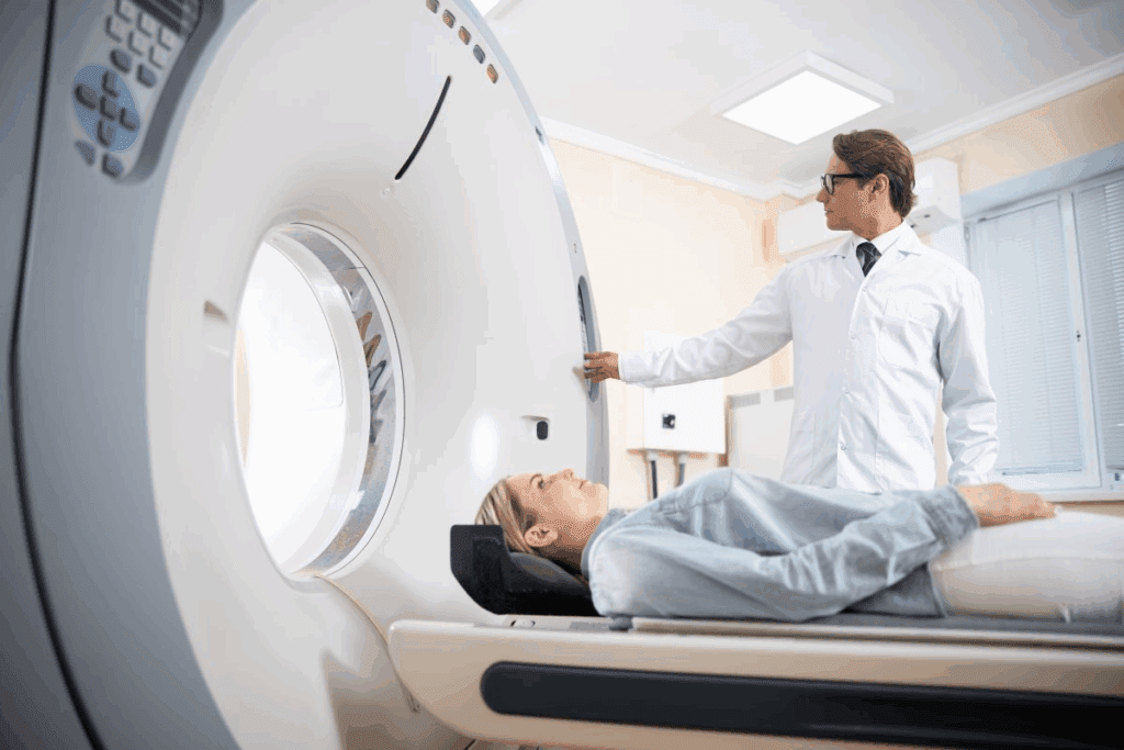
Pet radiology scan is a powerful tool for detecting diseases like cancer. In medical imaging, Positron Emission Tomography (PET) scans measure FDG uptake, which shows how active the body’s cells are.
High FDG uptake on a pet radiology scan often points to possible cancer activity — but that’s not always the case. Some non-cancerous conditions can also cause increased FDG uptake, which sometimes leads to false alarms and unnecessary concern.
Understanding how FDG uptake works in both cancerous and non-cancerous conditions is crucial. With a better grasp of what a pet radiology scan reveals, doctors can make more accurate diagnoses and plan the most effective treatment for each patient.
This article explores why pet radiology scans sometimes show high cell activity even when it’s not cancer.
Key Takeaways
- PET scans measure metabolic activity through FDG uptake.
- Increased FDG uptake is not exclusive to cancerous conditions.
- Benign processes can cause heightened metabolic activity.
- Understanding benign causes of FDG uptake is key for accurate diagnosis.
- Correctly reading PET scans is vital for good treatment plans.
What is FDG Uptake and How Does it Work?
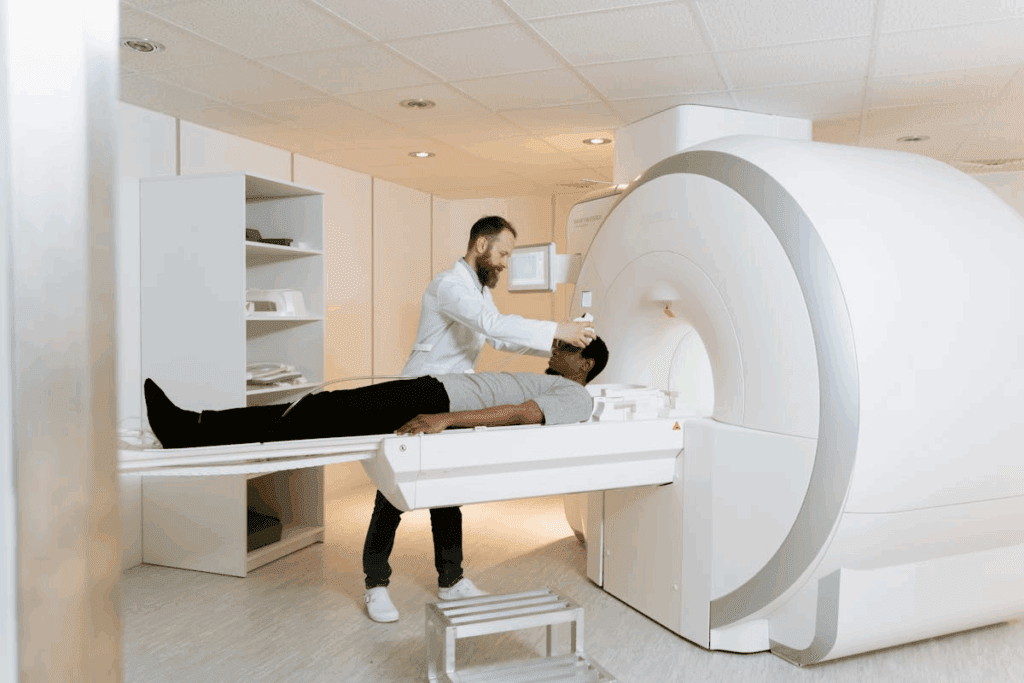
Fluorodeoxyglucose (FDG) uptake is key in PET scans, showing how cells work.Explains, “FDG uptake refers to the amount of radiotracer uptake.” This idea is vital for understanding PET scans and their use in medicine.
The Science Behind Fluorodeoxyglucose
FDG acts like glucose, which cells take in. Inside the cell, FDG gets stuck because it can’t leave. This lets PET scans measure how much FDG is there. The more FDG, the more active the cell is.
Glucose Metabolism and Cellular Activity
Cells that use a lot of energy, like tumor cells, need more glucose. They also take in more FDG. This is why PET scans are good at finding tumors and checking if treatments are working. How cells use glucose and FDG is key to reading PET scan results.
| Cell Type | Glucose Metabolism | FDG Uptake |
| Normal Cells | Low to Moderate | Low |
| Tumor Cells | High | High |
| Inflammatory Cells | Moderate to High | Moderate |
The Fundamentals of PET Scan Technology
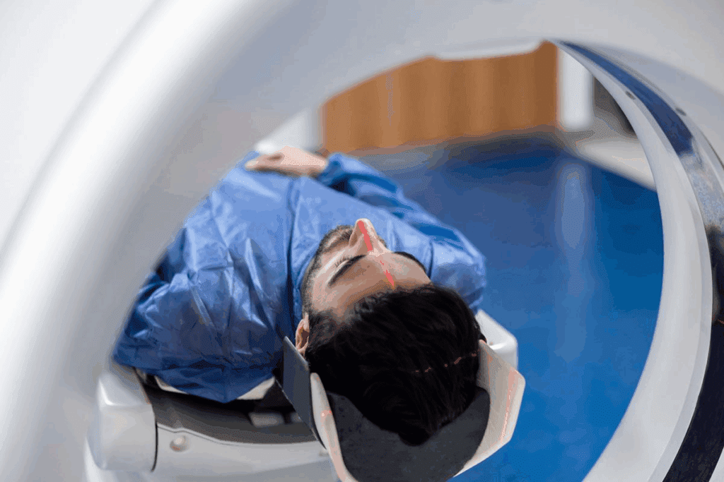
Understanding PET scan technology is key to seeing its value in oncology imaging and cancer staging. PET scans are vital in diagnosing and managing cancer. They show how active cells are metabolically in the body.
How PET Scans Detect Metabolic Activity
PET scans use a radioactive tracer, like fluorodeoxyglucose (FDG), to find active areas. Cancer cells take up more glucose, making them easy to spot. The PET scanner picks up the radiation from the tracer, showing where the activity is.
Patient Preparation and Scanning Protocols
Getting ready for a PET scan is important for good results. Patients usually fast for hours before to lower blood sugar. Then, they get the tracer injected and wait for it to spread in the body.
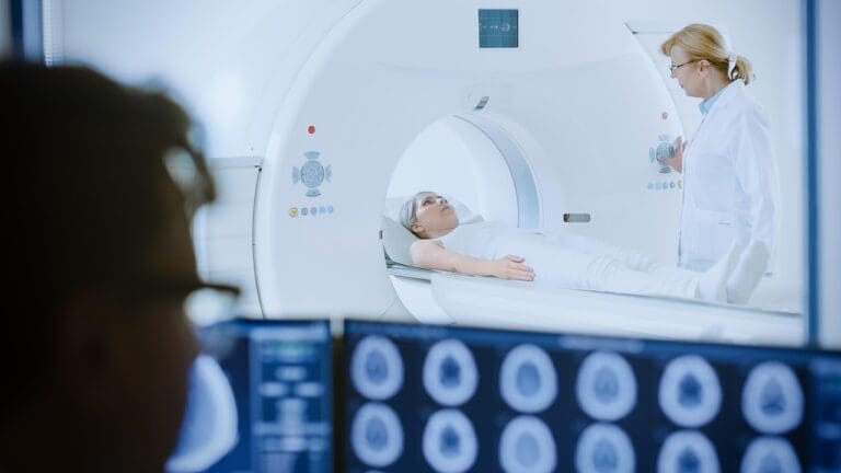
After that, they lie in the PET scanner. It captures the data on metabolic activity.
Normal Physiological Patterns of FDG Uptake
It’s important to know what’s normal when looking at PET scan results. FDG uptake can change a lot from person to person. Knowing these changes helps doctors make the right diagnosis.
Expected Distribution in Healthy Individuals
In healthy people, FDG uptake is seen in some organs and tissues. The brain uses a lot of glucose, so it shows a lot of FDG uptake. The heart and liver also take up FDG, but how much can change based on things like fasting and insulin levels.
| Organ/Tissue | Expected FDG Uptake |
| Brain | High |
| Heart | Variable |
| Liver | Moderate |
Variations in Normal Uptake Patterns
Even though there are usual patterns of FDG uptake, things can change. For example, brown adipose tissue might take up more FDG, often in cold weather or with certain health issues.
Knowing about these changes helps doctors tell normal from abnormal uptake. This is important for finding tumors and checking for abnormal cell activity.
The Meaning of FDG Avidity in Clinical Context
Knowing about FDG avidity is key to understanding PET scan results. It shows how much Fluorodeoxyglucose (FDG) a tissue or lesion takes up. This helps doctors see how active the body’s cells are.
Defining “FDG-Avid” in Medical Terminology
“FDG-avid” means tissues or lesions take up a lot of FDG. This usually means they are very active, like in cancers, infections, or inflammation. “FDG avidity” helps doctors figure out what’s going on and how to treat it.
But, not all high FDG uptake is cancer. Some non-cancerous things can also take up a lot of FDG. So, knowing the context is very important for correct diagnosis.
Standardized Uptake Value (SUV) Measurements
Standardized Uptake Value (SUV) measures how much FDG a tissue or lesion takes up. It’s a number that shows how active the tissue is compared to the rest of the body. This helps doctors understand how active the tissue is.
| SUV Parameter | Description | Clinical Significance |
| SUVmax | Maximum SUV value within a region of interest | Indicates the highest metabolic activity |
| SUVmean | Average SUV value within a region of interest | Provides an overall assessment of metabolic activity |
Doctors use FDG avidity and SUV to check how active the body’s cells are. This helps them make better decisions for patient care.
Common Benign Causes of Increased FDG Uptake
It’s important to know the benign causes of increased FDG uptake for accurate PET scan readings. Fluorodeoxyglucose (FDG) is a glucose analog that builds up in cells that are very active. This makes it great for finding and tracking different conditions, like cancer. But, not all high FDG avidity is from cancer; many benign conditions can also show it.
Inflammatory and Infectious Processes
Inflammatory and infectious processes often cause increased FDG uptake. Things like abscesses, pneumonia, and granulomatous diseases can show a lot of FDG avidity. This is because immune cells in these conditions use a lot of glucose, leading to high FDG uptake. It’s key to look at the whole picture and other imaging results to tell these conditions apart from cancer.
A study in The Journal of Nuclear Medicine showed FDG uptake in benign conditions like infections and inflammation. This highlights the importance of carefully reading PET scan results.
Post-Treatment Changes and Healing Tissues
After treatments like surgery, chemotherapy, or radiation, the body’s healing can cause increased FDG uptake. For example, inflammation and healing after surgery can lead to FDG accumulation. This might look like cancer if not seen in the context of recent treatments.
It’s vital to recognize these patterns for accurate PET scan readings in treated patients. The term FDG avid includes both benign and malignant causes. This means we need to look at the whole clinical picture to understand the results.
Benign Conditions That Mimic Malignancy on PET Scans
Some non-cancerous conditions can look like cancer on PET scans. Says it’s important to be careful when reading these scans. This helps tell the difference between harmless and harmful conditions.
Autoimmune Disorders and Their Imaging Features
Autoimmune diseases can show up as cancer on PET scans. For example, rheumatoid arthritis can cause avid FDG uptake in joints. It’s key to understand the situation and the scan’s findings to avoid mistakes.
- Rheumatoid arthritis
- Lupus
- Sjögren’s syndrome
These diseases can make PET scans hard to read. More tests and doctor’s opinions are often needed to figure out what’s going on.
Granulomatous Diseases and Sarcoidosis
Granulomatous diseases, like sarcoidosis, can also show up as FDG-avid on scans. This makes it tricky to tell if it’s cancer or not. Sarcoidosis can look like lymphoma or other cancers because it forms granulomas that take up FDG.
The fdg avidity in these diseases shows we need a detailed approach to diagnosis. When looking at PET scans, it’s important to think about harmless conditions that might look like cancer. This helps avoid unnecessary treatments.
Does fdg-avid always mean cancer? No. Knowing the patient’s history and using other tests helps us understand what the scan means.
Physiological Variants of FDG Uptake
It’s important to understand the different ways FDG uptake can happen in the body. These variations are normal and happen for many reasons. Knowing these patterns helps doctors tell the difference between safe and dangerous uptake.
Brown Adipose Tissue Activation
Brown adipose tissue (BAT) is a special fat that burns a lot of energy. It can make FDG uptake look higher. This usually happens when it’s cold outside.
It can look like cancer in places like the neck or spine. But, it’s actually a normal response to cold. Knowing this can prevent mistakes in diagnosis.
Muscle Activity and Exercise Effects
Exercise can also change how FDG uptake looks. When you work out, muscles use more glucose, showing up on scans. This is common in muscles of the neck, shoulders, and legs.
Seeing this pattern helps doctors know it’s not a disease. For example, if uptake matches muscle areas, it’s likely from exercise, not illness.
Knowing about these normal changes helps doctors make better diagnoses. This means patients get the right treatment faster and more accurately.
Iatrogenic and Medication-Related FDG Uptake
It’s key to know how medical actions and some drugs affect FDG uptake for correct PET scan reading. Iatrogenic factors are issues caused by medical treatments. These can change how glucose is used in the body, affecting FDG uptake.
Drug-Induced Changes in Glucose Metabolism
Some drugs can change how glucose is used, leading to different FDG uptake patterns. For example, G-CSF can make bone marrow more active, causing more FDG uptake there. Also, some drugs can lead to inflammation or infection, impacting FDG uptake.
Examples of medications that can influence FDG uptake include:
- Insulin and other diabetes medications, which can alter glucose metabolism
- G-CSF, which stimulates bone marrow activity
- Certain antibiotics and anti-inflammatory drugs, which can cause changes in inflammatory processes
Interventional Procedures and Their Imaging Sequelae
Interventional actions like biopsies, surgeries, and injections can also change FDG uptake. These actions can cause inflammation and higher glucose use in the affected areas. This leads to higher FDG uptake in those areas.
It’s vital to look at the patient’s medical history and recent treatments when reading PET scans. This helps avoid mistaking iatrogenic changes for disease.
Organ-Specific Benign FDG Uptake Patterns
It’s key to know how different organs take up FDG for PET scans. Benign conditions can look like cancer, so spotting specific patterns is important.
Thyroid and Endocrine System Findings
The thyroid gland can show different levels of FDG uptake. This is often because of benign issues like thyroiditis or adenomas. Thyroiditis can lead to widespread FDG uptake, while adenomas show up as single spots. Knowing these patterns helps tell benign from cancerous conditions.
The endocrine system’s glands, like the adrenal glands, can also have benign FDG uptake. This is usually because of hyperplasia or adenomas.
Gastrointestinal and Genitourinary Variations
The gastrointestinal tract shows various benign FDG uptake patterns. This is often because of inflammation, infection, or normal processes. For example, inflammatory bowel disease can increase uptake in the intestines. Normal bowel activity can also cause uptake.
In the genitourinary system, benign uptake is seen in the kidneys and uterus. This is due to FDG being excreted in urine and menstrual cycle changes.
It’s important to define FDG avidity and its role in medical settings. FDG-avid spots don’t always mean cancer; some benign conditions can also show high uptake. Spotting these patterns is key for correct diagnosis and treatment plans.
Differentiating Between Benign and Malignant FDG Uptake
It’s key to tell apart benign and malignant FDG uptake in PET scans. Getting it right is vital for good patient care and treatment plans.
Pattern Recognition in PET Interpretation
Recognizing patterns is essential in PET scans. “Pattern recognition is key in PET scans. It helps spot specific uptake patterns linked to different conditions.” Benign conditions usually have certain patterns, like diffuse or focal uptake, which differ from the irregular patterns of cancer.
Inflammatory issues might show a spread-out uptake, while cancer tends to have uneven uptake. Knowing these patterns takes a deep dive into the disease’s nature and typical imaging signs.
The Importance of Clinical Correlation
Linking PET scan results with a patient’s medical history is also critical. Stresses, “linking PET scan results with a patient’s medical history is vital. It gives a full picture of the patient’s health.” This means looking at the patient’s past health, symptoms, and lab tests to understand the PET scan better.
For example, someone with a history of infection might show high FDG uptake, which could be mistaken for cancer without the right context. On the other hand, a cancer patient might show benign uptake that looks like a normal finding without knowing their cancer history.
By using pattern recognition and clinical correlation together, doctors can better understand PET scans. This leads to more accurate diagnoses and better care for patients.
Advanced Imaging Techniques for Characterizing FDG Uptake
Advanced imaging techniques have changed how we look at FDG uptake. They give us deeper insights into how cells work. This is key for telling apart harmless and harmful changes, making diagnosis better.
Dual-Time-Point Imaging and Kinetic Analysis
Dual-time-point imaging takes PET scans two times after FDG is given. It helps spot the difference between cancer and non-cancer by seeing how FDG uptake changes. Cancer usually takes up more FDG over time, while non-cancer stays the same or goes down.
Kinetic analysis of FDG uptake gives detailed info on tissue metabolism. It lets doctors understand the disease better, helping to identify lesions more accurately.
Multimodality Approaches: PET/CT and PET/MRI
Multimodality imaging mixes PET’s functional info with CT or MRI’s detailed pictures. PET/CT is popular for its ability to pinpoint FDG uptake in the body, boosting confidence in diagnosis. PET/MRI offers even better soft-tissue contrast and functional details, ideal for some cases.
Combining PET with CT or MRI gives a full view of disease spread and metabolic activity. This approach helps better understand FDG uptake, leading to more informed decisions.
Alternative Radiotracers Beyond FDG
Many radiotracers are being used to improve PET imaging. These tracers target different parts of tumor biology or other diseases. They help us understand diseases better.
Proliferation Markers and Amino Acid Tracers
FLT (Fluorothymidine) shows how fast cells are growing. This is useful for seeing how aggressive tumors are. FET (Fluoroethyltyrosine) and Methionine measure how much cells are making proteins. They are great for brain tumor imaging, helping tell tumors apart from treatment effects.
Receptor-Targeted Imaging Agents
Receptor-targeted agents bind to specific receptors on cells, like cancer cells. For example, Gallium-68 DOTATATE finds neuroendocrine tumors with somatostatin receptors. These agents make diagnoses more accurate, leading to better treatment plans.
The use of these new radiotracers is growing PET imaging’s role in healthcare. It helps doctors make better choices for their patients.
Clinical Case Studies of Benign FDG-Avid Conditions
Clinical studies have shown that some benign conditions can look like cancer on PET scans. This makes it important to carefully read these scans. Getting the diagnosis right is key to not mistake a harmless condition for cancer.
Inflammatory Pseudotumors and Organizing Pneumonia
Inflammatory pseudotumors and organizing pneumonia are two benign conditions that can show up as cancer on PET scans. Inflammatory pseudotumors are caused by inflammation in a specific area. Organizing pneumonia is a lung injury that can also show up as increased FDG uptake.
- Inflammatory pseudotumors can happen in different organs like the lungs, liver, and spleen.
- Organizing pneumonia is often linked to infections or autoimmune diseases.
Tuberculosis and Other Infectious Mimics
Tuberculosis (TB) is a classic example of an infectious disease that can show up as cancer on PET scans. TB can look like cancer, mainly when there’s a single lesion or swollen lymph nodes.
Key features of TB on PET scans include:
- High FDG uptake in affected lymph nodes or lesions.
- Variable patterns of uptake, including diffuse or focal activity.
Other infections, like fungal diseases or abscesses, can also show up as increased FDG uptake. This highlights the need for careful clinical correlation and diagnostic evaluation.
Interpreting Negative or Low FDG Uptake Results
Negative or low FDG uptake results on PET scans are tricky to understand. The lack or little presence of fluorodeoxyglucose (FDG) can mean different things. It’s important to know the reasons and the bigger picture of the patient’s health.
What “No FDG-Avid” Findings Mean Clinically
“No FDG-avid” findings on a PET scan usually mean there’s little to no abnormal glucose use. This is often good news, showing there’s no cancer activity. But, it’s key to look at the bigger picture and other tests to avoid false-negative results.
Things like the type of cancer, its growth rate, and the patient’s health can affect FDG use.
- Low-grade tumors may not show up well on PET scans.
- Some cancers just don’t use a lot of glucose.
- How the patient is prepared and technical aspects can change scan results.
Low-Grade Malignancies with Minimal Uptake
Low-grade malignancies are hard to spot because they don’t use much FDG. This can lead to missing or misreading these cancers. It’s vital to understand these cancers and match PET scan results with other tests for accurate diagnosis.
Important things to think about include:
- Matching PET scan results with CT or MRI.
- Looking at the patient’s medical history and symptoms.
- Using more tests or follow-up scans when needed.
Conclusion: The Nuanced Interpretation of FDG Uptake in Clinical Practice
Understanding FDG uptake in PET scans is complex. It involves knowing about tumor detection, oncology imaging, and cancer staging. This article has shown how important it is to interpret FDG uptake carefully. Clinicians must look at many factors when they see FDG-avid lesions.
Getting a correct diagnosis is key. It’s about telling the difference between benign and malignant FDG uptake. This is a big challenge in oncology imaging and cancer staging. Healthcare professionals need to know about normal patterns, conditions that look like cancer, and how medicine affects FDG uptake.
New imaging methods, like dual-time-point imaging and using different imaging types together, help a lot. As we learn more, using other radiotracers and understanding FDG uptake better will be very important. This will help a lot in finding tumors and managing cancer.
FAQ
What does FDG-avid mean in the context of PET scans?
FDG-avid means cells take up Fluorodeoxyglucose (FDG), showing high activity. This is often seen in cancer but can also happen in non-cancerous conditions like inflammation or infection.
Is FDG uptake always indicative of cancer?
No, FDG uptake doesn’t always mean cancer. Other things like inflammation, infections, and some normal body functions can also show high FDG uptake.
How does FDG relate to glucose metabolism?
FDG acts like glucose and is taken up by cells. Cells that are very active, like cancer cells, use more glucose and FDG. This makes FDG uptake a good way to see how active cells are.
What is the role of PET scan technology in detecting FDG uptake?
PET scan technology uses FDG to see how active cells are. It helps find areas with abnormal activity, which is important for diagnosing diseases.
What are some common benign causes of increased FDG uptake?
Benign causes include inflammation, infections, changes after treatment, and some normal body functions. These can all show up as high FDG uptake on scans.
How can benign conditions be differentiated from malignant ones on PET scans?
To tell the difference, look at the pattern of uptake and the patient’s overall health. Advanced imaging and combining different scans can also help.
What is the significance of SUV measurements in assessing FDG uptake?
SUV measurements show how much FDG is taken up by tissues. This helps doctors understand how active the disease is and how well it’s responding to treatment.
Can medications and iatrogenic factors influence FDG uptake?
Yes, some medicines and medical procedures can change how cells use glucose. This can affect FDG uptake and might lead to wrong conclusions if not considered.
Are there alternative radiotracers used in PET imaging beyond FDG?
Yes, there are other tracers like markers for cell growth, amino acids, and targeted agents. They give different views of cell activity and are used for specific tests.
What does a negative or low FDG uptake result indicate?
A low FDG uptake might mean there’s little to no disease activity. But, some cancers might show very little uptake. So, it’s important to look at all the information together.
Reference
- Love, C., Tomas, M. B., Tronco, G. G., & Palestro, C. J. (2005). FDG PET of infection and inflammation. Radiographics, 25(5), 1357–1368. https://pubmed.ncbi.nlm.nih.gov/16244225/


