
What is Nuclear Medicine ?
Nuclear medicine is a field of medicine in which radioactive materials are used and includes various medical imaging and treatment procedures.
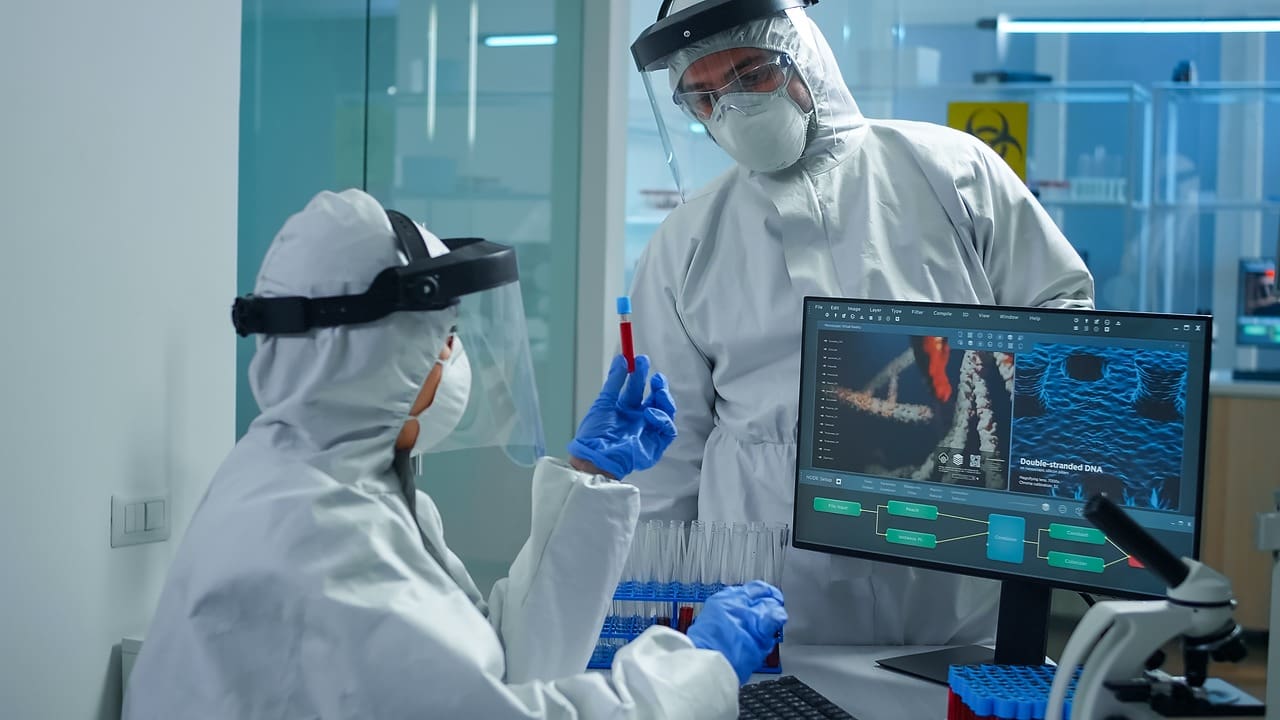
Nuclear medicine is a branch of medical imaging that uses small amounts of radioactive materials to help diagnose and treat diseases. Unlike X rays or MRIs, it shows how the body is working, which helps doctors find problems early.
Doctors give patients a small amount of radioactive material, called a radiotracer, by injection, swallowing, or breathing it in. The radiotracer gives off signals that a special camera picks up to make images of organs and tissues. This safe method helps find problems like cancer, heart disease, and some brain disorders.
Nuclear medicine plays a crucial role in modern healthcare, offering both diagnostic and therapeutic options. For patients, understanding what nuclear medicine entails can alleviate concerns and enhance their experience during procedures.

How Does Nuclear Medicine Work?
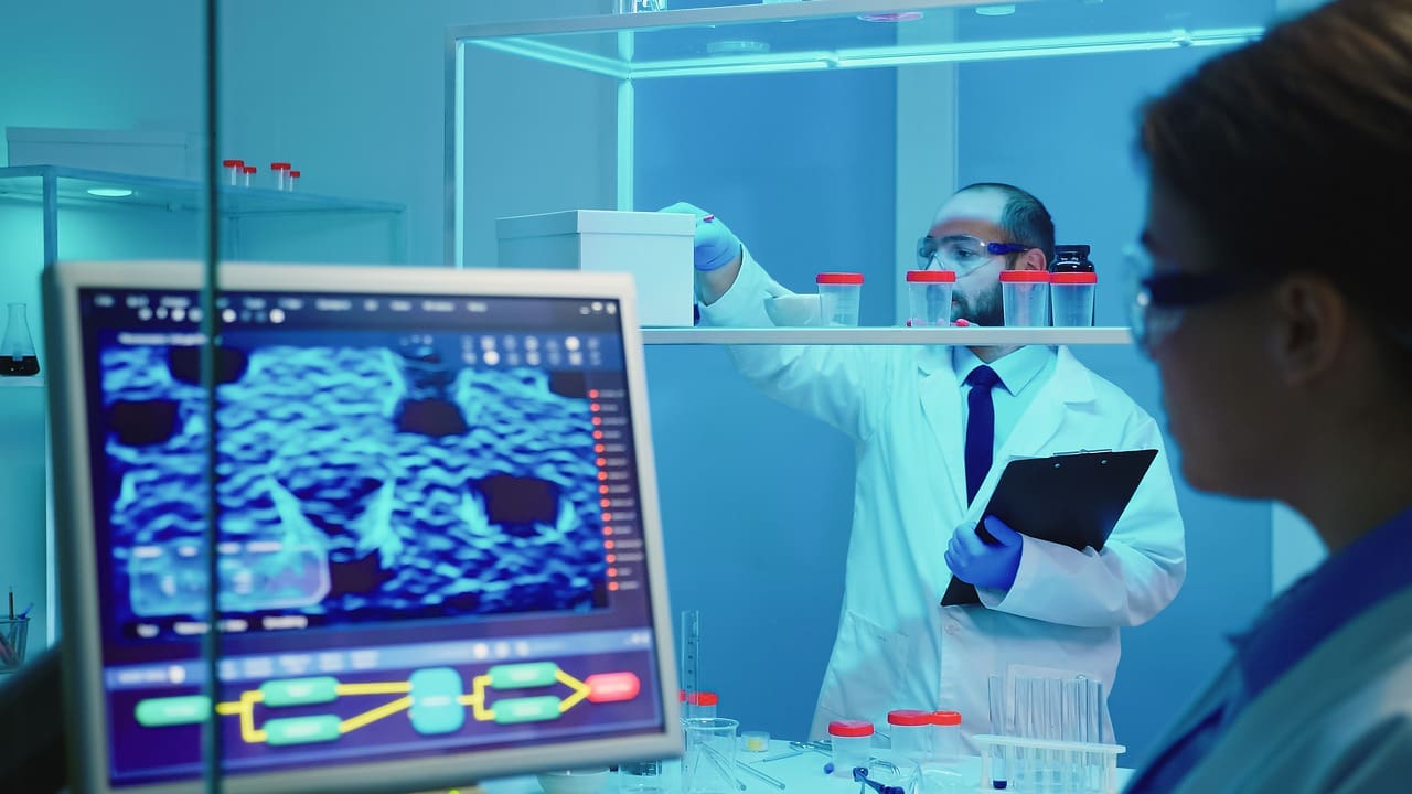
Nuclear medicine uses radioactivity and special cameras to take pictures inside the body. After a radiotracer is given, it collects in certain organs or tissues, making it easier to see areas that might be affected by illness.
Doctors choose the right radiotracer based on what they are looking for. For example, bone scans often use technetium-99m, which goes to areas where the bones are more active, like spots with inflammation or tumors.
After the radiotracer is given, it sends out signals that a gamma camera can detect. The camera turns these signals into pictures, showing how organs are working, how blood is flowing, and if there are any problems.
This type of imaging helps doctors make better decisions about diagnosis and treatment, which can lead to better results for patients.
Which Diseases Does Nuclear Medicine Treat ?
Nuclear medicine encompasses a medical field used for the diagnosis, treatment and follow up of various diseases. Here are some diseases that nuclear medicine focuses on:
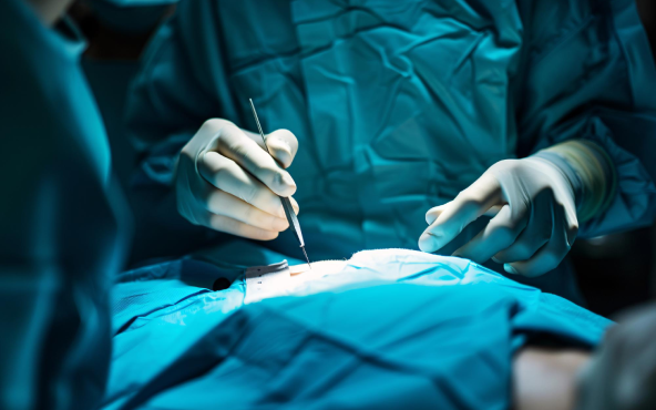
Cancer: Nuclear medicine is used for cancer diagnosis, evaluation of metastasis, and treatment of some types of cancer. Cancer cells can be identified using radioactive isotopes and monitored to follow up the response to treatment.
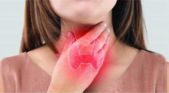
Thyroid Diseases: Nuclear medicine is used to evaluate the functions of the thyroid gland and determine thyroid nodules. Thyroid scintigraphy and radioactive iodine therapy are widely used applications for the diagnosis and treatment of thyroid diseases.
Bone Diseases: Nuclear medicine is used for the evaluation of bone structures with methods such as bone scintigraphy. Bone scans can be used for diagnosis and evaluation of bone metastases, infections or traumas.
Cardiovascular Diseases: Myocardial scintigraphy is a nuclear medicine method used for the evaluation of coronary artery disease. With this method, the blood flow and perfusion of the heart muscle are examined.
Lung Diseases: Lung perfusion scintigraphy and lung ventilation scintigraphy can be used for the evaluation of lung function and the diagnosis of pulmonary embolism.
Neurological Diseases: Nuclear medicine can help in the evaluation and determination of neurological diseases such as Alzheimer's disease, Parkinson's disease and epilepsy.
What Does a Nuclear Medicine Technologist Do? Roles and Responsibilities
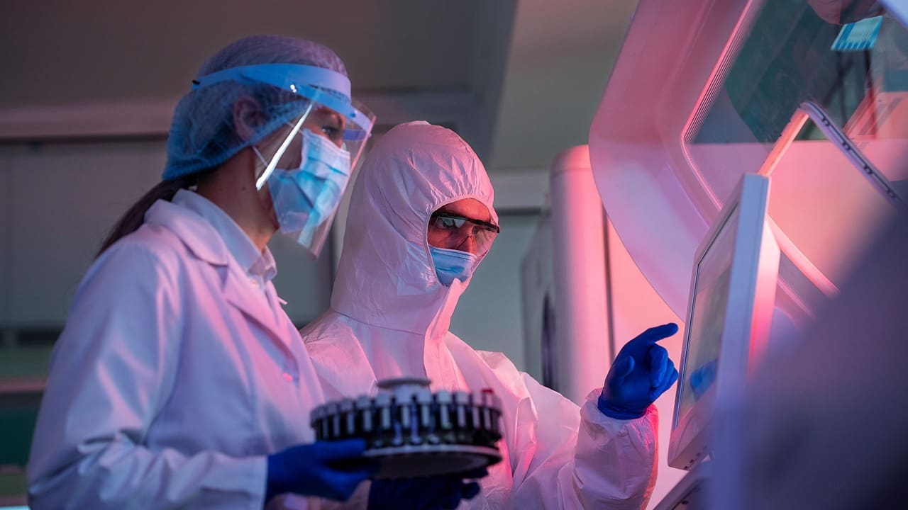
A nuclear medicine technologist is a trained healthcare worker who gets the radiotracers ready and gives them to patients. They make sure the imaging process is safe and works well.
Key responsibilities of a nuclear medicine technologist include:
- Patient Preparation: Technologists explain the procedures to patients, addressing any concerns and ensuring they understand the process.
- Radiopharmaceutical Administration: They prepare and administer the appropriate radiotracer, ensuring proper dosage and adherence to safety protocols.
- Imaging Procedures: Technologists operate gamma cameras and other imaging equipment to capture high quality images of the patient's organs and tissues.
- Patient Monitoring: During the procedure, they monitor patients for any adverse reactions and ensure their comfort.
- Image Analysis: After the procedure, technologists may assist in analyzing the images and preparing preliminary reports for physicians.
Nuclear medicine technologists play a key role in helping doctors make accurate diagnoses and in taking good care of patients.
What are Nuclear Medicine Tests? Types and Purposes
Nuclear medicine tests use radiotracers to check for different health problems. These tests show how organs are working and can help find diseases early. Common types of these tests include:
- Bone Scans: Used to identify bone diseases, infections, or fractures by highlighting areas of increased or decreased bone activity.
- Thyroid Scans: These tests assess thyroid function and can help diagnose conditions such as hyperthyroidism or thyroid cancer.
- PET Scans (Positron Emission Tomography): PET scans are commonly used in oncology to detect cancer and monitor treatment response by visualizing metabolic activity.
- Myocardial Perfusion Scans: These tests evaluate blood flow to the heart muscle, helping to diagnose coronary artery disease and assess heart function.
- Lung Scans: Used to evaluate lung function and detect conditions such as pulmonary embolism or chronic obstructive pulmonary disease (COPD).
Each nuclear medicine test has its own purpose and gives doctors important information to help plan treatment.
Is Nuclear Medicine Safe?
Minimal Radiation Exposure: The amount of radiation used in nuclear medicine tests is typically low and well within safety limits. The benefits of accurate diagnosis often outweigh the risks associated with radiation exposure.
- Monitoring and Protocols: Nuclear medicine facilities adhere to strict safety protocols to protect both patients and staff. This includes monitoring radiation levels and using protective equipment when necessary.
- Patient Considerations: Before undergoing a nuclear medicine test, patients should inform their healthcare provider of any medical conditions, allergies, or medications they are taking, as these factors may influence safety and effectiveness.
Knowing about the safety steps taken can help patients feel less worried about having a nuclear medicine procedure.
What is a Nuclear Medicine Bone Scan?
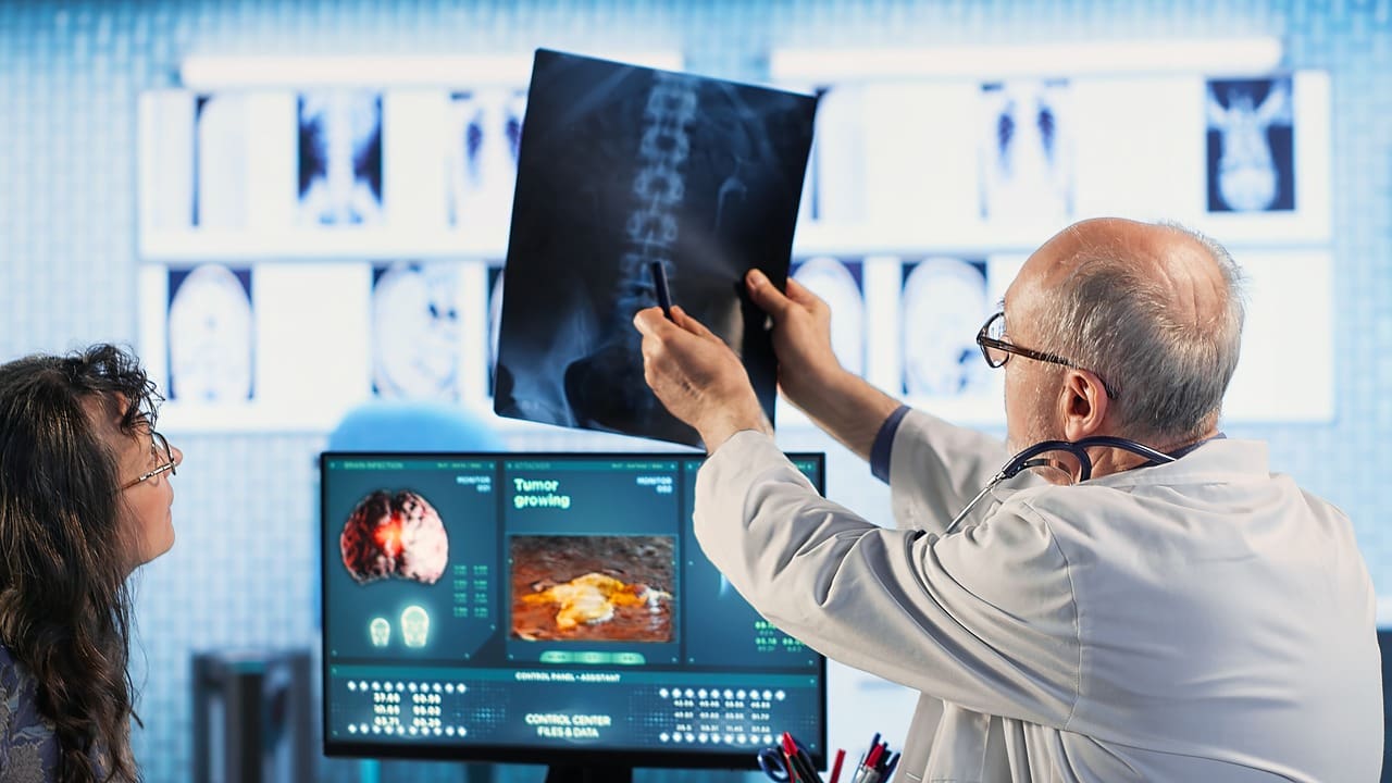
A nuclear medicine bone scan is a test that checks bone health and looks for problems. It is especially helpful for finding fractures, infections, tumors, or arthritis.
For a bone scan, a small amount of radioactive material is injected into a vein. It moves through the blood and collects in active parts of the bones. After waiting a bit, a special camera takes pictures of the bones.
Patients can expect the following during a nuclear medicine bone scan:
Duration: The entire process, including the injection and imaging, usually takes a few hours. Patients may need to wait for the radiotracer to circulate before imaging begins.
Comfort: The procedure is generally painless, although some patients may experience slight discomfort during the injection.
Post Scan Instructions: After the scan, patients are encouraged to drink fluids to help flush the radiotracer from their system.
What to Expect During a Nuclear Medicine Procedure
If you are having a nuclear medicine procedure, here are the main steps you can expect:
- Arrival and Check In: Upon arrival at the facility, patients will check in and may need to complete paperwork regarding their medical history.
- Preparation: A nuclear medicine technologist will explain the procedure, answer any questions, and prepare the patient for the scan.
- Radiotracer Administration: The technologist will administer the radiotracer, usually through an injection. Patients may feel a slight pinch during this process.
- Waiting Period: After administration, patients typically wait for a specified time to allow the radiotracer to circulate and accumulate in the targeted area.
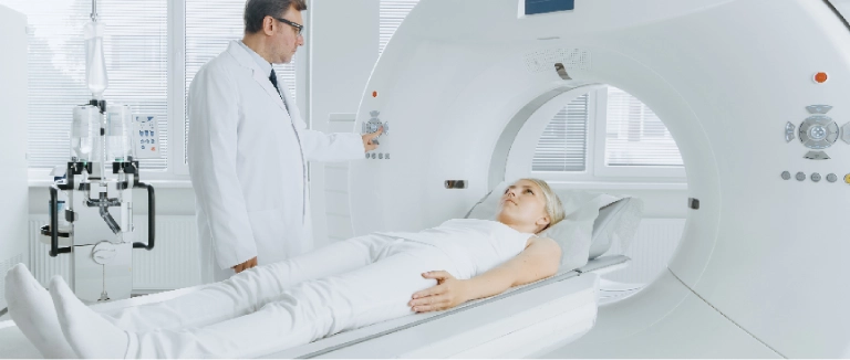
- Imaging: The patient will lie down on a table while the gamma camera captures images. This part of the procedure usually takes 20 to 60 minutes, depending on the test.
- Post Procedure Care: After the imaging is complete, patients can resume normal activities, although they may be advised to drink fluids to help eliminate the radiotracer.
Liv Hospital Nuclear Medicine
As in many other areas within Liv Hospital, a safe and effective service is provided in the Nuclear Medicine unit with competent personnel and state of the art devices.
Methods in the unit in this field:
They are sensitive and purpose specific methods. There are no allergies and side effects. They provide functional information at the physiological, metabolic level. Since the information is also numerical, it can be compared with each other when repeated. Due to their non invasive nature, they do not cause discomfort and harm to the patient. In Nuclear medicine applications, the patient receives very low levels of radiation.
Diagnostic Tests:
- PET/CT
- Gamma Camera
- C14 Device (Survey Meter)
- Gamma Probe
- Gamma counter Hot Lab (Department where radioactive materials are prepared) Effort test unit There are outpatient clinic units.
In the inpatient treatment unit;
- Treatment applications other than radioactive iodine
- Radioactive iodine applications (Two lead lined rooms)
- Radioactive material application and preparation unit are available.
For more information about our academic and training initiatives, visit Liv Hospital Academy.
Frequently Asked Questions for Nuclear Medicine
What should I do if I have concerns about radiation exposure?
If you have concerns about radiation exposure, discuss them with your healthcare provider. They can provide information about the safety measures in place and help you understand the risks and benefits of the procedure.
How long does the radiotracer stay in my body?
The radiotracer typically leaves the body within a few hours to a few days, depending on the specific substance used. Drinking fluids can help expedite the elimination process.
Are there any side effects of nuclear medicine?
Most patients experience minimal side effects from nuclear medicine procedures. However, some may have allergic reactions to the radiotracer. It's important to discuss any allergies or concerns with your healthcare provider before the procedure.
Should I stay away from people after the examination?
Since the amounts of radioactive substances used in the examinations are at very low levels, there is no need to stay away from people in general. However, it is recommended to stay away from children and pregnant women as a precautionary measure for 24 hours after the injection.
Are the radioactive materials used harmful?
In nuclear medicine applications, patients are exposed to very low levels of radiation. Applied radiopharmaceuticals are radioactive substances with low radiation and short life, and they do not accumulate in the body. They are excreted from the body by urine, sweat and intestines, as well as getting reduced by physical halving on their own. These substances can be safely administered to patients and children of all ages with doses determined according to their age and weight.
How often can nuclear medicine tests be performed?
In cases where the desired result cannot be achieved, when it is necessary to repeat the same examination, or in cases where several nuclear medicine examinations must be performed consecutively, scintigraphy can be performed at intervals of 2-3 days.
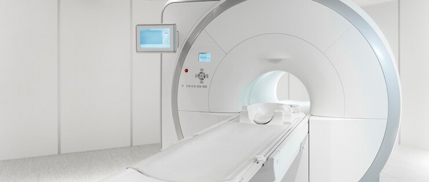
How is imaging performed in Nuclear Medicine?
Imaging is done with devices called Gamma Camera or PET-CT. The four sides of the devices are open and the imaging time varies according to the test.