
Lung cancer is a major cause of death worldwide. Early detection is key to effective treatment. A PET scan of the lungs is a vital tool for this purpose.
A pulmonary PET scan is a non-invasive test. It gives doctors detailed images of the lungs’ activity. This helps in diagnosing and managing lung conditions.
A lung PET scan shows areas of abnormal activity. It helps find lung cancer, infections, and inflammatory diseases. This allows for quick action.
PET scans are key in lung imaging. They use radioactive tracers to show how active the lungs are. This tech has changed how we diagnose and treat lung diseases.
A PET (Positron Emission Tomography) scan uses a special sugar molecule to find active areas in the body. For pulmonary imaging, it spots lung areas that are very active. This can mean lung problems, like cancer.
PET scans work by using a tracer that goes to active areas. This tracer sends out positrons. The PET scanner catches these positrons to make clear lung images.
For lung imaging, PET scans use Fluorodeoxyglucose (FDG) tracers. These tracers go to cells based on how active they are. In the lungs, cancer or infection areas take up more tracer, showing up on scans.
The tracer is injected into the blood. It goes to lung tissues and is picked up. The PET scanner then finds the positrons from the tracer. This makes a map of lung activity.
PET scans can spot metabolic activity because sick tissues work differently than healthy ones. In the lungs, this means they can find tumors, inflammation, or infections.
By measuring activity, PET scans help doctors diagnose and plan treatments. This info is key for better treatment plans and patient care.
PET scan technology has seen a lot of growth, mainly in lung imaging. The journey of PET scans in lung disease diagnosis and management has been marked by significant technological advancements and innovations.
The early days of PET technology for lung imaging were all about finding better diagnostic tools. The first PET scans used Fluorodeoxyglucose (FDG), a glucose molecule with a radioactive tracer, to see metabolic activity in lung tissues. This early technology was the start of what we have today.
As PET technology got better, so did its use in pulmonology. Researchers started looking at different radioactive tracers to make PET scans better. This led to new tracers that could focus on specific parts of lung disease, making diagnosis more accurate.
There have been big steps forward in PET scan technology for lung imaging. One big leap was the creation of PET-CT hybrid imaging. This combines PET’s functional info with CT’s anatomical details. It has made lung disease diagnosis and staging more precise.
Another big step was better image resolution and reconstruction algorithms. Today’s PET scanners can show clearer images. This helps spot small lung nodules and accurately measure metabolic activity.
Today, PET scan technology keeps getting better, with new tracers and imaging techniques being researched. Advanced PET scanning methods now let doctors see lung function and disease activity in great detail. These advanced methods are key for diagnosing and managing complex lung conditions.
The use of artificial intelligence (AI) and machine learning in PET scan analysis is also growing. AI can help doctors read PET images better and faster. This could lead to quicker diagnosis and treatment of lung diseases.
As PET scan technology keeps improving, it will play a bigger role in lung disease diagnosis and management. The ongoing development of PET scanning methods promises to improve patient care and outcomes in pulmonology.
PET scans of the lungs are key in diagnosing and managing lung diseases. They show detailed images of lung function and metabolism. This helps doctors identify and stage lung diseases more accurately.
Lung cancer is a common use of PET scans. PET scans for lung cancer help find tumors, check lymph nodes, and spot metastases. This info is vital for lung cancer staging and treatment planning.
Tristan Rogers is an example of how PET scans help. PET scans can spot metabolic changes in cancer cells early. This is better than traditional imaging.
Pulmonary nodules are common on chest scans. PET scans for pulmonary nodules help tell if they’re benign or malignant by looking at their metabolic activity.
Nodules with high metabolic activity are likely malignant. Those with low activity are usually benign. This helps decide if a biopsy is needed or if more imaging is required.
PET scans are also useful for inflammatory and infectious lung diseases. They help diagnose conditions like sarcoidosis, tuberculosis, and pneumonia.
PET scans can show how active inflammatory processes are. This helps doctors track disease activity and treatment response. It’s key for managing chronic conditions and adjusting treatments.
PET scans give a detailed look at lung metabolism and inflammation. They help doctors diagnose and manage lung conditions more accurately.
PET scans are key in lung health checks. But how do they stack up against CT and MRI? Knowing the differences helps doctors make better diagnoses and plans.
CT scans and PET scans have different roles in lung imaging. CT scans show the body’s structure, while PET scans reveal metabolic activity. A PET-CT lung scan combines these, giving a full picture of structure and function.
CT scans spot structural issues like tumors or inflammation. Yet, they can’t always tell if something is cancerous. That’s where lung imaging with PET scan shines. It highlights areas of high metabolic activity, pointing to cancer or other diseases.
MRI is another tool for lung imaging, showing soft tissues well without radiation. But for lung imaging with PET scan, PET is better at spotting metabolic changes in lung diseases.
When comparing pet scan vs MRI, PET is top for lung lesion metabolic activity. MRI is better for soft tissue and some inflammatory conditions. The choice between PET and MRI depends on the clinical question.
PET-CT hybrid imaging combines PET and CT, changing lung diagnostics. It gives both anatomical and functional info at once, boosting accuracy and confidence.
For lung conditions, PET-CT lung scan has big benefits. It helps detect and stage lung cancer better, characterizes nodules, and assesses treatment response. This combo helps doctors make better care decisions.
In summary, lung imaging with PET scan has unique benefits for metabolic activity checks. Knowing the differences between PET, CT, and MRI is key for choosing the right imaging for each patient.
To get the most out of your pulmonary PET scan, it’s key to prepare well. Good preparation means the scan will give accurate and helpful images for diagnosis.
Before your PET scan, you’ll need to follow certain dietary rules. It’s usually advised to avoid eating or drinking anything except water for a while before the scan. Your healthcare provider will tell you all about what to avoid, which might include:
Also, tell your doctor about any medications you’re taking. Some might need to be changed or stopped before the scan.
On the day of your PET scan, wear comfortable, loose-fitting clothing without metal parts. You’ll need to remove any jewelry, glasses, or metal objects before the scan. It’s wise to:
To make sure your PET scan is safe and effective, share important medical info with your healthcare provider. This includes:
By sharing this info and following the prep guidelines, you can help make sure your PET scan is a success. It will give the needed diagnostic information.
Getting a PET scan for lung imaging is a detailed process. It helps doctors find and track lung diseases. This tool is key for spotting metabolic activity in the lungs, which can show diseases like cancer. The whole process is set up to get accurate and trustworthy results.
The first step is injecting a radioactive tracer, like Fluorodeoxyglucose (FDG), into a vein. This tracer goes to cells with high activity. The body absorbs it, and it spreads out over 30 minutes to an hour.
While waiting, patients are asked to rest quietly. This helps the tracer spread evenly. “
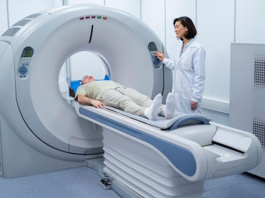
After the tracer spreads, the patient gets on a scanning table. The technologist makes sure they’re comfortable and in the right spot. The scan takes about 30 minutes, and the patient must stay very quiet.
The PET scanner uses the tracer’s gamma rays to make detailed lung images.
After the scan, patients can usually go back to their normal day right away. The tracer leaves the body in a few hours. Drinking lots of water helps get rid of it faster.
Some might feel a bit uncomfortable where the tracer was injected. But serious problems are very rare. “
After the scan, a radiologist checks the images. The results go to the doctor who ordered the scan. Patients are usually free to leave after the scan, unless told not to.
Knowing what to expect during your lung PET scan can make it less scary. A PET scan is a tool doctors use to check lung health and find problems.
You’ll lie on a table that slides into a big scanner for the PET scan. It’s usually comfortable and painless. You might feel a bit uncomfortable because you have to stay very quiet for a long time. But the scan itself doesn’t hurt.
The scanner might feel a bit tight, which can worry some people. But it’s not fully closed, and you can talk to the technologist through an intercom.
The whole PET scan usually takes 30 minutes to an hour. But, getting ready and waiting for the tracer to work can add more time.
You’ll talk to trained medical technologists during the scan. They’ll help you get comfortable and in the right spot.
Before starting, you can ask any questions. The technologists will also tell you how to stay quiet and follow any breathing instructions.
Overall, the PET scan aims to be as comfortable and stress-free as it can be. By knowing what to expect, you can get ready better.
Interpreting PET scan results is key in diagnosing and treating lung diseases. Patients often wonder what their results mean and how they’ll affect their care.
Radiologists examine PET scan images to see metabolic activity in the lungs. Bright spots on the scan may show tumors or inflammation. Their skill is vital in telling if an area is benign or could be cancerous.
Key factors in interpreting PET scans include:
SUV (Standardized Uptake Value) is a key metric in PET scan analysis. It shows how much radioactive tracer an area takes up compared to the body’s average. Higher SUV values often mean more aggressive disease, while lower values suggest less serious conditions.
Understanding SUV values is essential for:
Patients usually wait a few hours to days for their PET scan results. The wait time depends on the facility and the case’s complexity. The radiologist interprets the images, writes a report, and sends it to the doctor. The doctor then talks to the patient about the results.
Patients can prepare for their results discussion by:
PET scans are a game-changer in lung disease diagnosis. They show detailed images of how cells are working. This helps doctors spot and treat lung problems better than ever before.
PET scans are great at finding lung diseases early. They spot changes in lung cells that might not show up in symptoms yet. Catching diseases early can really help patients live longer and recover better.
Using PET scans for lung disease helps doctors find patients who need help fast. This leads to better care and treatment for lung problems.
PET scans are key in figuring out how far lung cancer has spread. They give doctors the details they need to plan the best treatment. This makes treatments more effective for patients.
What PET scans show helps doctors decide on treatments like surgery or chemo. This means patients get the right treatment for their needs.
PET scans also track how well treatments are working. They show changes in cell activity over time. This helps doctors adjust treatments for better results.
For lung cancer and other chronic lung diseases, PET scans are super useful. They help doctors quickly change treatments to keep patients healthy and happy.
PET scans are a valuable tool for diagnosis, but they come with risks. It’s important for patients and healthcare providers to know these risks. This ensures safe and effective use of PET scans.
PET scans use small amounts of radioactive tracers. These tracers emit radiation that helps create images of the body’s activity. To reduce this risk, the dose of tracer is kept as low as possible while maintaining image quality.
Strategies to minimize radiation exposure include:
Allergic reactions or complications can occur with PET scans. These reactions can be mild or severe. Patients are closely monitored during and after the scan to quickly address any issues.
Precautions to minimize the risk of allergic reactions include:
Healthcare facilities have strict safety protocols for PET scans. These include guidelines for handling radioactive tracers and monitoring patients. These steps help minimize risks.
By understanding risks and taking steps to reduce them, patients and healthcare providers can ensure PET scans are used safely. This is key for diagnosing and managing lung conditions.
Knowing the cost of a PET scan is key for those thinking about it for lung issues. The price can change a lot based on different things.
PET scans for lung imaging in the U.S. can cost between $1,000 and $5,000 or more. This depends on where you are and what the scan needs. Regional variations in price are big, with cities usually being pricier than rural areas.
But, prices can drop to $800 in some places or go over $6,000 in others.
Most health insurance in the U.S. covers PET scans for lung imaging if it’s needed. But, preauthorization is often needed. It’s important to check your insurance’s rules.
For those without good insurance, there are financial assistance programs out there. Some hospitals and imaging centers offer discounts or payment plans.
By knowing the costs and what help is out there, patients can make better choices for their lung health care.
PET scan technology has changed a lot for diagnosing and treating lung diseases. New innovations have made these scans more accurate and helpful. This has improved how we manage lung diseases.
New tracers have been key in improving PET scans for lung diseases. These tracers target specific cells or proteins. This makes imaging of lung conditions more precise.
Fluorodeoxyglucose (FDG) is a common tracer, but new ones are being made. They focus on lung cancer and other diseases. This helps doctors find and track lung diseases better.
PET scans now show more detail, thanks to new technology. This is important for accurate diagnoses and understanding lung diseases. Modern scanners use better materials and algorithms for clearer images.
Combining PET with CT and MRI scans has also helped. PET-CT hybrid imaging is very useful. It gives both functional and anatomical info in one scan.
Artificial Intelligence (AI) is now used in analyzing PET scans. AI helps doctors read images better and faster. It spots things that humans might miss.
AI helps find lung nodules and understand lung lesions better. This leads to better treatment plans. As AI gets better, it will play an even bigger role in PET scans for lung diseases.
Different patients getting PET scans for lung imaging face unique challenges. Healthcare providers must address these to ensure safe and effective scans.
For pet scan for pediatric patients, special steps are taken. The dose of the radioactive tracer is adjusted based on the child’s weight and age. Also, sedation or anesthesia might be needed to keep the child from moving during the scan.
Healthcare providers must look at the child’s medical history and any past reactions to contrast agents or sedatives. PET scans in kids are used only when the benefits outweigh the risks.
The use of pet scan during pregnancy is rare due to radiation risks. If a scan is needed, the dose is carefully chosen to protect the fetus. This ensures quality images while keeping radiation low.
For breastfeeding mothers, there’s a worry about the tracer in breast milk. Guidelines suggest stopping breastfeeding for 24 hours after the scan to protect the baby.
PET scans for elderly patients or those at high risk need careful planning. Elderly patients might have conditions that affect scan results or make them more sensitive to the tracer.
High-risk patients, like those with diabetes or kidney disease, might need special preparation. For example, diabetic patients might need to adjust their medication or blood sugar levels before the scan.
By customizing PET scan procedures for different patients, healthcare providers can improve diagnostic results while reducing risks.
Choosing the right place for a lung PET scan is important. It affects how accurate and reliable the results will be. Look beyond convenience to the quality of care and technology used.
Check if the facility is accredited by a known organization. Accreditation means the facility meets high standards for patient care and imaging quality. Look for the American College of Radiology (ACR) or Intersocietal Accreditation Commission (IAC) accreditation.
Quality matters too. This includes the facility’s experience, technology, and staff qualifications. A team with experience in pulmonary imaging can give you better results.
Talk to your healthcare provider before the scan. Ask important questions like:
Prepare for your consultation by making a list of questions and sharing your medical history. Discuss any worries or fears you have about the scan. After the scan, talk to your provider about the results and what comes next.
Choosing the right facility and being prepared for your PET scan ensures a smooth process. Remember, the quality of your care depends on your choices about where and how you get tested.
PET scan technology is set to improve how we diagnose and treat lung diseases. With ongoing advancements, we’ll see more precise lung images. This will help doctors make better choices for their patients.
New tracers and imaging methods are being researched. They could help find lung cancer sooner and track how treatments work. Artificial intelligence is also being explored to improve PET scan analysis.
As technology advances, patients will see better care and outcomes. The future of PET scans in lung medicine looks very promising. It’s set to greatly impact how we treat patients.
When picking a place for your PET scan, look at their standards and staff expertise. Your doctor can help you find a good one.
PET scans can be used for these groups, but special care is needed. Talk to your doctor about any concerns you have.
Yes, new tracers and better technology have improved PET scans. Artificial intelligence is also being used to make them even better for lung health.
PET scans might expose you to some radiation and could cause allergic reactions. But these risks are small, and safety steps are taken to keep them low.
Doctors look at the scan pictures to find any problems. They use a number called SUV to see how much dye is in certain areas.
During the scan, you’ll get a dye and lie on a table. The scan takes 30-60 minutes. You must stay very quiet and calm.
To get ready for a PET scan, you might need to eat less, skip some medicines, and wear loose clothes. Your doctor will tell you what to do.
PET scans can spot lung diseases early and track how well treatments work. They’re great for finding and checking on lung cancer and other lung issues.
A PET scan uses a dye that goes into the body and lights up where it’s needed most. This dye helps the scanner make clear pictures of the lungs.
A PET scan of the lungs is a test that uses a special dye to see how lung tissues work. It helps doctors find and treat lung problems.