Last Updated on November 26, 2025 by Bilal Hasdemir

At Liv Hospital, we know how vital diagnostic imaging is in heart care. Angiography is key for seeing blood vessels and finding heart disease.
Our video guide shows you the angiogram process. It highlights the skill and care our doctors use in this minimally invasive method.
We aim to offer world-class healthcare. We focus on patient care and support services.
Key Takeaways
- Understanding the importance of angiography in cardiovascular diagnosis
- Step-by-step guide to the angiogram process
- Expertise and care in performing angiograms
- Minimally invasive nature of the procedure
- Comprehensive support services for international patients
Understanding Angiography: Purpose and Clinical Applications

Angiography is key in modern vascular medicine. It gives us deep insights into blood vessels. We use it to diagnose and treat many vascular conditions. It’s a vital tool in our work.
Definition and Diagnostic Value
Angiography lets us see inside blood vessels. We use contrast media and X-rays to get clear images. This helps us spot problems like atherosclerosis, peripheral arterial disease, and brain aneurysms.
Its value comes from giving us detailed images. These images help us make better treatment plans. Whether it’s checking blockages or planning angioplasty, angiography is key.
Types of Angiography Procedures
There are many types of angiography, each for different needs:
- Coronary Angiography: Looks at the heart’s arteries to find heart problems.
- Cerebral Angiography: Checks the brain’s blood vessels for issues like aneurysms.
- Peripheral Angiography: Examines blood vessels outside the heart and brain, often for peripheral arterial disease.
Each type has its own way of helping diagnose and treat vascular issues.
Statistical Overview: Global Utilization and Outcomes
Angiography is used all over the world. Its use shows how important it is in medicine. Millions of procedures are done every year, helping many patients.
Research shows angiography helps in diagnosis and treatment planning. This leads to better care for patients. As technology improves, we’ll see more use of angiography in vascular medicine.
Essential Equipment and Team Requirements

To ensure a safe and effective angiography operation, it’s vital to have the right equipment and team. The angiography procedure is complex. It needs specialized imaging technology and a skilled medical team.
Angiography Suite Setup
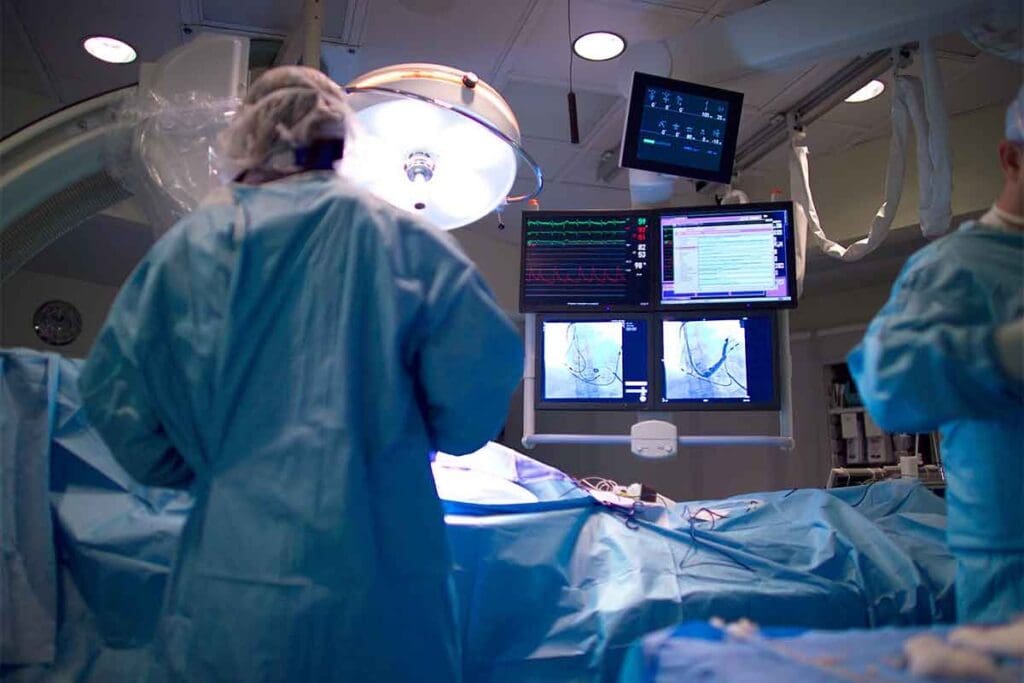
The angiography suite is a specially designed room. It houses the imaging equipment needed for the procedure. This includes a high-resolution X-ray system for detailed images of blood vessels.
The suite also has monitors for real-time imaging. There are patient monitoring systems to track vital signs during the procedure.
Required Medical Personnel and Qualifications
Performing an angiography operation needs a multidisciplinary team of healthcare professionals. This team includes cardiologists or radiologists who specialize in angiography. They also have radiographers or technologists who operate the imaging equipment.
Nurses assist with patient care before, during, and after the procedure. Each team member must have the right qualifications and experience. This ensures the procedure is conducted safely and effectively.
Critical Imaging and Monitoring Equipment
The imaging equipment used in angiography is key to the procedure’s success. This includes the X-ray system and contrast media injectors. These injectors administer the contrast material needed to visualize blood vessels.
Patient monitoring equipment tracks heart rate, blood pressure, and other vital signs in real-time. This ensures the patient’s safety throughout the procedure.
Pre-Procedure Patient Preparation
We prepare patients for angiography with a detailed process. This includes evaluation, testing, and education. It ensures a smooth procedure for everyone.
Patient Assessment and Contraindications
We start by assessing each patient thoroughly. We look at their medical history, current meds, and allergies, like to contrast media. Patients with kidney disease or diabetes need extra care because of contrast risks.
We also check the patient’s vascular access sites. This helps us plan the procedure and avoid complications.
Laboratory Tests and Imaging Prerequisites
Before an angiography, several lab tests are needed. These include:
- Blood urea nitrogen (BUN) and creatinine levels to check kidney function
- Complete blood count (CBC) to check blood cell counts
- Coagulation studies to assess bleeding risks
- Electrocardiogram (ECG) for those with heart disease
We might also ask for imaging tests like a chest X-ray or previous scans before the angiography.
Informed Consent and Patient Education
Getting informed consent is key. We make sure patients know the risks, benefits, and options. We explain contrast media, possible complications, and what to expect.
Teaching patients is also important. We tell them to fast before the procedure and about their meds. Clear instructions and addressing concerns help reduce anxiety and ensure compliance.
By following this detailed preparation, we make sure our patients are ready for a successful angiography.
Anesthesia and Sedation Protocols
Angiography procedures need careful thought about anesthesia and sedation for safety and comfort. The choice between local and general anesthesia depends on the procedure type, patient health, and procedure complexity.
Local vs. General Anesthesia Considerations
Local anesthesia is often used in angiography. It numbs the area where the catheter goes in, usually with lidocaine. This lets patients stay awake and alert, lowering risks from general anesthesia.
General anesthesia is for more complex cases or when patients can’t cooperate. It needs close watch by an anesthesiologist.
“The choice of anesthesia should be tailored to the individual patient’s needs and the specific requirements of the angiography procedure.” – Expert in Interventional Radiology
Sedation Management During Angiography
Sedation is used with local anesthesia to help patients relax. Sedation levels can vary from mild to deep, where patients almost fall asleep.
Monitoring sedation levels is key to avoid too much sedation. This can cause breathing problems and other issues. We use sedation scales to check levels and adjust as needed.
Monitoring Patient Comfort and Vital Signs
We watch patients’ comfort and vital signs closely during angiography. We track heart rate, blood pressure, oxygen levels, and breathing rate. This lets us quickly address any issues.
By managing anesthesia and sedation well, we ensure angiography is safe and comfortable for patients. Good patient comfort management is essential for a successful procedure.
The Angiography Operation: A Detailed Guide
We’ll walk you through the angiography operation, a detailed process for diagnosing vascular conditions. It involves several key steps, from initial access to contrast media administration.
Initial Access: Femoral vs. Radial Approach
The first step is accessing the vascular system. We usually use the femoral or radial artery for this. The choice depends on the patient’s anatomy and the procedure’s needs.
| Access Site | Advantages | Disadvantages |
| Femoral Artery | Larger access site, easier to maneuver catheters | Higher risk of bleeding complications, requires longer bed rest |
| Radial Artery | Lower risk of bleeding, allows for earlier mobilization | Smaller access site, potentially more challenging for catheter navigation |
Catheter Selection and Insertion Technique
After gaining access, we choose the right catheter for the patient’s anatomy and target vessels. We then insert it with a precise technique to avoid complications.
Catheter Selection Criteria:
- Size and flexibility
- Shape and tip design
- Material and coating
Navigation to Target Vessels
We guide the catheter to the target vessels under real-time X-ray. This step needs precision and knowledge of vascular anatomy.
Contrast Media Administration and Timing
The last step is giving contrast media to see the vascular structures. The timing of contrast administration is key for clear images.
Contrast Media Administration Guidelines:
- Determine the appropriate volume and rate of contrast injection
- Monitor the patient’s response to contrast media
- Adjust the timing of image acquisition based on contrast flow
Real-Time Imaging and Interpretation Techniques
Real-time imaging is key for diagnosing and treating vascular conditions through angiography. We use advanced imaging to guide catheters through blood vessels. This ensures precision and safety during the procedure.
Fluoroscopic Guidance Methodology
Fluoroscopic guidance is essential in angiography. It lets us see the catheter’s movement in real-time. A fluoroscope, which emits a continuous X-ray beam, creates images on a monitor.
We use this technology to navigate through blood vessels. We make adjustments as needed to reach the target area.
Key aspects of fluoroscopic guidance include:
- Continuous X-ray imaging for real-time feedback
- Contrast media injection to enhance vascular visibility
- Precise catheter navigation through complex vascular anatomy
Image Acquisition Protocols and Optimization
Optimizing image acquisition protocols is vital for high-quality angiographic images. We adjust parameters like frame rate, X-ray dose, and contrast media timing. This ensures we get detailed images for accurate diagnosis.
The optimization process involves:
- Adjusting the frame rate to match the flow of contrast media
- Modulating the X-ray dose to balance image quality and patient safety
- Timing contrast media injections to coincide with image acquisition
Reading and Interpreting Angiographic Images
Interpreting angiographic images requires knowledge of vascular anatomy and pathology. We analyze images for signs of stenosis, aneurysms, or other vascular abnormalities. Accurate interpretation is critical for diagnosing conditions and planning treatments.
Key elements in interpreting angiographic images include:
- Identifying vascular stenosis or occlusions
- Detecting aneurysms or malformations
- Assessing blood flow and vascular resistance
By combining advanced real-time imaging techniques with expert interpretation, we can provide accurate diagnoses and effective treatment plans for patients undergoing angiography.
Specialized Angiography Operation Techniques
We use advanced angiography methods to see and check different blood vessel areas. These special techniques help us find and treat many blood vessel problems. We will look at coronary, cerebral, and peripheral vascular angiography in this section.
Coronary Angiography Specifics
Coronary angiography is key for checking heart artery disease. This process involves putting a catheter into the heart arteries to add contrast media. This lets us see the heart’s blood flow clearly. It needs careful navigation to see the heart arteries well.
- Choosing the right catheter is important, with different shapes and sizes for different heart anatomies.
- How and when we give contrast media is critical to catch the heart’s blood flow.
- We use special image settings for clear pictures of the heart arteries.
Cerebral Angiography Considerations
Cerebral angiography helps find and treat brain blood vessel problems. This method needs careful thought about the brain’s blood vessels and risks. Choosing the right path for the catheter is key for accurate diagnosis and treatment plans.
- We check the patient’s brain blood vessel map before starting.
- Getting the catheter through the brain’s blood vessels needs great precision.
- After the procedure, we watch for any brain problems.
Peripheral Vascular Angiography Approaches
Peripheral vascular angiography is used for diseases in the blood vessels outside the heart. The method changes based on the area being looked at. For example, angiography of the lower legs needs access to see the blood vessels there.
- Picking the right spot for access is key for successful peripheral vascular angiography.
- We adjust how much and how fast we give contrast media based on the area.
- We tailor image taking to show the specific blood vessel anatomy.
Understanding these special angiography methods helps us see how complex and detailed vascular diagnosis and treatment are. Each method needs a custom approach, blending technical skill with medical knowledge.
Troubleshooting Common Challenges During Procedures
Angiography procedures can face several challenges that need quick action. We will talk about common issues and how to solve them.
Vascular Access Difficulties
Vascular access problems are common in angiography. These can be due to many reasons like anatomical issues, vascular disease, or past treatments. We use special techniques like ultrasound-guided access to solve these problems.
Managing Contrast Reactions
Contrast reactions are another issue in angiography. It’s important to spot these reactions early. We have a plan for managing them, including emergency meds and watching patient vital signs.
- Recognize the signs of a contrast reaction
- Administer emergency medications as needed
- Monitor patient vital signs closely
Addressing Technical Equipment Issues
Technical problems can happen during angiography too. Keeping equipment in good shape is key to avoid failures. We have steps for fixing common tech issues, so procedures can go smoothly.
Being ready for these challenges helps us succeed in angiography. It ensures we give our patients the best care.
Post-Procedure Care and Patient Monitoring
After angiography, taking good care of patients is key for their safety and best results. We move from the procedure to watching over patients closely. Our goal is to avoid problems and help them recover well.
Immediate Post-Angiography Observation
Right after angiography, patients stay in a recovery area for a while. This watchful period is very important. We check their vital signs like blood pressure and heart rate to make sure they’re okay.
We also keep an eye on the spot where the procedure was done for any bleeding. Patients stay there until they’re fully awake and recovered from any medicine they got.
Vascular Access Site Management
It’s very important to take care of the spot where the procedure was done. We use pressure or special devices to stop bleeding. This depends on the procedure and the patient.
We check the site often for any problems. We also tell patients how to care for it at home. We teach them what to watch for and when to get help.
Discharge Criteria and Patient Instructions
Before leaving, we make sure patients are doing well. They need to have stable vital signs and no complications. We also make sure they can manage their pain.
We give them clear instructions on how to care for themselves. This includes wound care and taking their medicine. We also tell them what to watch for and when to get help.
| Discharge Criteria | Patient Instructions |
| Stable vital signs | Wound care and dressing changes |
| Absence of complications | Medication management |
| Adequate pain management | Follow-up appointments |
| Ability to ambulate | Recognizing signs of complications |
By following these steps and giving good care, we lower the chance of problems. This helps patients do well after angiography.
Conclusion: Best Practices in Modern Angiography
Angiography needs precision, skill, and a deep understanding of modern techniques. By using advanced imaging and refined protocols, we boost the value and safety of these procedures.
Staying current with video angiography advancements is key. This means using high-quality images, preparing patients well, and having skilled staff. We must also focus on patient comfort and safety, managing sedation and watching vital signs closely.
By following these best practices and improving our methods, we can better help patients. As the field grows, it’s vital to keep delivering top-notch care. This includes supporting patients from around the world.
FAQ
What is an angiogram, and how is it performed?
An angiogram is a way to see inside blood vessels. It helps find problems with blood flow. We do it by putting a thin tube into a blood vessel. Then, we use special images to guide it to the right spot.
How is angiography done, and what are the different types?
Angiography uses special dye to see blood vessels. There are many types, like for the heart, brain, and legs. Each one is used for different reasons and in different ways.
What is the difference between local and general anesthesia during angiography?
Local anesthesia just numbs the area where the tube goes in. General anesthesia makes you sleep. We pick one based on how simple or complex the procedure is.
How do you manage patient comfort and vital signs during angiography?
We keep a close eye on how you’re feeling and your vital signs. We might give you something to relax if needed.
What are the common challenges during angiography procedures, and how are they addressed?
Sometimes, it’s hard to get into the blood vessel, or you might have a reaction to the dye. We might try a different spot or give you medicine. We also check the equipment.
How is post-procedure care managed after an angiography operation?
After it’s done, we watch you for a bit. We take care of the spot where the tube was and tell you what to do at home before you go.
What are the benefits of using real-time imaging during angiography?
Seeing everything in real-time helps us place the tube more accurately. It makes the procedure safer and more precise.
How is contrast media administered during angiography?
We use the tube to give you the dye. We control how much and when to get the best pictures and avoid problems.
What are the discharge criteria after an angiography procedure?
You can go home when your vital signs are stable, the access site is okay, and you can follow the instructions.
Can I watch a video of an angiogram procedure?
Yes, we have videos to help you understand what happens during an angiogram.
How is patient education handled before an angiography procedure?
We teach you about the procedure, its risks, and benefits. We make sure you understand and agree before we start.
What is the role of fluoroscopic guidance in angiography?
It lets us see the tube and blood vessels as we move it. This helps us place it correctly and navigate.
Are there different techniques for coronary, cerebral, and peripheral vascular angiography?
Yes, each type has its own way of doing things. We adjust our approach based on what’s best for you.
Reference
- Omeh, D. J. (2023). Angiography. In StatPearls. StatPearls Publishing. Retrieved October 20, 2025, from https://www.ncbi.nlm.nih.gov/books/NBK557477/


