Last Updated on November 27, 2025 by Bilal Hasdemir
Diagnostic imaging is key in today’s medicine, and PET scans and CT scans are two of the most valuable tools. They help detect and understand many health issues, but they work in different ways. A common question patients ask is, “Does scar tissue show up on a PET scan? In most cases, PET scans highlight areas of unusual activity, so active cancer cells will appear clearly, while scar tissue”being inactive”usually does not light up the same way. However, sometimes inflammation in scar tissue can mimic activity, which is why often use PET scans along with CT or MRI for a more accurate diagnosis.
About half of surgeries lead to scar tissue. This makes it important to find and track scar tissue. The big question is: Can these scans really spot scar tissue? Knowing the difference between PET scan and CT scan is vital for both patients and .
Key Takeaways
- PET scans and CT scans serve different diagnostic purposes.
- The ability of these scans to detect scar tissue varies.
- Understanding the difference between PET and CT scans is essential for accurate diagnosis.
- PET-CT scans combine the advantages of both technologies.
- The choice between PET and CT scans depends on the specific medical condition.
The Science of Medical Imaging

Medical imaging has rapidly evolved, transforming the way we diagnose diseases. It lets see inside the body in great detail. This is key in today’s medicine.
Evolution of Diagnostic Imaging Technologies
Diagnostic imaging has changed a lot over time. It started with X-rays and now includes advanced tools like PET and CT scans. ‘s article says these new tools have made diagnosing diseases better.
CT scans came out in the 1970s, letting see the body in slices. Later, PET scans showed how active the body’s cells were. Together, these tools help plan treatments better.
The Role of Imaging in Modern Medicine
Medical imaging is vital today. It helps find, understand, and track diseases like cancer. PET and CT scans are key because they show both how the body works and its structure.
PET scans are key in cancer care, showing how active tumors are. CT scans give detailed pictures of the body’s shape. This helps see structural changes.
Using PET and CT together makes diagnosing even better. This mix gives a full picture of diseases. It’s a big help in offices.
What Is a PET Scan?
PET scans are a key part of medical imaging. They help see how the body works by looking at metabolic processes. A PET scan, or Positron Emission Tomography, is a tool that shows what’s happening inside the body.
The Science Behind Positron Emission Tomography
Positron Emission Tomography works by finding positrons from radioactive tracers. These tracers go into the body and light up areas that are very active. The PET scanner picks up these positrons and makes detailed pictures of what’s happening inside.
The most used tracer is Fluorodeoxyglucose (FDG). It’s a sugar molecule with a radioactive tag. Cancer cells use more sugar than normal cells, so FDG lights up cancer during a PET scan.
How PET Scans Work
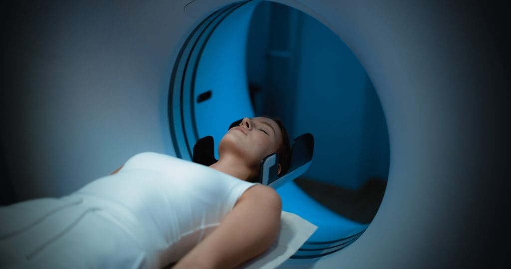
Getting a PET scan involves a few steps. First, the patient gets the radioactive tracer. Then, they lie on a table that slides into the PET scanner. The scanner picks up signals from the tracer, and a computer turns these into images.
PET scans can be used alone or with CT scans. This combo, called PET/CT, gives both metabolic and anatomical info. It helps see more clearly by combining metabolic data with detailed body images.
Common Uses of PET Scans in Practice
PET scans are often used to check for cancer. They help find cancer, see how far it has spread, and check if treatments are working. They’re also used in brain and heart studies.
PET scans are very useful in medicine. They show how the body’s metabolic processes work. This makes them a key tool in today’s healthcare.
What Is a CT Scan?
A CT scan, or Computed Tomography, is a way to see inside the body. It uses X-rays and computers to make detailed images. These images show organs, bones, and blood vessels clearly.
Understanding Computed Tomography Technology
CT scans use a rotating X-ray tube and a detector. They measure how much X-rays are absorbed by the body. Then, a computer turns this data into detailed images.
Technology has improved a lot. Now, scans are faster and the images are clearer.
The main parts of a CT scanner are:
- The gantry, which houses the X-ray tube and detector
- A moving table that allows the patient to be slid into the gantry
- A computer system to reconstruct the images
How CT Scans Create Detailed Images
During a CT scan, X-rays pass through the body. The detector catches the X-rays that aren’t absorbed. This info goes to the computer, which makes images.
The result is detailed cross-sectional images. These can be viewed alone or turned into 3D models.
Key benefits of CT scan imaging include:
- High-resolution images of internal structures
- Ability to detect a wide range of conditions, from injuries to cancers
- Quick scan times, often just a few minutes
Primary Applications of CT Scans
CT scans are used in many ways in medicine. They help diagnose and stage cancer. They also check for internal injuries and guide procedures like biopsies.
They are great in emergencies because they are fast and show important details.
PET Scan vs CT Scan: Key Differences
PET scans and CT scans are both important for finding out what’s wrong inside the body. They work in different ways and give us different kinds of information. Knowing how they differ helps make better plans for treatment.
Fundamental Technological Differences
The main difference is in how they work. PET scans use positron emission tomography. This means they inject a special tracer that shows up in areas that are very active. CT scans, on the other hand, use X-rays to show detailed pictures of what’s inside based on how dense it is.
What Each Scan Actually Measures
PET scans look at how active the body’s cells are. This makes them great for finding cancer because cancer cells are very active. CT scans, on the other hand, look at the density of tissues. They give clear pictures of the body’s structure.
Diagnostic Capabilities Comparison
PET scans are super good at finding active tissues, which is why they’re great for tracking cancer. They help see how well treatments are working and if cancer is coming back. CT scans, with their detailed pictures, are better for finding structural problems like injuries or infections.
In short, both PET and CT scans are vital for diagnosing, but they’re used for different things. Knowing their differences helps use them to their fullest advantage in caring for patients.
How PET Scans Detect Tissue Abnormalities
PET scans look at how cells work in the body. They help find problems, like cancer. This is key for diagnosing and treating many diseases.
Metabolic Activity Detection Mechanisms
PET scans find problems by looking at how cells use energy. They use a special sugar that cells with high energy use more. This makes cancer cells show up on scans.
The steps are simple:
- The patient gets a small dose of a special sugar, Fluorodeoxyglucose (FDG).
- This sugar goes to cells that use a lot of energy, like cancer cells.
- A PET scanner finds this sugar and shows where it is, highlighting active areas.
FDG Uptake Patterns in Different Tissues
How much FDG a tissue uses tells us a lot. For example:
- Cancerous tissues use a lot of FDG because they are very active.
- Inflammatory tissues also use more FDG because they are busy.
- Normal tissues use less FDG, but some like the brain and heart use more.
Knowing these patterns helps understand PET scan results better.
Limitations in Tissue Characterization
Even though PET scans are great, they have some downsides. For example:
- They can’t always tell if a problem is cancer or something else that’s active.
- They might miss small problems because they’re not that detailed.
say PET scans work best when used with other tests like CT or MRI.
“The combination of functional information from PET with anatomical information from CT or MRI enhances diagnostic accuracy.”
Knowing what PET scans can and can’t do helps make better choices for patients.
Does Scar Tissue Show Up on a PET Scan?
Scar tissue’s visibility on PET scans depends on its metabolic activity. Scar tissue forms as the body heals. It can have different characteristics and metabolic levels.
Biological Characteristics of Scar Tissue
Scar tissue is made of fibrous tissue that replaces damaged tissue. Its visibility on PET scans depends on several factors:
- Collagen content: Scar tissue has a lot of collagen, which can change its metabolic activity.
- Vascularity: Whether or not it has blood vessels.
- Inflammatory cells: The presence of these cells can raise metabolic activity.
Metabolic Activity in Scar Tissue
Scar tissue usually has lower metabolic activity than cancerous tissues. But, some conditions can increase this activity:
- Infection or inflammation in the scar.
- Recent surgery or injury.
This increased activity can sometimes make scar tissue visible on PET scans.
PET Scan Results for Different Types of Scars
The visibility of scar tissue on PET scans varies by type:
- Post-surgical scars: These may show more uptake because of recent damage.
- Chronic scars: Usually have low metabolic activity and might not show up.
- Inflamed scars: Can have higher uptake because of inflammation.
Challenges in Interpreting PET Results for Scarring
Interpreting PET scans for scar tissue is tricky because of:
- Variable metabolic activity.
- Similarities with other conditions.
- The need for a full understanding of the patient’s history and situation.
Getting it right requires knowing the patient’s medical history and the context of their condition.
How CT Scans Visualize Scar Tissue
CT scans show scar tissue by looking at the changes after an injury. Scar tissue forms as the body heals. Its look on a CT scan can change a lot.
Density and Structural Changes in Scar Tissue
Scar tissue has different density and structure than normal tissue. It can have more collagen deposition, tissue contraction, and calcifications. CT scans can spot these differences because they show different tissue densities.
The healing process changes scar tissue’s density. This is because of collagen and other proteins. These changes make scar tissue show up on CT scans.
CT Imaging Characteristics of Scarring
Scarring on CT scans looks different based on the scar’s location, age, and type. Scar tissue often shows up as distortion or architectural distortion. The attenuation of scar tissue can also tell us a lot. Some scars are less dense, while others are more dense than the tissue around them.
Limitations of CT in Scar Tissue Evaluation
CT scans are good for seeing scar tissue, but they have limits. It’s hard to tell scar tissue apart from tumors because they can look similar. This makes it tough to diagnose.
Also, CT scans might not catch all scar tissue, like when it’s not very different from the tissue around it.
| Characteristics | Scar Tissue | Normal Tissue |
| Density | Variable, often increased | Consistent with tissue type |
| Structural Changes | Distortion, contraction | Normal architecture |
| CT Attenuation | Hypo or hyperattenuation | Typical for tissue type |
Combined PET-CT Scans: The Best of Both Worlds
PET and CT scans together have made diagnosing better. This mix, called PET-CT, uses the best of both to understand tissue problems better.
Why Combine These Technologies?
PET and CT scans together have big benefits. PET scans show how tissues work, while CT scans show their shape. Together, they give a full picture of tissues.
Using both PET and CT scans fixes their individual problems. For example, PET scans might not always show where things are, but CT scans can help with that.
Benefits of Hybrid Imaging for Scar Tissue Assessment
Hybrid PET-CT imaging is great for looking at scar tissue. It lets see how scar tissue works and what it looks like. This is key to telling scar tissue apart from other problems, like cancer.
Key benefits of PET-CT for scar tissue assessment include:
- Improved diagnostic accuracy
- Enhanced characterization of tissue abnormalities
- Better differentiation between scar tissue and other lesions
Applications of PET-CT
PET-CT scans are used in many ways, not just for scar tissue. They help in finding, staging, and checking how well cancer treatments work. They are also used in heart and brain health for diagnosis.
PET-CT scans are very useful in medicine today. They help make better treatment plans and improve patient care.
Cancer Detection: PET Scan vs CT Scan
PET and CT scans are key in finding cancer. They use different ways to spot tumors. Each tool gives unique views of the body’s inner workings and how it uses energy.
How Each Technology Detects Malignancies
PET scans find cancer by looking at where energy is used a lot. They use a sugar molecule that cancer cells love more than healthy ones. This makes PET scans great for spotting cancer cells all over the body.
CT scans, on the other hand, use X-rays to show detailed pictures of inside the body. They find cancer by seeing changes in body shape, like tumors. They’re good at measuring how big and where tumors are.
The Critical Challenge: Differentiating Cancer from Scar Tissue
It’s hard to tell cancer from scar tissue, which is a big challenge. Scar tissue can look like cancer on scans, leading to wrong results.
PET scans help by looking at how active tissues are. Scar tissue usually doesn’t use energy as much as cancer. But, it’s not always easy to tell them apart.
“The ability to differentiate between scar tissue and active cancer is critical for accurate diagnosis and treatment planning.”
- Oncologist
Comparative Accuracy for Different Cancer Types
The accuracy of PET and CT scans changes with the type of cancer. For example, PET scans work well for lymphomas and some lung cancers because they use a lot of energy.
- PET scans are great for cancers that use a lot of energy.
- CT scans show detailed body pictures, helping to see tumor size and location.
- Using both PET and CT scans together gives a better view than either one alone.
In summary, both PET and CT scans are important for finding cancer. Each has its own strengths and weaknesses. Knowing how well they work for different cancers helps make better plans for treatment.
Preparing for Your Scan: What to Expect
Knowing what to do before a PET or CT scan is key for a good experience. These scans help see inside your body. But, they need different preparations and follow different steps.
Before Your PET Scan
For a PET scan, you’ll likely need to fast for a few hours. This means no food or drinks except water. Wear comfy, loose clothes and avoid metal items like jewelry.
Medication and Special Instructions: Tell your about any meds you’re on. Some might need to stop before the scan. If you’re diabetic, you’ll get special instructions for your insulin and meals.
Before Your CT Scan
CT scan prep changes based on the body part scanned. For the abdomen or pelvis, you might drink a contrast material. You’ll also need to skip eating or drinking for a few hours beforehand.
Contrast Material: Drinking a contrast material might make you feel warm or taste metallic. This is normal and goes away quickly. Let your know if you have any allergies or concerns.
During and After the Procedure
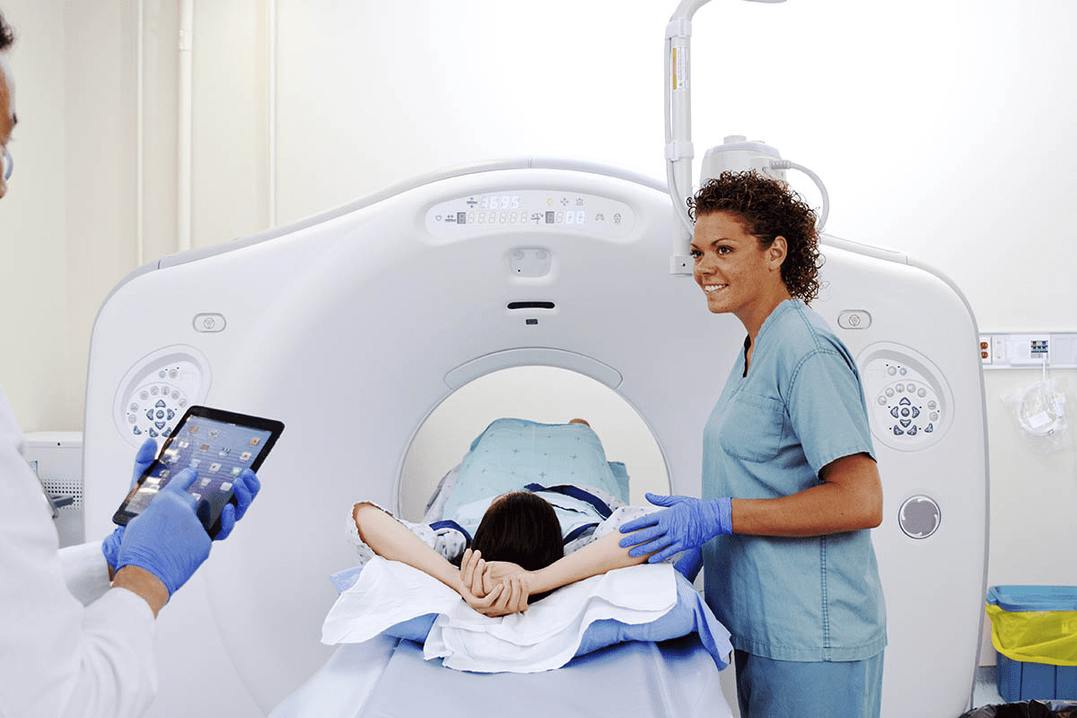
During both scans, you’ll lie on a table that moves into a big machine. The scans are painless and last from 15 to 60 minutes. This depends on the scan type and area being checked.
After the Scan: You can usually go back to normal activities right after. If you got a contrast material or a radioactive tracer, your body will get rid of it. Drinking water helps this process.
Always follow your healthcare provider’s or imaging center’s specific instructions. They might have extra steps based on your needs and the scan type.
How Long Does It Take to Get PET Scan Results?
Knowing when you’ll get your PET scan results is important. It helps manage what you expect. The process includes the scan and the specialist’s review of the results.
Typical Timeframes for Results Processing
The wait for PET scan results can change based on a few things. These include how busy the facility is and how complex the scan was. Usually, people wait from 24 hours to a few days.
Typical Timeframes:
| Facility Type | Average Wait Time |
| Hospital | 2-5 days |
| Private Imaging Center | 1-3 days |
| Specialized Diagnostic Center | 24 hours |
Factors That May Delay Results
Several things can make you wait longer for your PET scan results. These include:
- The need for more imaging or tests
- Scans that need extra analysis
- Busy schedules at imaging centers
- Consultations with other
Understanding Your Imaging Report
The PET scan report is detailed. It shows the scan’s findings and the thoughts. It’s key to talk about this report with your to understand what it means.
Key Components of a PET Scan Report:
- Patient information and scan details
- Description of findings
- Comparison with previous scans (if applicable)
- Interpretation and recommendations
By understanding your PET scan report, you can better understand your diagnosis and treatment plan.
When Choose PET Over CT (or Vice Versa)
PET and CT scans are used for different things in medical imaging. pick one over the other based on the patient’s condition, disease stage, and health. This choice is key for accurate diagnosis and treatment.
Decision-Making Factors
look at several factors when choosing between PET and CT scans. They consider the tissue’s metabolic activity, the need for detailed anatomy, and the patient’s medical history. PET scans are great for showing metabolic activity, which is why they’re often used for cancer diagnosis. CT scans, on the other hand, are better for seeing structural details.
In some cases, might choose a PET-CT scan. This combines both metabolic and anatomical information. It helps in making a more precise diagnosis and planning treatment.
Specific Conditions and Preferred Imaging Methods
Some conditions are better suited for PET or CT scans. For example, PET scans are often used for cancer detection and staging because they show high metabolic activity in tumors. CT scans are better for injuries, vascular diseases, and infections because they show detailed anatomy.
The choice between PET and CT scans can also depend on monitoring disease progression or treatment response. For instance, PET scans can measure how well a tumor responds to chemotherapy by looking at metabolic changes.
When Sequential or Combined Imaging Is Necessary
In many cases, using PET and CT scans together or one after the other is best. For example, a CT scan might first find a structural issue, then a PET scan might check the tissue’s metabolic activity. This approach helps in making a more accurate diagnosis and treatment plan.
Hybrid PET-CT scanners are also becoming more common. They allow for both metabolic and anatomical data to be collected at the same time. This is very useful in complex cases where detailed information about both function and structure is needed.
Future Developments in Medical Imaging
Medical imaging is getting better with new technologies. These advancements will make diagnostic procedures more precise and effective. Key improvements include better PET and CT scans, artificial intelligence in interpreting scans, and better detection of scar tissue.
Emerging Technologies in PET and CT
Medical imaging is seeing big changes in PET and CT technologies. Some new trends include:
- High-resolution imaging: New PET scanners can show more detailed images of the body’s metabolic activities.
- Advanced CT scanners: Next-generation CT scanners will have higher resolution images and use less radiation.
- Hybrid imaging techniques: Combining PET and CT scans is becoming common. It gives a more complete view of the body’s structures and functions.
Artificial Intelligence in Scan Interpretation
Artificial intelligence (AI) is being used more in medical imaging. It helps make scan interpretation more accurate and efficient. AI can:
- Detect abnormalities: AI finds patterns that humans might miss, improving accuracy.
- Quantifying disease extent: AI helps measure how much disease is present, which is key for treatment plans.
- Streamlining workflow: AI automates some image analysis, freeing up radiologists to focus on harder cases.
Improvements in Scar Tissue Detection and Characterization
Finding and understanding scar tissue is a big challenge in medical imaging. Future improvements will help with this. They include:
- Advanced imaging techniques: New methods and technologies will better distinguish scar tissue from other tissues.
- Enhanced image analysis software: Better algorithms in software will help accurately detect and understand scar tissue.
These advancements in medical imaging will greatly improve patient care. They will help make more accurate diagnoses and create better treatment plans.
Conclusion
Medical imaging is key in today’s healthcare. It helps find and treat many health issues. The choice between PET scans and CT scans depends on what information is needed.
PET scans are great at showing how active cells are. This makes them perfect for finding cancer and checking how treatments work. On the other hand, CT scans give detailed pictures of the body’s structure. They are good for seeing changes and finding scar tissue.
Knowing what each scan can do is important for making the right diagnosis and treatment plan. As imaging tech gets better, using both PET and CT scans will be more important than ever. This will help give the best care to patients.
In short, the debate between PET scans and CT scans shows how important it is to pick the right scan for each health issue. This ensures patients get the right diagnosis and treatment.
FAQ
How do I prepare for a PET or CT scan?
To prepare for a PET or CT scan, follow certain diet rules and avoid some medicines. Also, remove any metal items. The exact steps can vary based on the scan type and the place.
Can PET scans detect cancer more accurately than CT scans?
PET scans are great for finding cancer because they look at cell activity. But, they might not work for all cancers, and scar tissue can affect their accuracy. CT scans are useful for getting more information.
What factors influence the choice between a PET scan and a CT scan?
Choosing between a PET scan and a CT scan depends on the health issue. It also depends on whether you need to see how cells work or the body’s structure. Sometimes, both are needed.
How long does it take to get PET scan results?
Getting PET scan results can take a few hours to days. It depends on the place and how complex the case is.
How does a CT scan visualize scar tissue?
A CT scan can spot scar tissue by looking at density and structure. Scar tissue can be different from other tissues, making it stand out.
Does scar tissue show up on a PET scan?
Scar tissue might not show up on a PET scan. This is because scar tissue doesn’t have much activity. So, it’s less likely to be seen.
What is the main difference between a PET scan and a CT scan?
A PET scan looks at how cells work, while a CT scan shows the body’s structure. This means PET scans focus on activity, and CT scans on details.


