Last Updated on November 18, 2025 by Ugurkan Demir

At Liv Hospital, we know how important pictures are for teaching patients about complex surgeries like ACL reconstruction. Every year, over 100,000 ACL reconstructions happen in the US. We see the need for clear explanations of this common surgery.
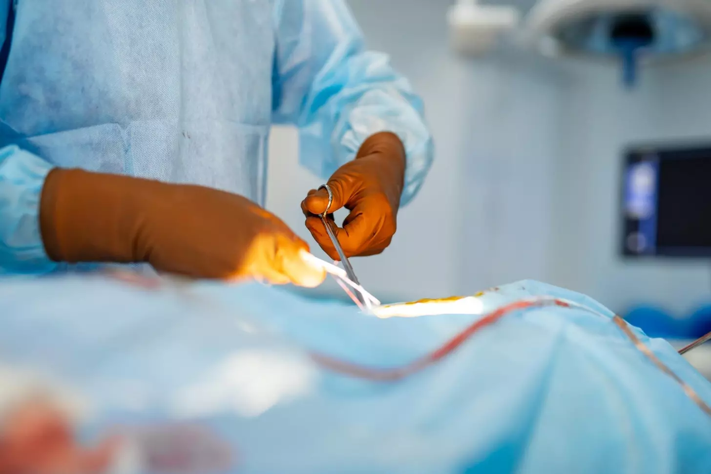
We think ACL surgery images and photos are key for patients to understand ACL reconstruction. Looking at these pictures helps patients know what to expect before, during, and after the surgery.
We’re dedicated to top-notch healthcare and supporting international patients. We want to give patients all the information they need to make good choices about their health.
ACL surgery pictures are key in teaching patients about their treatment. At our place, we see how important pictures are. They help patients understand their options and what surgery is like.
Visuals are essential for teaching patients. They show what surgery is and what it can do. ACL reconstruction surgery pictures make it easier for patients to understand.
Images of ACL surgery help patients get it. They show the surgery steps and what to expect. This helps calm fears and makes patients feel more ready for surgery.
Photography is key in medical records. It keeps a record of a patient’s health and treatment. For ACL surgery, photographic documentation helps track progress and educate patients. We use top-quality images for the best care and understanding.
Using acl surgery pictures in teaching patients improves their understanding and happiness. Our focus on visuals shows our commitment to caring for patients fully.
Diagnostic imaging is key for checking the ACL’s health. At Liv Hospital, we use top-notch imaging to accurately assess ACL injuries. This is vital for choosing the right treatment.
These images show how much damage the ACL has. They help doctors understand the injury and explain it to patients.
Normal ACL pictures help us see what a healthy ACL looks like. They are important for comparing with injured ACLs to see how much damage there is.
A normal ACL looks like a tight band on MRI scans. It shows the ligament is strong and attached well to the bones, meaning it’s healthy.
Images of torn ACLs are critical for figuring out how bad the injury is. They show where and how big the tear is. This helps doctors decide the best way to treat it.
| Characteristics | Normal ACL | Torn ACL |
|---|---|---|
| Appearance on MRI | Continuous, taut band | Discontinuous, with signs of disruption |
| Ligament Integrity | Intact | Compromised |
| Treatment Approach | Conservative management or preventive measures | Surgical intervention, such as ACL reconstruction |
Looking at torn ACL images helps doctors decide on the best treatment. At Liv Hospital, we use these images to make treatment plans that fit each patient’s needs.
At Liv Hospital, we use pre-operative ACL surgery photos to plan ACL reconstruction surgeries carefully. These images help us understand the knee’s condition and choose the best surgical approach.
Clinical examination documentation is key in pre-operative planning. High-quality images of the knee during examination help us see how severe the injury is. We can also spot any extra challenges.
Visualizing the surgery is a big part of pre-operative planning. By looking at detailed images of the knee, we can figure out the best surgical method. This includes where to place grafts and tunnels.
| Pre-Operative Planning Step | Description | Benefit |
|---|---|---|
| Clinical Examination Documentation | High-quality images of the knee during clinical examination | Assess extent of injury and identify complicating factors |
| Surgical Planning Visualizations | Detailed analysis of knee images to plan surgical technique | Optimize graft and tunnel placement, anticipate challenges |
Using pre-operative ACL surgery photos helps us give our patients the best care. These images are essential for our acl surgery planning. They help us get the best results and make our patients happy.
At Liv Hospital, we use arthroscopic ACL surgery for a less invasive option. This method uses small incisions and advanced tech to fix knee stability. Pictures of this surgery help us understand it better.
The setup of arthroscopic equipment is key for a successful ACL surgery. High-definition cameras and special tools help see and fix the damaged ACL. Photos show the care and precision in this step.
Portal placement is very precise. Images show how careful cuts are made for the tools. This is important for seeing and fixing the ACL well.
Intraoperative arthroscopic views give a close look at the ACL during surgery. These images are key for seeing the injury’s extent and the repair steps. For more on ACL reconstruction, see Premera’s medical policy on ACL reconstruction.
Looking at these ACL surgery photos helps patients understand the procedure and recovery. Arthroscopic techniques have made ACL surgery more precise and less invasive.
Graft selection is key in ACL reconstruction surgery. It greatly affects the surgery’s success. At Liv Hospital, we focus on picking the right graft for each patient.
We look at different graft options. These include hamstring autografts, patellar tendon grafts, and quadriceps tendon grafts. Each has its benefits and is chosen based on the patient’s needs.
Hamstring autografts are often chosen for ACL reconstruction. The process takes a part of the hamstring tendon. This method is liked for its low risk of complications and strong graft.
Patellar tendon grafts are also a favorite. The graft comes from the middle of the patellar tendon. It’s carefully prepared for use in the surgery.
Quadriceps tendon grafts are an option for those needing a bigger graft. Allografts, from donors, are used in some cases, like revision surgeries. Advanced imaging helps check if the graft is right for the patient.
In summary, choosing and preparing the graft is vital in ACL reconstruction. By using different grafts and modern surgery, we aim for the best results for our patients.
ACL reconstruction surgery is a detailed process that needs skill and precision. We will show you the key steps, from start to finish.
Notchplasty is a key part of ACL surgery. It involves removing bone or soft tissue to make room for the graft.
The surgeon carefully removes any extra bone or damaged tissue during notchplasty. This is important for the graft to work right.
Making tunnels in the femur and tibia is a big part of ACL surgery. These tunnels hold the graft, which replaces the damaged ACL.
The surgeon drills these tunnels with great care. They make sure the tunnels are right for the graft to work best.
| Step | Description |
|---|---|
| Femoral Tunnel Drilling | Creating a tunnel in the femur to accommodate one end of the graft. |
| Tibial Tunnel Drilling | Drilling a tunnel in the tibia to secure the other end of the graft. |
After the tunnels are ready, the graft goes through them and is fixed in place. This is key for knee stability and function.
There are different ways to fix the graft, like screws or special devices. The choice depends on the graft and the surgeon’s preference.
After fixing the graft, the surgeon checks everything to make sure it’s good. They look at knee stability, movement, and graft health.
This final check is very important. It makes sure the ACL surgery was a success, giving the patient the best results.
Surgeons use different methods for ACL reconstruction, like single-bundle, double-bundle, and all-inside. The choice depends on the patient’s body, how active they are, and the surgeon’s style.
Single-bundle ACL reconstruction uses one graft. Double-bundle uses two, aiming to match the ACL’s natural structure. Double-bundle techniques might give better stability for younger, more active people.
All-inside ACL reconstruction fixes the graft inside the bone tunnels. This method causes less damage to the soft tissues around it.
Anatomic ACL reconstruction aims to restore the ACL’s natural position. Transtibial techniques drill through the tibia to make the femoral tunnel. Knowing the differences helps understand their benefits.
| Technique | Description | Advantages |
|---|---|---|
| Single-Bundle | Reconstructs ACL with a single graft | Simpler procedure, less graft harvest |
| Double-Bundle | Creates two bundles to replicate native ACL | Improved rotational stability |
| All-Inside | Graft fixed within bone tunnels without external devices | Less soft tissue trauma, potentially faster recovery |
Showing pictures of ACL surgery recovery helps patients learn a lot. At Liv Hospital, we know how key it is to get patients ready for the recovery phase.
Our photos show how the knee is wrapped to reduce swelling and support healing.
Many patients worry about how their knee will look right after surgery. Our images reassure them that swelling and bruising are common. We use these post-op acl surgery pictures to tell patients what to expect.
Early images of rehabilitation show the start of recovery, like the first exercises. These initial rehabilitation imaging examples show how patients start their recovery journey.
By giving these visual aids, we help our patients understand their recovery better.
Images of before and after torn ACL surgery show the recovery journey. At Liv Hospital, we know how important these pictures are. They help patients understand what to expect.
One week after surgery, patients often see swelling and bruising. These photos help them see the healing process. They know what to expect in the first days.
By one month, swelling goes down and knee mobility improves. Pictures at this time show how well the rehab plan works.
Between three to six months, knees get stronger and function better. Images from this time show how therapy helps. They show patients getting back to normal activities.
Photos at the return to activity milestone show when patients can do sports again. These pictures prove the surgery and rehab were successful.
Our team at Liv Hospital cares for patients from start to finish. We share before and after surgery images. We want to educate and reassure our patients.
Key milestones in ACL recovery include:
Seeing these milestones helps patients understand their recovery. It makes them more confident in their treatment.
Knowing how scars change after ACL surgery helps patients understand their recovery better. At Liv Hospital, we make sure our patients know what to expect from their surgery’s cosmetic results.
Right after ACL surgery, the cuts are closed with stitches or staples. The area is then covered with a dressing. It’s very important to take good care of the wound to avoid infection and help it heal. We give our patients clear instructions on how to do this.
How fast scars heal can differ from person to person. At first, scars might look red and swollen. But as time goes on, they usually fade and become less noticeable. We use pictures to show patients what to expect during their recovery.
By one week after surgery, scars are just starting to heal. By one month, they’ve improved a lot, looking flatter and lighter. By three to six months, scars keep getting better and are less noticeable.
The long-term look of scars after ACL surgery is usually very good. Most people find their scars barely noticeable and don’t worry about them. We say that how well scars heal can depend on things like skin type and overall health.
By knowing how scars heal and what to expect, patients can better enjoy the results of their ACL surgery. At Liv Hospital, we aim to give great surgical results and support every step of the way.
The images of ACL surgery in this article show big steps forward in orthopedic surgery. At Liv Hospital, we use these advances to give our patients top-notch care. This leads to the best results in ACL reconstruction.
Success in ACL surgery often comes from the skill of orthopedic surgeons. New methods like arthroscopic surgery, choosing the right graft, and using new ways to fix the ACL have changed how we treat injuries. We stay up-to-date with the latest methods and tools to ensure our patients get the best care.
Visuals play a big role in helping patients understand their treatment. Looking at ACL surgery photos helps patients learn about their condition and the surgery. This knowledge helps them make better choices about their care. As we keep improving in orthopedics, showing pictures will be key to achieving great results for our patients.
ACL surgery photos are key in teaching patients about their surgery. They show what the procedure does and how recovery goes. At Liv Hospital, we use these images to make sure our patients know what to expect.
Images like normal ACL ligament pictures and torn ACL diagnostic images help us see how bad the injury is. These are vital for figuring out the right treatment at Liv Hospital.
Pre-operative ACL surgery photos help us plan the surgery. They let our surgeons at Liv Hospital choose the best approach for each patient. This ensures the best results for our patients.
There are several graft options for ACL reconstruction, like hamstring autograft and patellar tendon graft. Our surgeons at Liv Hospital explain each option and why they choose it.
Post-operative ACL surgery photos show what happens right after surgery. They include bandaging, the knee’s appearance, and the start of rehabilitation. These images help patients know what to expect right after surgery and in the early recovery stages.
Before and after images of torn ACL surgery show how patients get better over time. They cover stages like one week, one month, and three to six months. These images show how knee function improves and the recovery journey.
ACL surgery scar evolution images show how scars change over time. They include the immediate look of incisions, how scars heal, and the long-term look. These images help patients understand what their scars might look like.
Arthroscopic ACL surgery is less invasive, leading to less scarring and quicker healing. Images of this procedure show the precision and care used at Liv Hospital.
ACL surgery photos show the detailed work of ACL reconstruction surgery. They highlight the progress in modern orthopedics. At Liv Hospital, we use the latest in orthopedic surgery to get the best results for our patients.
ACL reconstruction uses various techniques, like single-bundle and double-bundle methods. Images show these methods, helping patients understand their options and why certain techniques are chosen.
Subscribe to our e-newsletter to stay informed about the latest innovations in the world of health and exclusive offers!