Last Updated on November 5, 2025 by Bilal Hasdemir

A cerebral arteriovenous malformation (AVM) is a rare birth defect. It involves tangled connections between arteries and veins in the brain. These connections skip the capillaries.
This setup creates a direct, high-flow circuit. It risks rupture, leading to intracranial hemorrhage. We know AVMs are complex and can be life-threatening. They need quick and expert care.
At Liv Hospital, we focus on patient care. We aim to give the best treatment for ruptured AVMs in the brain. Our advanced care ensures international patients get top-notch treatment.

Cerebral arteriovenous malformations are a serious brain disorder. They happen when arteries in the brain connect directly to veins without capillaries. We’ll look into what this condition is, how it affects the brain, and why it happens.
A cerebral arteriovenous malformation (AVM) is when arteries in the brain connect to veins without capillaries. This can happen in different sizes and places in the brain.
Without capillaries, an AVM creates a high-flow, low-resistance shunt. This can harm the brain tissue around it. AVMs are rare, affecting less than 1 percent of people.
The exact reason AVMs form is not known, but genetics might play a big part. It’s thought that errors in vascular development cause these abnormal connections.
| Characteristics | Normal Vasculature | Cerebral AVM |
|---|---|---|
| Capillary Presence | Present | Absent |
| Blood Flow | Regulated | High-flow |
| Vascular Resistance | Normal | Low-resistance |

Studies have shown us how common brain AVMs are and who they affect. They are rare but can cause serious brain damage if they rupture. Knowing about them is key to treating them.
About 1 in 100,000 adults have cerebral AVMs. But, how many are found can change. Young people, mostly between 15 and 20, are more likely to be diagnosed.
Here are some important facts about brain AVMs:
| Age Group | Prevalence Rate | Common Presentation |
|---|---|---|
| 15-20 years | Higher detection rate | Rupture or hemorrhage |
| 20-40 years | Moderate detection rate | Seizures or headaches |
| 40+ years | Lower detection rate | Incidental findings or neurological deficits |
Brain AVMs affect people differently. Some groups are at higher risk. For example, symptoms can include headaches, seizures, and sudden stroke-like symptoms if they rupture.
AVMs are present at birth but symptoms can appear at any age. Ruptures often happen in people 15 to 20, but can also occur later.
It’s important to understand who is at risk. This helps doctors find and treat AVMs early. By doing so, they can lower the chance of rupture and its serious effects.
It’s important to know what causes cerebral arteriovenous malformations (AVMs) to manage them well. While we don’t know all the reasons AVMs form, research has found some clues.
Most AVMs are present at birth, suggesting they are congenital. But the exact role of genetics is not fully understood. Studies suggest that genetic mutations might contribute to AVMs, even though there’s no single “AVM gene.” Sometimes, AVMs can run in families, pointing to a genetic link.
Researchers are working hard to understand how genetics might lead to AVMs. They’re looking into how genetic factors might increase the risk of AVMs or make them more likely to rupture.
Some genetic syndromes and conditions raise the risk of getting cerebral AVMs. For example, Hereditary Hemorrhagic Telangiectasia (HHT), or Osler-Weber-Rendu syndrome, is a genetic disorder that often leads to AVMs in the brain. People with HHT are more likely to have cerebral AVMs.
Other conditions might also increase the risk of AVMs, but more research is needed. We’re learning more about how these conditions might be linked to AVMs and their risk of bleeding.
AVM rupture is a big concern, causing about half of all cases. Each AVM has a 1-3 percent chance of bleeding each year. Knowing these risks and what causes them is key to managing AVMs.
Intracranial vascular malformations are abnormal blood vessels in the brain. They can affect brain function and health. This depends on their size and location.
We will look at the different types of cerebral vascular malformations. We will discuss their characteristics and how they impact the brain.
Cerebral arteriovenous malformations (AVMs) involve an abnormal connection between arteries and veins in the brain. This can lead to neurological problems.
Cerebral AVMs can happen anywhere in the brain. They may cause seizures, headaches, or neurological issues, depending on where they are.
Cerebellar arteriovenous malformations are AVMs in the cerebellum. The cerebellum controls coordination and balance. Cerebellar AVMs can affect motor control and cause symptoms like ataxia or coordination problems.
The cerebellum’s role in motor control makes AVMs in this area very significant. They can impact daily activities.
There are other types of intracranial vascular malformations, like cavernous malformations and venous malformations. Each has its own characteristics and risks.
For example, cavernous malformations have large blood vessel cavities. They can cause seizures or hemorrhages. Knowing the type of malformation is key to finding the right treatment.
We know each type of cerebral vascular malformation needs a specific approach. By understanding these malformations, healthcare providers can create effective treatment plans.
Cerebral arteriovenous malformations (AVMs) can show up in many ways. They can be found without symptoms or cause severe bleeding. How a brain AVM presents can greatly affect a patient’s outcome.
Many people with cerebral AVMs don’t show symptoms until they’re found by chance during scans. About half of brain AVMs are found this way. Asymptomatic AVMs are tricky because they might not be found until something serious happens.
When a brain AVM ruptures, it can lead to hemorrhagic stroke. This causes sudden and severe symptoms. These symptoms can include:
AVMs can also cause neurological deficits. These deficits can get worse over time if not treated. Symptoms may include:
Some people with cerebral AVMs have seizures or chronic headaches. Seizures can be mild or severe and might be the first sign of an AVM. Headaches can be caused by the AVM’s size, location, or if there are aneurysms.
In conclusion, the symptoms of cerebral AVMs vary a lot. Understanding these symptoms is key for early diagnosis and treatment. We stress the importance of getting medical help if symptoms don’t go away or get worse.
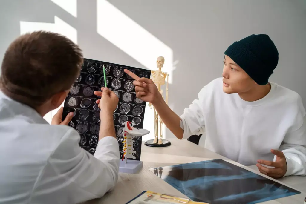
Diagnosing cerebral arteriovenous malformations (AVMs) requires advanced imaging and clinical checks. These steps help us find AVMs and understand their details. This is key for choosing the right treatment.
Several imaging methods are used to spot cerebral AVMs. These include:
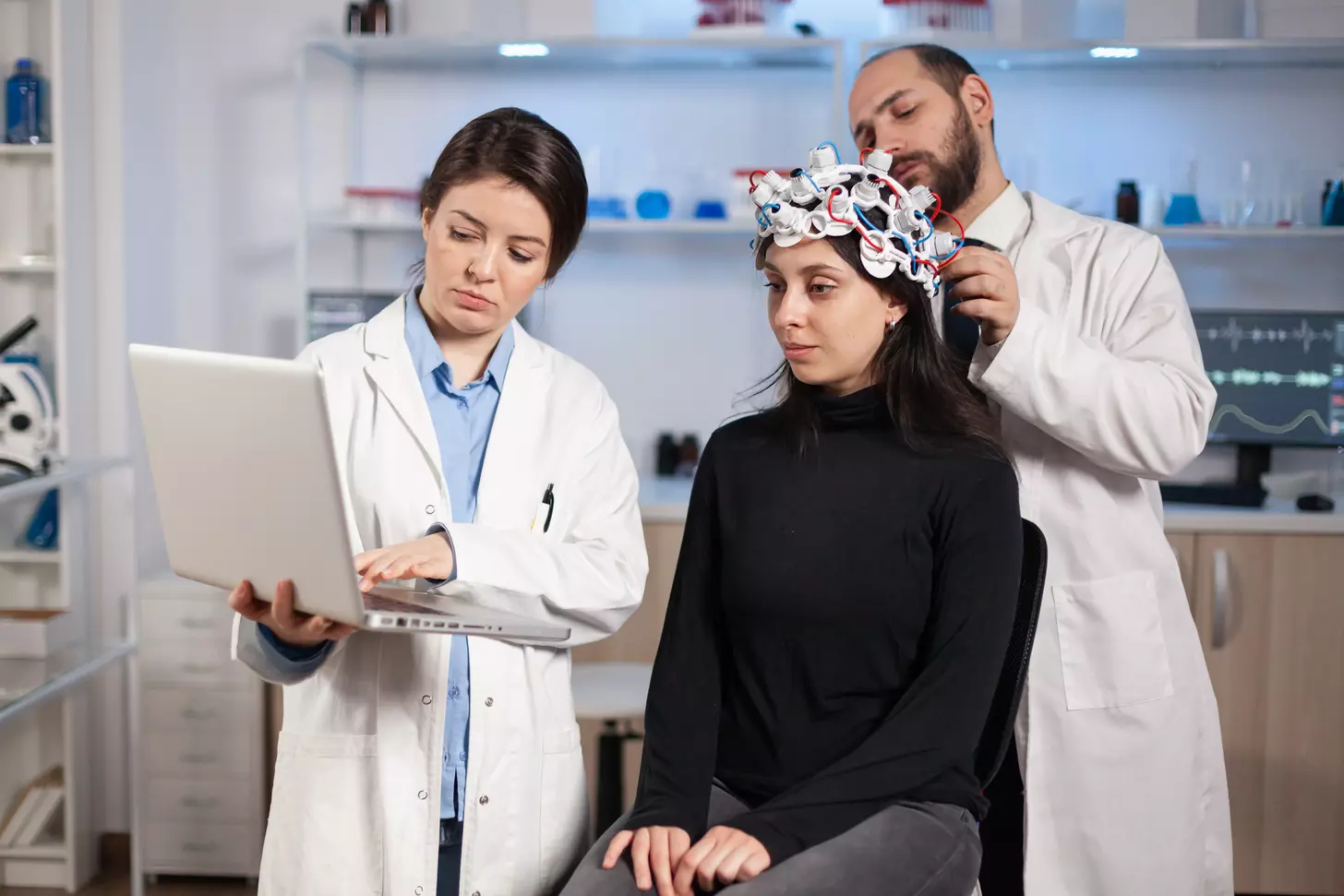
Imaging isn’t the only thing we do. A detailed check-up is also vital. We do a physical exam, focusing on the nervous system and symptoms like seizures or headaches. An electroencephalogram (EEG) might be done to check brain electrical activity.
We also talk to patients about their symptoms and health history. This helps us understand how the AVM might affect them. With this info, we create a treatment plan that fits the patient’s needs.
It’s important to know what increases the risk of AVM rupture. This is key for managing and treating AVMs. The risk of bleeding from a cerebral arteriovenous malformation (AVM) is a big worry. We need to look at several important factors to assess this risk.
The chance of bleeding from an AVM is about 2-3% each year. But, this number can change based on different things. Some main factors that increase the risk of AVM rupture include:
Each of these factors adds to the patient’s risk profile. Knowing their impact is vital for making the right treatment choices.
Many predictive models and scoring systems help figure out the risk of AVM rupture. These models use the risk factors mentioned earlier. They give a score that shows how likely bleeding is. For instance, the Spetzler-Martin grading system helps predict surgical risks and gives clues about AVM characteristics that might affect rupture risk.
Other models, like the ARUBA study, look at the natural history of unruptured AVMs. They explore the risks tied to different treatment plans. By using these models, doctors can better understand the risk of AVM rupture for each patient. This helps them make treatment plans that fit each person’s needs.
When an AVM is found before it ruptures, picking the right treatment is tough. Doctors look at many things, like the AVM’s size and the patient’s health. They also think about the risks and benefits of each treatment.
For some unruptured AVMs, doctors might choose not to treat right away. This is if the AVM isn’t causing problems and isn’t likely to burst. They watch it closely with scans to see if it changes.
Key aspects of conservative management include:
Removing the AVM surgically is a sure way to treat it. Doctors do this for AVMs at high risk of bursting or causing symptoms. They consider many things, like where the AVM is and the patient’s health.
The benefits of microsurgical resection include: getting rid of the AVM right away, lowering the chance of future bleeding, and possibly improving brain function.
Endovascular embolization is a less invasive way to treat AVMs. It involves blocking the AVM’s blood vessels with special materials. This method can be used alone or with other treatments to make them work better.
Advantages of endovascular embolization include:
Stereotactic radiosurgery (SRS) is a precise radiation therapy for AVMs. It helps the AVM shrink over time. SRS is good for AVMs that can’t be removed by surgery because of their location or size.
The benefits of SRS include its non-invasive nature, the chance for complete AVM disappearance, and its use for AVMs in hard-to-reach places.
A ruptured brain AVM is a medical emergency. It needs quick action to stop bleeding and prevent more problems. The main goal is to stop the bleeding, control seizures, and remove the AVM to avoid future ruptures.
First, we stabilize the patient and treat the bleeding. This includes managing pressure in the brain, controlling seizures, and making sure the brain gets enough blood. Quick action is key to stop more bleeding and protect the brain.
We might use external ventricular drainage to relieve pressure from blood or fluid. We also give anticonvulsants to stop seizures, which can make things worse.
There are different ways to treat a ruptured AVM. Microsurgical resection is a direct surgery to remove the AVM. It works well for AVMs that are easy to reach.
Endovascular embolization is a less invasive method. It uses materials to block the AVM. This can be done alone or with other treatments like surgery or radiosurgery.
Stereotactic radiosurgery is a non-surgical method. It uses precise radiation to shrink the AVM over time. It’s not for immediate treatment but for long-term management.
After treatment, a detailed rehabilitation program is needed. It helps patients recover from the damage caused by the AVM rupture. This includes physical, occupational, and speech therapy, customized for each patient.
Rehab aims to improve the patient’s function and quality of life. We use a team approach, including neurologists and rehabilitation experts. This helps the patient on their recovery path.
The outcome after an AVM rupture can change a lot. It depends on how bad the rupture was and how well treatment worked. We’ll look at these points to help you understand what might happen.
What happens if an AVM rupture isn’t treated? Some AVMs might stay the same, even if they don’t cause problems until later. But, if someone has a big brain bleed, it can be very serious. Sadly, some might not make it.
Others might have seizures or brain problems that don’t go away. The chance of bleeding again is a big worry. Knowing this helps doctors talk to patients and decide on treatment.
How well a patient does after treatment can vary a lot. Different treatments, like surgery, embolization, or radiosurgery, work differently. We’ll look at what affects these results, like:
How well a patient feels after an AVM rupture is very important. We look at physical, emotional, and mental health. Things that affect how well a patient does include:
Understanding these helps doctors give better care. We aim to give top-notch care to patients from around the world. We meet their special needs and worries.
In short, the future after an AVM rupture is complex. By looking at the natural history, treatment results, and quality of life, we can help patients better. We want to give the best care possible.
Cerebral arteriovenous malformations (AVMs) are complex vascular lesions. They need a deep understanding of their definition, symptoms, diagnosis, and treatment options. This article has covered the many sides of AVMs, including their causes, risk factors, and management strategies.
Knowing about AVMs is key to spotting risks and planning treatments. We talked about how doctors diagnose AVMs and the treatments available. These include microsurgical resection, endovascular embolization, and stereotactic radiosurgery.
In summary, managing AVMs needs a team effort. Each patient’s situation is unique. By covering the main points about AVMs, we aim to give a clear summary. This should help those looking to understand this complex condition, providing a valuable avm summary.
A cerebral arteriovenous malformation is a rare brain condition. It happens when arteries and veins connect directly, skipping the usual capillary network.
Symptoms can vary. They might include seizures, headaches, or neurological problems. Some people might not show any symptoms at all.
Doctors use imaging like MRI, CT scans, or cerebral angiography. These help see the AVM and understand its details.
The biggest risk is rupture, which can lead to bleeding. This can cause serious harm or even death. Other risks include seizures and neurological problems.
Doctors look at the AVM’s size, location, and how it drains blood. They also consider the patient’s health and medical history.
Treatment depends on the AVM and the patient. Options include watching it, surgery, embolization, or radiosurgery. The choice is based on the AVM’s features and the patient’s health.
First, doctors focus on stabilizing the patient. Then, they treat the AVM with surgery or embolization. After that, the patient needs rehabilitation.
Recovery depends on the severity of the rupture and the treatment’s success. Some patients recover well, while others face ongoing challenges.
Most AVMs are not inherited. But, some cases might be linked to genetic syndromes or family history. This suggests a possible genetic factor.
Cerebral AVMs are in the cerebrum, while cerebellar AVMs are in the cerebellum. Both have abnormal blood connections. But, they differ in location and might have different symptoms and treatments.
There are no known ways to prevent cerebral AVMs. Their causes are not fully understood.
Doctors make treatment choices based on the AVM’s details, the patient’s health, and the risks and benefits of each option.
Subscribe to our e-newsletter to stay informed about the latest innovations in the world of health and exclusive offers!
WhatsApp us