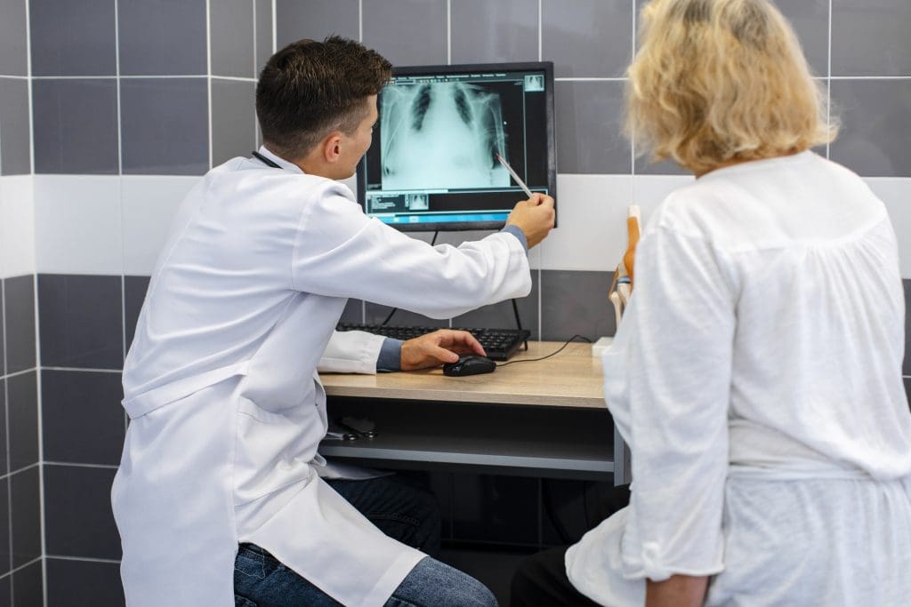
Recent studies in European Radiology show that FDG PET scans are key in finding and checking lung cancer. They can spot cancer cells at a molecular level. This makes them very sensitive and specific.
FDG PET scans have greatly helped in treating lung cancer by catching it early. Studies prove that how accurate these scans are is very important. It helps doctors decide the best treatment.

Key Takeaways
- FDG PET scans play a vital role in diagnosing and staging lung cancer.
- The accuracy of FDG PET scans is critical in determining patient outcomes.
- Recent studies have highlighted the sensitivity and specificity of FDG PET scans.
- Early detection and treatment of lung cancer improve patient outcomes.
- FDG PET scans offer a high degree of accuracy in detecting cancerous cells.
Understanding FDG PET Scan Technology
To understand FDG PET scans, we need to look at the technology behind them. These scans are key in finding and managing cancers, like lung cancer.
What is an FDG PET Scan?
An FDG PET scan uses a special sugar molecule, Fluorodeoxyglucose (FDG), to see how active cells are in the body. It’s great for finding cancer because cancer cells use more sugar than normal cells.
How FDG PET Scans Work
First, FDG is injected into the patient. It’s like sugar, so cells that are very active take it in. Cancer cells, being very active, take in more FDG.
The PET scanner then finds the FDG and makes pictures of where it is. This shows doctors where cancer might be.
The Role of Fluorodeoxyglucose (FDG)
Fluorodeoxyglucose (FDG) is the key to PET scans. It shows which cells are most active. Doctors use it to tell cancer cells apart from normal cells.
Knowing how FDG PET scans work helps us see their strengths and weaknesses. As technology gets better, these scans will help doctors more in fighting cancer.
Measuring Accuracy in Medical Imaging
Medical imaging accuracy is key for making good treatment decisions. The precision of tests like FDG PET scans greatly affects patient outcomes.
Sensitivity and Specificity Explained
Sensitivity is about a test’s ability to find those with the disease. Specificity is about its ability to find those without the disease.
A sensitive test catches most actual cases, avoiding false negatives. A specific test correctly rules out most people without the condition, avoiding false positives.
Positive and Negative Predictive Values
The Positive Predictive Value (PPV) shows the chance a positive test means you really have the disease. The Negative Predictive Value (NPV) shows the chance a negative test means you don’t have the disease.
These values depend on how common the disease is in the tested group.
Standardized Uptake Value (SUV)
The Standardized Uptake Value (SUV) measures PET scan tracer uptake in tissues. It shows metabolic activity in lesions.
| Metric | Description | Clinical Significance |
| Sensitivity | True Positive Rate | Ability to detect disease |
| Specificity | True Negative Rate | Ability to rule out disease |
| PPV | Probability of disease given positive test | Influenced by disease prevalence |
| NPV | Probability of no disease given negative test | Influenced by disease prevalence |
| SUV | Quantifies tracer uptake in tissues | Assesses metabolic activity |
Overall FDG PET Scan Accuracy Rates
Knowing how accurate FDG PET scans are is key for both patients and doctors. These scans are vital for managing lung cancer well.
General Diagnostic Accuracy
FDG PET scans are very good at finding different cancers, including lung cancer. Research shows they are reliable.
Diagnostic Accuracy Metrics
| Metric | Value |
| Sensitivity | 85-90% |
| Specificity | 80-85% |
| Positive Predictive Value | 75-80% |
| Negative Predictive Value | 90-95% |
Factors Affecting Accuracy
Many things can change how accurate FDG PET scans are. These include how well the patient is prepared, the technology used, and the tumor’s details.
- Patient Preparation: It’s important to fast and manage blood sugar well.
- Scanner Technology: Newer PET scanners are more accurate.
- Tumor Characteristics: The tumor’s activity and size matter for detection.
Comparing Accuracy Across Different Generations of PET Scanners
PET scanner technology has gotten better over time. This has made scans more accurate.
Comparison of PET Scanner Generations
| Scanner Generation | Sensitivity | Resolution |
| First Generation | 70-75% | 6-8 mm |
| Current Generation | 90-95% | 2-4 mm |
How Accurate is a PET Scan for Lung Cancer?
Understanding PET scans for lung cancer is key for diagnosis and treatment. PET scans are a vital tool in oncology. They show how active tumors are metabolically.
Detection Rates for Primary Lung Tumors
PET scans are good at finding primary lung tumors. They highlight areas with high metabolic activity. Studies show that PET scans can detect lung cancer with a sensitivity of around 90%.
But, the accuracy can change based on tumor size and location. Larger tumors are easier to spot, while smaller ones can be harder. Newer PET scanner technology helps find smaller tumors better.
Accuracy in Different Types of Lung Cancer
PET scan accuracy varies with lung cancer types. For example, adenocarcinoma and squamous cell carcinoma have different metabolic profiles. This affects how well PET scans can detect them.
PET scans work well for non-small cell lung cancer (NSCLC), which includes adenocarcinoma and squamous cell carcinoma. But, they might not be as accurate for small cell lung cancer (SCLC) because of its unique biology.
Statistical Evidence from Clinical Studies
Many studies have looked into PET scan accuracy for lung cancer. A study in the Journal of Clinical Oncology found that PET scans had a sensitivity of 96.8% and a specificity of 77.8% for detecting NSCLC.
| Study | Sensitivity (%) | Specificity (%) |
| Journal of Clinical Oncology | 96.8 | 77.8 |
| Radiology | 93.2 | 85.1 |
| European Journal of Nuclear Medicine | 90.5 | 82.3 |
The evidence shows PET scans are a reliable tool for lung cancer diagnosis. They have high sensitivity and specificity rates in many studies.
Sensitivity of PET Scans in Lung Cancer Detection
PET scans are key in finding lung cancer. They show both how the body works and its structure. This makes them vital for diagnosing and understanding lung cancer.
Detection of Small Lung Nodules
PET scans are great at spotting small lung nodules. They can find nodules as small as 8-10 mm. But, how well they work depends on the nodule and the scanner.
Early detection is key. It means doctors can act fast. A study found that early use of PET scans can change treatment plans and improve patient outcomes.
Identifying Early-Stage Lung Cancer
PET scans are also good at finding early lung cancer. They can spot changes in cancer cells early. This is important because early tumors are easier to treat.
“PET scans offer a high degree of sensitivity in detecting lung cancer at an early stage, which is critical for effective treatment and improved survival rates.”
Factors Affecting Sensitivity in Lung Cancer
PET scans are generally specific for lung cancer; however, there are instances where they may misidentify inflammation or other conditions as cancer.
- The type and stage of lung cancer
- The size and location of the tumor
- The patient’s blood glucose levels
Knowing these factors helps doctors understand PET scan results. This is important for making the best care plans for patients.
Specificity of PET Scans for Lung Cancer
PET scans are key in telling apart cancerous from non-cancerous growths in the lungs. This is important because it helps doctors decide the best treatment. It also affects how well a patient will do.
Distinguishing Malignant from Benign Lesions
PET scans look for high activity in cells, which often means cancer. But, not all high activity is cancer. Some non-cancerous conditions can also show high activity.
Key factors for telling cancer from non-cancer include:
- The intensity of FDG uptake, measured by the Standardized Uptake Value (SUV)
- The size and location of the lesion
- Clinical correlation with patient history and other diagnostic findings
Reducing False Positive Results
False positives can cause a lot of worry and extra tests. They can also lead to wrong treatments. To avoid this, doctors look at the whole picture and use other scans too.
Some common causes of false positives include:
- Inflammatory processes
- Infectious diseases
- Granulomatous diseases
Clinical Implications of High Specificity
When PET scans show a growth is cancerous, doctors are very sure. This surety is key for choosing the right treatment. It could be surgery, chemo, or radiation.
Research shows PET scans are very good at finding lung cancer. They work best when used with other tests. This accuracy is important for giving patients the right care and avoiding unnecessary steps.
Patient Preparation and Its Impact on FDG PET Scan Accuracy
Getting ready for an FDG PET scan is key to getting good results. It helps doctors find and understand cancer, like lung cancer, better. How well you prepare matters a lot.
Fasting Requirements
Fasting is a big part of getting ready for an FDG PET scan. You need to not eat or drink anything for 4 to 6 hours before. This helps the FDG tracer work better.
Fasting Guidelines:
- Avoid eating or drinking anything except water for 4 to 6 hours before the scan.
- Some medications may be taken with a small amount of water.
- Inform your doctor about any diabetes medications or conditions.
Blood Glucose Level Management
Keeping blood sugar levels in check is important for the scan. High blood sugar can mess up the FDG uptake. This might lead to wrong results.
| Blood Glucose Level | Action |
| Below 200 mg/dL | Proceed with the scan |
| 200 mg/dL or higher | Reschedule or adjust protocol |
Activity Restrictions Before Scanning
Some activities can mess with FDG distribution in the body. This might affect the scan’s accuracy. Avoid hard exercise or activities that strain muscles for a while before the scan.
Following these steps helps make sure your FDG PET scan is accurate. This is important for finding and treating lung cancer effectively.
FDG PET Scan Accuracy in Cancer Staging
FDG PET scans are key for doctors to see how far cancer has spread. This helps them create treatment plans that fit each patient. Staging cancer means finding out how big the tumor is and if it has spread to other parts of the body.
Accuracy in Determining Tumor Extent
FDG PET scans are very good at showing how big tumors are. They use a special sugar that cancer cells eat, making them stand out. This gives doctors a clear picture of the tumor’s size and where it is.
Studies have shown that these scans work well for many cancers, like lung cancer. Knowing this helps doctors decide on surgery or radiation therapy.
Lymph Node Involvement Assessment
Checking if cancer has spread to lymph nodes is very important. FDG PET scans are good at finding cancer in these nodes. This helps doctors figure out the cancer’s stage and what treatment to use.
| Cancer Type | Sensitivity of FDG PET in Detecting Lymph Node Involvement | Specificity of FDG PET in Detecting Lymph Node Involvement |
| Lung Cancer | 85% | 90% |
| Breast Cancer | 80% | 85% |
| Colorectal Cancer | 88% | 92% |
Distant Metastasis Detection
FDG PET scans are also great for finding cancer that has spread far away. This is important for accurate staging. Knowing where the cancer has spread helps doctors plan the best treatment.
Clinical studies have shown that these scans can find distant metastasis very accurately. This changes how doctors manage patients and affects their outlook.
False Positives in FDG PET Scans
It’s key to know about false positives in FDG PET scans. False positives happen when a scan shows cancer when there isn’t any. This can lead to extra tests and worry for patients.
Common Causes of False Positives
Several things can cause false positives in FDG PET scans. These include:
- Inflammatory conditions
- Infections
- Granulomatous diseases
- Recent surgical procedures
Inflammatory conditions are a big reason for false positives. They can look like tumors because of how they use energy.
Inflammatory Conditions Mimicking Cancer
Inflammatory conditions can make FDG PET scans show false positives. This is because they can look like tumors. For example, pneumonia, sarcoidosis, and rheumatoid arthritis can all cause false positives.
| Condition | Characteristics | Potential for False Positive |
| Pneumonia | Increased FDG uptake in lung tissue | High |
| Sarcoidosis | Granulomatous inflammation | High |
| Rheumatoid Arthritis | Inflammation in joints | Moderate |
Strategies to Minimize False Positive Results
To cut down on false positives, it’s important to look at the patient’s history and other tests. Ways to do this include:
- Combining PET with CT or MRI for better anatomical correlation
- Using quantitative measures like SUV to assess FDG uptake
- Considering alternative diagnoses that could explain FDG uptake
By knowing why false positives happen and how to avoid them, doctors can make FDG PET scans more accurate.
False Negatives in FDG PET Scans
It’s important to know about false negatives in FDG PET scans. This is because they can lead to delayed diagnosis and wrong treatment plans. False negatives happen when a PET scan misses cancer that is actually there.
Types of Cancers Frequently Missed
Some lung cancers are hard to spot with FDG PET scans. This is because their metabolic activity is low. For example, adenocarcinoma in situ and minimally invasive adenocarcinoma might not show up well because they don’t take up much FDG.
Size Limitations in Detection
The size of a tumor affects how easy it is to find with FDG PET scans. Tumors that are small, under 8-10 mm, are often missed. Newer PET scanners can see smaller tumors better, thanks to better technology.
Metabolic Factors Affecting Detection
How a tumor uses energy affects if it shows up on FDG PET scans. Tumors that don’t use much glucose are harder to spot. Also, high blood sugar can make tumors harder to see, leading to false negatives.
| Factors Affecting False Negatives | Description | Impact on Detection |
| Tumor Size | Smaller tumors are harder to detect. | High risk of being missed if below scanner resolution. |
| Metabolic Activity | Tumors with low glucose metabolism. | Less likely to be detected due to low FDG uptake. |
| Blood Glucose Levels | High levels can inhibit FDG uptake. | Potential for false negatives due to reduced tumor visibility. |
Knowing about these factors helps doctors understand FDG PET scan results better. This knowledge is key for making the right decisions about further tests or treatment.
PET Scans vs. Other Imaging Tests for Lung Cancer
PET scans are generally specific for lung cancer; however, there are instances where they may misidentify inflammation or other conditions as cancer.
PET scans have a unique advantage over CT scans because they provide metabolic information that helps distinguish between cancerous and non-cancerous tissues.
CT scans are common for lung cancer diagnosis. They give detailed views of the body’s structure. Yet, they might not always tell the difference between cancer and non-cancerous tissues.
PET scans, though, show how active tumors are metabolically. This helps spot cancerous tissues more clearly.
PET scans are better at finding cancer in lymph nodes and other organs. They can change treatment plans by revealing more disease than CT scans alone.
PET vs. MRI
PET scans are generally specific for lung cancer; however, there are instances where they may misidentify inflammation or other conditions as cancer.
- PET scans are more sensitive for detecting lung cancer metastases.
- MRI is useful for specific cases, such as evaluating superior sulcus tumors or spinal cord involvement.
Integrated PET/CT Advantages
PET/CT scanners combine PET and CT technologies. PET/CT offers both the metabolic info from PET and the detailed anatomy of CT. This gives a full view of the disease.
“The use of PET/CT in lung cancer staging has been shown to improve diagnostic accuracy and reduce the number of unnecessary surgeries.”
Source: Journal of Clinical Oncology
PET/CT has many benefits. It improves diagnosis, staging, and treatment planning. This hybrid imaging is essential in lung cancer management.
PET Scan Accuracy in Treatment Monitoring
PET scans are key in managing lung cancer. They help doctors see how well treatment is working. This lets them decide if they should keep or change the treatment plan.
Evaluating Response to Therapy
PET scans are essential for checking how lung cancer responds to treatment. Doctors compare scans before and after treatment. This helps them see if the cancer is getting smaller, staying the same, or growing.
Key benefits of using PET scans for evaluating response to therapy include:
- Accurate assessment of tumor response
- Early detection of treatment efficacy
- Guidance for adjustments in treatment plans
Detecting Recurrence
PET scans also help find lung cancer that comes back. After treatment, cancer can return. PET scans can spot this early, when it’s easier to treat.
Early detection of recurrence is vital for better treatment. PET scans are great at catching cancer return because they can see changes in the body.
Timing Considerations for Accurate Assessment
When to do PET scans is very important. Scans too soon after treatment might not show the true effect of treatment. This is because of inflammation or other changes.
Scans for checking treatment response are best done after a good amount of time after treatment. This time lets inflammation and other changes go away. It gives a clearer view of how well the treatment worked.
By planning when to do PET scans, doctors can use them best. This helps in monitoring lung cancer treatment and improves patient results.
Interpreting PET Scan Results for Lung Cancer
Understanding PET scan results is key for lung cancer diagnosis and treatment. It involves knowing how SUV values, visual checks, and clinical info affect the results.
Understanding SUV Values
The SUV measures how much a tracer is taken up by the body. Higher SUV values often mean more aggressive tumors. But, SUV values can change based on blood sugar, time of scanning, and scanner settings.
“SUV values should be interpreted with caution, as they can vary between different scanners and institutions,” a study in the Journal of Nuclear Medicine says. Clinical context and other imaging findings should always be considered when interpreting SUV values.
Visual Assessment Techniques
Visual checks involve looking at PET scan images for abnormal tracer uptake. Experienced radiologists and nuclear medicine physicians use their skills to tell benign from malignant lesions based on uptake pattern, intensity, and location. This method is not always exact and can depend on the observer’s experience.
- Pattern of uptake: Focal or diffuse
- Intensity of uptake: Compared to background or reference tissues
- Location of uptake: Anatomical correlation with other imaging modalities
Importance of Clinical Correlation
Clinical correlation is essential when looking at PET scan results. Patient history, symptoms, and other diagnostic findings should be combined with PET scan results for better decisions. For example, a high SUV value in a lung nodule is more significant when considering the patient’s smoking history and other clinical factors.
“The integration of PET scan results with clinical information enhances diagnostic accuracy and aids in personalized treatment planning for lung cancer patients.”
By using SUV values, visual checks, and clinical correlation, healthcare providers can make more accurate diagnoses. This helps in creating effective treatment plans for lung cancer patients.
Conclusion: The Future of FDG PET Scans in Lung Cancer Diagnosis
FDG PET scans are key in finding lung cancer. They help spot tumors, check lymph nodes, and find cancer spread. With new research, these scans will get even better.
New studies aim to make FDG PET scans more accurate. They want to find small tumors and cancer early. New PET scanner tech, like PET/CT and PET/MRI, will also help a lot.
Artificial intelligence and machine learning will soon help read FDG PET scans. This will lead to more precise and personal diagnoses. The future of FDG PET scans in lung cancer looks very promising.
FAQ
How accurate is a FDG PET scan for diagnosing lung cancer?
FDG PET scans are very accurate for lung cancer diagnosis. Their sensitivity and specificity vary by study and patient. They are highly effective in finding primary lung tumors.
What is the sensitivity of PET scans in detecting lung cancer?
PET scans are generally specific for lung cancer; however, there are instances where they may misidentify inflammation or other conditions as cancer.
PET scans are generally specific for lung cancer; however, there are instances where they may misidentify inflammation or other conditions as cancer.
PET scans are generally specific for lung cancer; however, there are instances where they may misidentify inflammation or other conditions as cancer. They can sometimes mistake inflammation or other conditions for cancer. More tests and doctor’s review are needed to confirm.
What factors can affect the accuracy of FDG PET scans?
Several things can change how accurate FDG PET scans are. These include how well the patient prepares, blood sugar levels, and inflammation. Scanner quality and image processing also play a role.
How do PET scans compare to other imaging tests for lung cancer diagnosis?
PET scans have a unique advantage over CT scans because they provide metabolic information that helps distinguish between cancerous and non-cancerous tissues. They show how tumors work, not just their size. But, they’re often used with CT or MRI for a full picture.
Can PET scans detect small lung nodules?
PET scans are generally specific for lung cancer; however, there are instances where they may misidentify inflammation or other conditions as cancer.
How are PET scan results interpreted for lung cancer diagnosis?
Doctors look at PET scan results in two ways: visually and with numbers like SUV values. They also consider the patient’s medical history and lab results.
What is the role of FDG PET scans in cancer staging?
FDG PET scans are key in cancer staging. They show how far the cancer has spread, including to lymph nodes and distant sites. This helps doctors decide on treatment and predict outcomes.
Can PET scans be used to monitor treatment response in lung cancer?
Yes, PET scans can track how lung cancer responds to treatment. They watch for changes in tumor activity over time. This helps doctors know if treatment is working or if they need to try something else.
What are the limitations of FDG PET scans in lung cancer diagnosis?
FDG PET scans have some downsides. They can give false positives or negatives, and technical issues can affect results. Doctors often need to use other tests to confirm findings.


