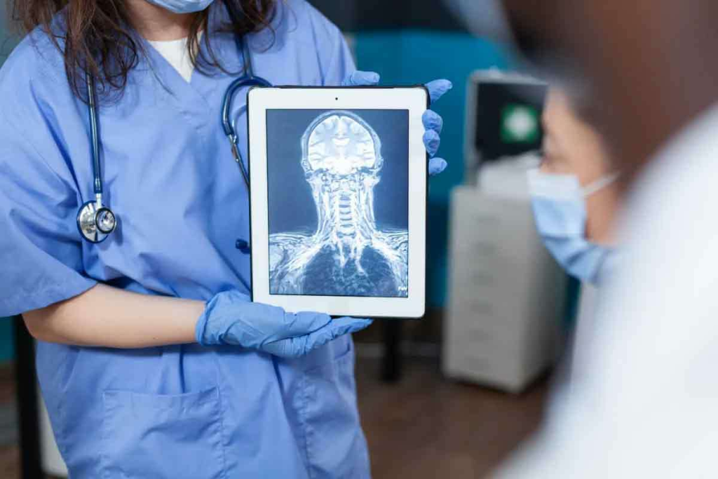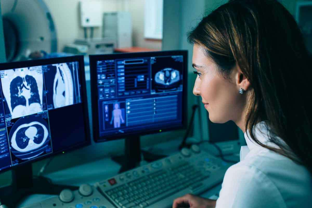
Getting a correct diagnosis is key when treating chest diseases. A chest CT scan is vital for spotting many conditions, including both harmless and harmful tumors, infections, and other structural issues. Detecting an abnormal computed tomography result is important for identifying these problems and providing the best treatment. Liv Hospital is dedicated to top-notch care and imaging for the chest, ensuring patients receive the best care possible. With about half of chest CT scans showing an abnormality, accurate diagnosis is very important.

Advanced CT technology has changed how we diagnose and treat diseases. Chest CT scans are now key in modern medicine. They give a detailed look at the chest area.
CT technology has come a long way. From single-slice scanners to multi-detector CT (MDCT) systems, we’ve made big strides. These changes mean faster scans, clearer images, and 3D views of the body.
Key advancements include:
These updates have made chest CT scans better at finding problems. They can spot issues that regular X-rays miss.
CT scans are now used more for patients with breathing problems. About 50% of these scans find important issues. This shows how vital CT scans are for diagnosing and treating patients.
| Study | Detection Rate | Clinical Implication |
| Study A | 45% | Significant abnormalities detected, leading to changes in patient management |
| Study B | 52% | High detection rate of abnormalities, stressing the CT’s role in diagnosis |
| Study C | 48% | CT scans found critical issues not seen on X-rays |

Radiologists use advanced methods to spot abnormal computed tomography patterns in chest scans. They carefully look at CT scan images for small signs of health problems.
Radiologists have a checklist to check every part of the chest scan. This careful method helps find issues that might be missed. They often use Radiology Assistant for detailed guidelines on chest CT scans.
Top hospitals like Liv Hospital use the latest chest CT scan methods. These methods include special software to find problems accurately. This makes diagnoses more precise and helps plan better treatments.
Thanks to these advanced techniques, doctors can give more accurate diagnoses. This is key for better patient care. The mix of technology and skill in chest CT imaging is a big step forward in medicine.
CT scans often find pulmonary nodules. It’s important to know what they are and why they matter.
Pulmonary nodules are small, round tissue masses seen on chest CT scans. They might be harmless or could be cancer. Often, they’re found by accident during scans for other reasons.
Some features of pulmonary nodules hint at cancer. Look for irregular or spiculated margins, large size, and calcifications or fat.
Checking these details is key to figuring out if a nodule might be cancerous.
| Morphological Feature | Malignancy Risk |
| Irregular/Spiculated Margins | High |
| Large Size (>1 cm) | Moderate to High |
| Calcifications or Fat Presence | Low to Moderate |
Handling nodules found by accident involves a few steps. These include looking at the patient’s health, imaging follow-ups, and sometimes biopsies.
Guidelines help decide how to manage these nodules. They look at the nodule’s size, the patient’s risk for cancer, and if they have symptoms.
Knowing these guidelines is vital for doctors to make the best choices for patients with nodules found by accident.
Ground-glass opacities are often seen on abnormal chest CT scans. They can point to different health issues. It’s important to understand what they mean to help patients.
These opacities can be caused by many things, like infections, inflammation, or cancer. Infectious etiologies like pneumonia and inflammatory conditions like acute interstitial pneumonia can show up as ground-glass opacities.
When you see ground-glass opacities on a chest CT, think about the patient’s history and symptoms. Look at other imaging too. The way the opacities spread and look can give clues.
Knowing the patient’s situation well is key to understanding the findings.
The way ground-glass opacities change can tell you a lot about the cause. In cases like pneumonia, they might go away with treatment. But in chronic diseases like interstitial lung disease, they might stay or get worse.
| Condition | Typical Progression Pattern |
| Infectious Pneumonia | Resolution with treatment |
| Interstitial Lung Disease | Persistence or progression |
| Adenocarcinoma in situ | Potential for slow growth or stability |
Watching how ground-glass opacities change on follow-up CT scans is important. It helps see how well treatment is working and how the disease is changing.
Understanding ground-glass opacities on abnormal computed tomography scans is very important. It helps doctors make better decisions and improve patient care.
Looking at lymphadenopathy on chest CT scans is complex. It needs a good grasp of lymph node sizes and where they are. Enlarged lymph nodes can mean different things, like infections, cancers, or inflammation.
Lymph nodes play a big role in our immune system. When they get bigger, it might mean our body is fighting something off. On a chest CT scan, how big and shaped a lymph node is tells us if it’s normal or not. Usually, if a lymph node is over 1 cm in diameter, it’s seen as abnormal.
Table: Criteria for Evaluating Lymph Node Size
| Lymph Node Location | Normal Size | Abnormal Size |
| Mediastinal | <1 cm | >1 cm |
| Hilar | <1 cm | >1 cm |
| Axillary | <1.5 cm | >1.5 cm |
Where enlarged lymph nodes are located can give us hints about what’s going on. For example, if lymph nodes are big on both sides of the chest, it might be sarcoidosis. But if they’re in the middle of the chest, it could be lymphoma or cancer spreading.
It’s key for doctors and radiologists to know about lymphadenopathy on abnormal chest CT scans. This helps them guess what might be wrong and plan the right treatment. By looking at size, location, and pattern, they can make better guesses and help patients get the right care.
Chest CT scans often show pleural abnormalities like effusions and thickening. These signs need careful checking. They can point to many conditions, from simple to serious.
The pleura, a thin layer around the lungs, can get affected by many diseases. It’s key to correctly identify these issues on CT scans for the right treatment.
Pleural effusion is when fluid builds up in the pleural space. It’s a common finding on chest CT scans. The amount of fluid can be small or large, affecting breathing.
Measuring the fluid’s volume is important. Also, knowing its density on CT can tell us what it is, like a transudative or exudative effusion.
| Characteristic | Transudative Effusion | Exudative Effusion |
| Density on CT | Typically | Often > 10 HU, may be heterogeneous |
| Causes | Congestive heart failure, cirrhosis | Infection, malignancy, inflammation |
Pleural-based masses and focal abnormalities need close attention. They can be benign, like pleural plaques from asbestos, or cancerous, like mesothelioma.
Any focal pleural thickening or masses on CT scans need more tests. Often, a biopsy is needed for a clear diagnosis. Other signs, like calcification or lymphadenopathy, can help in diagnosing.
In summary, pleural abnormalities on chest CT scans include effusions and focal masses. Accurate assessment and measurement of these issues are vital for proper treatment and better patient care.
It’s key to spot the unique signs of interstitial lung disease for the right diagnosis and care. This group of lung issues shows different patterns on chest CT scans. These patterns help doctors understand and treat the disease.
ILD is split into two main types based on CT scans. Fibrotic patterns show signs of long-term lung scarring like reticulation and honeycombing. On the other hand, inflammatory patterns have ground-glass opacities, which mean there’s active inflammation.
Knowing the difference between these patterns is vital. It helps decide the best treatment and what to expect for the patient’s future. For example, fibrotic ILD might need treatments to stop scarring, while inflammatory ILD could be treated with drugs to reduce inflammation.
Where the lung issues appear on CT scans gives important hints. For example, subpleural distribution often points to idiopathic pulmonary fibrosis (IPF). A perilymphatic distribution is more common in sarcoidosis.
Grasping these patterns is critical for doctors and radiologists to make the right diagnosis. By combining what they see on scans with the patient’s symptoms, they can give a more accurate diagnosis. This helps them create a treatment plan that fits the patient’s needs.
CT angiography has changed how we diagnose vascular problems in the chest, like pulmonary embolism. This advanced imaging gives detailed views of the pulmonary vasculature. It helps doctors diagnose and treat conditions well. The role of CT angiography in finding vascular problems is huge.
Pulmonary embolism (PE) is a serious condition that needs quick diagnosis and treatment. CT angiography is the top choice for diagnosing PE, with high accuracy. In acute PE, CT angiography shows filling defects in the pulmonary arteries.
Key findings in acute PE include:
In chronic PE, findings are different, showing vascular remodeling and recanalization. CT angiography helps in understanding the chronicity of PE and guiding treatment.
Pulmonary hypertension is when pulmonary artery pressures are too high, causing vascular remodeling. CT angiography is key in spotting signs of pulmonary hypertension, like:
Experts say, “Diagnosing pulmonary hypertension involves clinical assessment, imaging, and hemodynamic measurements.”
“Early detection of vascular abnormalities is critical for preventing long-term damage and improving patient outcomes.”
Understanding these vascular problems through CT scans is key to accurate diagnosis and effective treatment. The detailed info from CT angiography helps tailor treatment plans to each patient’s needs.
Getting chest CT scans right is key for spotting many chest problems. It helps doctors decide on the best treatments. This is important for how well patients do.
Spotting things like lung spots, foggy lung areas, and swollen lymph nodes on a CT scan is vital. It lets doctors make smart choices about patient care.
Using CT scan results in treatment plans can really help patients. Finding problems early, like blood clots or lung cancer, can save lives. Radiologists and doctors working together can make treatment plans that work.
In short, reading chest CT scans well is critical for good patient care. Doctors need to know what they see on scans to give top-notch care. Working together, doctors and radiologists can make sure patients get the best care possible.
Spotting abnormalities on a chest CT scan is key for correct diagnosis and treatment planning. This is because up to 50% of scans for sudden symptoms show something unusual.
New CT technology has greatly improved at finding abnormalities. This has made thoracic imaging more accurate.
The size, shape, and density of pulmonary nodules can hint at cancer. This helps doctors decide on the next steps and diagnosis.
Ground-glass opacities are subtle signs of serious issues. Accurate reading is vital to understand their importance and guide further tests.
Radiologists look at the size, shape, and where lymph nodes are to tell if they’re normal or not. This helps pinpoint specific conditions.
Pleural abnormalities like effusions, thickening, and masses are often seen on CT scans. These can be measured and described, aiding in diagnosis.
CT scans spot patterns that help diagnose interstitial lung disease. They can tell if it’s fibrotic or inflammatory and narrow down possible causes.
CT angiography can spot signs of both acute and chronic pulmonary embolism. It shows vascular issues and signs of high blood pressure and changes in blood vessels.
Correctly identifying abnormalities on a chest CT scan is essential. It guides treatment and ensures the best care for patients.
An abnormal chest CT scan shows unusual findings in the chest. This includes things like nodules, swollen lymph nodes, or pleural issues.
Subscribe to our e-newsletter to stay informed about the latest innovations in the world of health and exclusive offers!
WhatsApp us