Last Updated on October 22, 2025 by mcelik

Did you know that over 5.8 million Americans are living with Alzheimers imaging? This condition slowly damages memory and thinking skills.
A SPECT (Single Photon Emission Computed Tomography) scan is a tool doctors use. It checks how well the brain works. This helps doctors figure out if someone has dementia and how bad it is.
Brain perfusion SPECT is great for spotting brain problems. It looks at how blood flows to the brain. This gives doctors clues about how dementia is getting worse.

SPECT scans are key in neuroimaging. They show how blood flows in the brain. This helps us understand brain function and diagnose neurological disorders.
A SPECT scan is a way to see brain activity through blood flow. It uses a tiny amount of radioactive tracer. This tracer goes into the bloodstream and builds up in the brain where blood flows.
This scan is special because it shows how the brain works. Other scans might just show what the brain looks like.
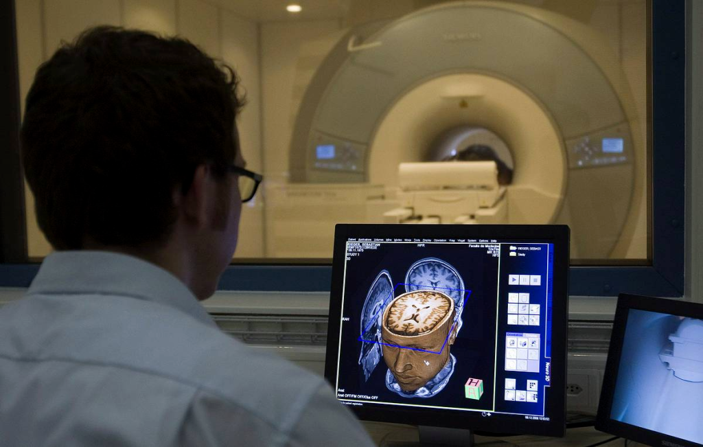
Getting a SPECT scan starts with a small injection of radioactive tracer. The tracer goes to parts of the brain with blood flow. Then, a SPECT camera picks up the gamma rays from the tracer.
This makes a 3D picture of brain activity. Doctors can use this to find and treat brain problems.

SPECT scans are different from MRI and PET scans. MRI shows the brain’s structure in detail. But SPECT scans look at brain function by measuring blood flow.
| Imaging Technique | Primary Use | Key Features |
| SPECT | Functional brain imaging | Measures blood flow, detects gamma rays |
| MRI | Structural brain imaging | High-resolution images, no radiation |
| PET | Functional brain imaging | Measures metabolic activity, uses positron emission |
Knowing how these scans differ helps doctors choose the best one for each patient.
Brain perfusion SPECT is a complex imaging method. It shows how blood flows in the brain, key for spotting dementia. In a normal brain, blood flows evenly, giving oxygen and nutrients to all parts.
A healthy brain has a special blood flow pattern. It has more blood in areas that control movement, senses, and thinking. SPECT scans can see these patterns, helping doctors compare them to sick brains.
Dementia, like Alzheimer’s, changes how blood flows in the brain. It leads to less blood in some areas, a sign of dementia. This change is linked to brain tissue loss and amyloid plaques.
Radiotracer compounds, like Technetium-99m HMPAO, are used in SPECT scans. They show blood flow in the brain. This lets doctors create detailed images of brain blood flow.
Knowing how brain perfusion SPECT works is key to its use in diagnosing dementia. It helps doctors understand brain changes and make better diagnoses.
SPECT scans are a key tool in finding dementia. They show special blood flow patterns in the brain. This helps doctors spot and tell apart different types of dementia early on.
Spotting dementia with SPECT scans means looking at brain blood flow. Each dementia type has its own blood flow pattern. For example, Alzheimer’s disease shows less blood flow in certain brain areas.
Doctors use these patterns to make accurate dementia diagnoses. They can tell Alzheimer’s disease, frontotemporal dementia, and vascular dementia apart.
SPECT scans are good at finding dementia, but not perfect. They work best when used with a doctor’s evaluation. How well they work can change based on the dementia type and diagnosis criteria.
SPECT’s ability to detect brain changes is a big plus. It’s not 100% accurate, but it adds valuable info. This info helps doctors make more accurate dementia diagnoses when used with other tools.
SPECT imaging is also great for catching dementia early. It can spot changes in brain blood flow that signal early dementia. This means doctors can diagnose before symptoms get worse.
Early detection is key for starting treatments early. SPECT scans help make this possible. They are a big part of diagnosing dementia well.
SPECT technology has changed how we diagnose Alzheimer’s. It shows detailed brain perfusion patterns. This makes SPECT scans key in diagnosing and managing Alzheimer’s.
SPECT scans show patterns linked to Alzheimer’s. Characteristic perfusion patterns include reduced blood flow in certain brain areas. These patterns help tell Alzheimer’s apart from other dementias.
The temporal and parietal lobes are hit hard by Alzheimer’s. SPECT scans show less blood flow in these areas. This matches the memory loss and sensory issues seen in Alzheimer’s patients.
Serial SPECT scans track Alzheimer’s disease over time. By comparing successive scans, doctors can see how the disease is growing. This helps in making treatment plans and adjusting them as needed.
In summary, SPECT scans are critical in Alzheimer’s imaging. They provide insights into the disease’s patterns and how it progresses. This information is vital for accurate diagnosis and effective management of Alzheimer’s.
Understanding SPECT scans can help doctors tell apart frontotemporal dementia (FTD) and Alzheimer’s disease. These two conditions are hard to diagnose and treat.
FTD shows distinctive perfusion patterns on SPECT scans. These patterns are different from Alzheimer’s. FTD often has less blood flow in the frontal and anterior temporal lobes.
The main features of FTD’s perfusion patterns are:
The abnormalities in the frontal and temporal lobes on SPECT scans match FTD’s symptoms and cognitive problems. For example, those with behavioral variant FTD often have less blood flow in the frontal lobe.
Some notable abnormalities are:
SPECT scans can help doctors accurately tell FTD apart from Alzheimer’s disease. The unique patterns and areas affected help make better decisions.
Several factors affect how accurate the diagnosis is:
By using SPECT scans and clinical evaluation together, doctors can better tell FTD from Alzheimer’s.
Diagnosing Lewy body dementia has become more accurate with DaTscan technology. DaTscan uses a radioactive tracer to show the dopamine system in the brain.
A dopamine transporter scan, or DaTscan, is a way to see the brain’s motor control system. It’s used for diseases like Parkinson’s and Lewy body dementia.
The scan involves injecting a radioactive tracer into the body. This tracer binds to dopamine transporters in the brain. A SPECT scanner then shows where the tracer is, giving a picture of dopamine levels.
Diagnosing dementia can be tricky, as it’s hard to tell Parkinson’s disease dementia from Alzheimer’s. DaTscan helps with this. Alzheimer’s mainly affects memory, not dopamine early on. But Lewy body dementia and Parkinson’s disease dementia do affect dopamine.
DaTscan shows if the dopamine system is working right. A normal scan means Alzheimer’s is more likely. But an abnormal scan points to Lewy body dementia or Parkinson’s disease.
DaTscan is very good at spotting Lewy body dementia. It shows the loss of dopamine transporters in the brain, a key sign of the disease.
| Diagnostic Parameter | DaTscan Performance |
| Sensitivity | 85-90% |
| Specificity | 80-85% |
| Positive Predictive Value | 80-90% |
| Negative Predictive Value | 85-90% |
DaTscan has greatly improved Lewy body dementia diagnosis. This means doctors can better manage and treat patients.
SPECT imaging is key in spotting vascular dementia. It shows special patterns of blood flow.
Vascular dementia happens when blood flow to the brain drops. This is due to many small strokes or tiny blood vessel problems. SPECT scans can show where blood flow is low.
These scans reveal multiple spots where blood flow is low. This matches areas where strokes or tiny blood vessel problems have happened.
The extent and where these problems are can change. For example, someone with many strokes might have many spots where blood flow is low. But, tiny blood vessel problems might show up as more subtle changes in blood flow.
It’s hard to tell if someone has vascular dementia or Alzheimer’s disease. SPECT scans help by showing blood flow patterns specific to each condition.
By looking at these patterns, doctors can better understand why someone’s thinking is getting worse.
Diagnosing vascular dementia gets better when SPECT scans are used with MRI or CT scans. This way, doctors can see both how the brain works and its structure.
For instance, someone thought to have vascular dementia might get a SPECT scan to check blood flow. They might also get an MRI to look at brain structure, like tiny strokes or damage to tiny blood vessels. This helps doctors make a more accurate diagnosis and plan the best treatment.
Key benefits of combined assessment include:
When it comes to diagnosing dementia, knowing the difference between FDG PET and SPECT is key. Both are important in brain imaging but have unique roles and benefits.
FDG PET and SPECT differ in their ability to spot dementia. FDG PET is more sensitive in catching early signs of Alzheimer’s, like in the temporal and parietal lobes. It can spot brain changes before symptoms show up.
SPECT, which looks at blood flow in the brain, is useful too. It might not be as good as FDG PET in some cases. Yet, SPECT helps identify blood flow patterns linked to different dementias, like Alzheimer’s and frontotemporal dementia.
Looking at cost and availability, SPECT is more common and cheaper than FDG PET. This is because SPECT cameras are found in more hospitals and clinics. Plus, the radioactive materials for SPECT are less pricey.
Even though FDG PET is pricier, it’s getting more common. This is because it’s useful for more than just dementia, like in cancer diagnosis.
The choice between FDG PET and SPECT depends on the situation. For example, FDG PET is best for looking at brain metabolism. This is key in diagnosing Alzheimer’s and tracking its progress.
SPECT is better for checking blood flow in the brain. This is helpful in spotting and differentiating various dementias, like vascular dementia.
Getting ready for a SPECT scan involves a few steps. Knowing these can make the process smoother and more successful for patients.
Before a SPECT scan, patients need to follow some guidelines. These include:
It’s also important to tell your healthcare provider about any medical conditions. This could be diabetes or kidney disease. These conditions might change how the procedure works or how the results are seen.
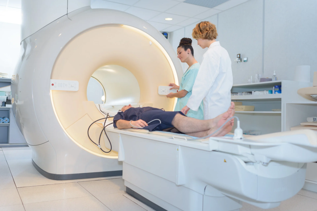
During the SPECT scan, you’ll lie on a table that slides into a scanner. The scanner will move around your head, taking images of your brain. The whole process is usually painless and takes about 30 minutes to an hour.
You’ll need to stay very quiet and not move during the scan. This helps make sure the images are clear and accurate. Sometimes, a mild sedative might be given to help you relax.
After the scan, you can usually go back to your normal activities. But, some people might feel a bit dizzy or nauseous. This is because of the radiotracer used in the scan.
Drinking lots of water is a good idea to help get rid of the radiotracer. Also, be sure to follow any care instructions given by your healthcare provider after the scan.
Key Takeaways:
Understanding SPECT scan results is complex. It involves looking closely at how blood flows through the brain. Doctors and radiologists team up to study these images. They gain insights into brain health and spot possible issues.
Neurologists search for specific patterns in SPECT scans. These patterns can show different types of dementia. They check for areas where blood flow is too high or too low.
They compare the scans to normal brain patterns. This helps them find any signs of dementia or other brain problems.
Doctors use both qualitative and quantitative methods to review SPECT scans. Qualitative means looking at the images themselves. They look for obvious problems in blood flow.
Quantitative uses software to measure blood flow levels. This gives more detailed information about blood flow issues.
| Assessment Type | Description | Advantages |
| Qualitative | Visual inspection by experienced clinicians | Quick, utilizes clinical expertise |
| Quantitative | Software-based measurement of radiotracer uptake | Precise, objective data |
Artificial Intelligence (AI) is changing how we read SPECT scans. AI can spot patterns in many images that humans might miss.
These systems make diagnoses more accurate and consistent. They help reduce the differences in opinion between doctors.
Key benefits of AI in SPECT interpretation include:
SPECT scans are key in diagnosing dementia. They show brain perfusion and help spot different types of dementia. But, they face several challenges.
One big issue with SPECT scans is false positives and negatives. False positives can happen due to patient movement, wrong radiotracer dosage, or scan artifacts. False negatives might occur in early dementia or when the disease hits areas SPECT can’t easily see.
To deal with these problems, strict imaging protocols are needed. Clinical correlation is also important. Experts say, “The accuracy of SPECT imaging depends on the scan’s technical aspects and the clinical context.”
“The interpretation of SPECT images requires a thorough understanding of brain anatomy, physiology, and the specific characteristics of the radiopharmaceutical used.” ” Expert in Nuclear Medicine
SPECT scans also involve radiation exposure. This exposure, though small, can slightly increase cancer risk. The dose from a brain SPECT scan is low but should be considered, mainly for younger patients or those getting many scans.
SPECT scans face barriers in accessibility and cost. Not all places have the right equipment or know-how. Insurance coverage also varies, making it hard for patients to get these scans.
To tackle these issues, we need:
By understanding and tackling these challenges, healthcare can better use SPECT scans. This will help improve patient care.
It’s important to know when SPECT scans are used for dementia diagnosis. SPECT scans are a key tool in neurology for checking patients with suspected dementia.
Doctors suggest SPECT scans for dementia in certain situations. This includes when patients show symptoms that are hard to diagnose with just a doctor’s evaluation. SPECT scans show specific blood flow patterns in the brain, helping doctors figure out what kind of dementia a patient has.
A SPECT scan can tell the difference between Alzheimer’s and frontotemporal dementia. It does this by showing how blood flows in different parts of the brain. This info is key for creating the right treatment plan for each patient.
Insurance for SPECT scans in the U.S. changes based on the provider and policy. Mostly, Medicare and many private plans cover SPECT scans for dementia when they’re needed. But, you might need to get approval first and show your doctor’s notes.
It’s important for patients and doctors to check insurance before getting a SPECT scan. You need to know what your policy covers and any costs you might face.
Choosing the right patients for SPECT scans is key for good results. Doctors look for people with unclear cognitive decline, unusual or fast dementia, and those hard to diagnose with usual tests.
SPECT scans also help track how a disease changes over time and if treatments work. By picking patients carefully, doctors can get the most out of SPECT scans in their practice.
SPECT is key in diagnosing and managing dementia. It shows how different types of dementia, like Alzheimer’s, affect the brain. This helps doctors understand the condition better.
Research is making SPECT even better. New radiotracers and ways to analyze images are improving its accuracy. Combining SPECT with other imaging methods will make it even more useful for diagnosing Alzheimer’s and dementia.
The future of SPECT in dementia care looks bright. It will help doctors diagnose and manage dementia more effectively. This will lead to better care for patients. As technology advances, SPECT’s role in dementia diagnosis will grow, giving doctors more tools to help their patients.
A SPECT scan is a way to see how different parts of the brain work. It helps doctors figure out if someone has dementia. This includes Alzheimer’s disease.
A SPECT scan uses a tiny bit of radioactive stuff. It goes into your blood and shows up in brain areas with more blood flow. The scan then makes a 3D picture of your brain’s blood flow.
SPECT scans look at blood flow in the brain. MRI and PET scans look at structure or how cells use sugar. SPECT is great for seeing how dementia changes blood flow.
Dementia can change how blood flows in the brain. SPECT scans can show these changes. For example, Alzheimer’s disease might show less blood flow in certain areas.
SPECT scans might spot dementia early by finding blood flow changes. But, how well they work can depend on the type of dementia and other health issues.
DaTscan is a SPECT scan that looks at dopamine in the brain. It helps doctors tell if someone has Lewy body dementia, not Alzheimer’s disease.
Each type of dementia shows its own pattern on SPECT scans. For example, frontotemporal dementia might show changes in the front and side parts of the brain. Vascular dementia might show signs of small strokes.
SPECT and FDG PET scans both show brain function, but in different ways. SPECT looks at blood flow, and FDG PET looks at sugar use. The choice depends on what the doctor needs to know.
During a SPECT scan, you’ll get a tiny amount of radioactive stuff injected. Then, you’ll sit for about 30 minutes to an hour while the scan works. You’ll need to stay very quiet.
Doctors look at SPECT results to see how the brain is working. They use both what they see and numbers to understand the scan. New tech like AI might help make these results even clearer.
SPECT scans have some downsides. They can sometimes give wrong answers, and there’s a small risk of radiation. Also, not everyone can get one because of cost or where it’s available.
SPECT scans are useful when it’s hard to tell what’s wrong with someone’s brain. Doctors look at how the person acts, their health history, and if they need more information to decide.
In the United States, insurance for SPECT scans varies. It usually covers them when they’re needed to help figure out dementia.
The future of SPECT scans looks bright. New tech and better tracers are coming. SPECT will likely keep being a key tool for checking how dementia affects the brain.
Ferrando, S., von Gunten, A., Amieva, H., & Allali, G. (2021). Brain SPECT as a biomarker of neurodegeneration. Frontiers in Neurology. https://www.ncbi.nlm.nih.gov/pmc/articles/PMC8141564/
Jagust, W. (2001). SPECT perfusion imaging in the diagnosis of Alzheimer’s disease. Neurology, 56(7), 950“956. https://doi.org/10.1212/WNL.56.7.950
Matsuda, H. (2007). Role of neuroimaging in Alzheimer’s disease, with emphasis on brain perfusion SPECT. Journal of Nuclear Medicine, 48(8), 1289“1300. https://jnm.snmjournals.org/content/48/8/1289
Papathanasiou, N. D., Tatsch, K., & Politis, M. (2012). Diagnostic accuracy of ¹ ² ³I-FP-CIT (DaTSCAN) in dementia with Lewy bodies: a systematic review and meta-analysis. European Journal of Nuclear Medicine and Molecular Imaging. https://www.sciencedirect.com/science/article/abs/pii/S1353802011003129
Subscribe to our e-newsletter to stay informed about the latest innovations in the world of health and exclusive offers!
WhatsApp us