Last Updated on November 26, 2025 by Bilal Hasdemir

At Liv Hospital, we use advanced imaging techniques in angiography interventional radiology. We diagnose and treat vascular conditions with great precision and little discomfort. Interventional radiology (IR) is a special field of medicine. It uses advanced imaging to perform treatments that are minimally invasive.
With IR angio procedures, we focus on patient care and achieving the best results. Our methods ensure top-notch angiography procedures for complex vascular diseases.
Key Takeaways
- Advanced imaging techniques are used in interventional radiology to diagnose and treat vascular conditions.
- IR angio procedures offer minimally invasive treatments with precision and minimal discomfort.
- Liv Hospital provides patient-centered care and achieves optimal outcomes through IR angio.
- World-class angiography procedures are designed to address complex vascular diseases.
- Minimally invasive treatments reduce recovery time and improve patient outcomes.
The Fundamentals of Angiography Interventional Radiology

Angiography is key in interventional radiology, showing blood vessels clearly. It’s vital for finding and treating vascular diseases. We’ll look at what angiography is, its non-invasive nature, and its big impact on vascular medicine.
Minimally Invasive Vascular Imaging Explained
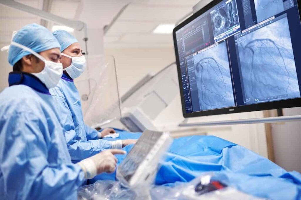
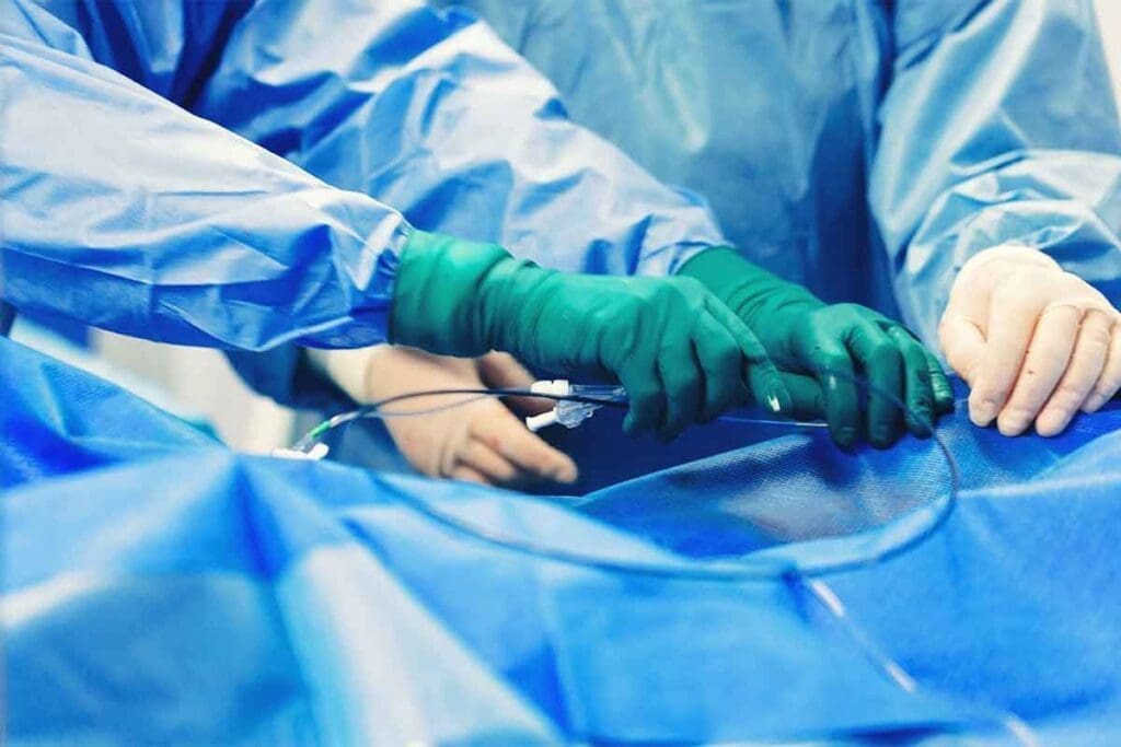
Angiography uses a catheter to inject contrast into blood vessels. Then, X-ray imaging shows these vessels. This minimally invasive method lets doctors see vascular structures without surgery.
This method has many benefits. It means less recovery time, fewer risks, and can be done as an outpatient. Interventional radiologists use their skills to guide through complex blood vessel paths.
How Angiographic Procedures Revolutionized Vascular Medicine
Angiographic procedures have changed vascular medicine a lot. They give clear images of blood vessels. This helps find blockages, aneurysms, and malformations accurately.
This clear view has changed how treatments are planned. It allows for more precise interventions.
| Condition | Diagnostic Capability | Treatment Planning |
| Blockages | Accurate visualization of stenoses | Angioplasty and stenting |
| Aneurysms | Detailed assessment of aneurysm size and morphology | Coiling or stent grafting |
| Malformations | Clear delineation of abnormal vascular structures | Embolization or surgical intervention |
The Role of IR Angiogram in Modern Healthcare
The IR angiogram is a big deal in healthcare today. It gives detailed images of blood vessels. This helps diagnose and treat many vascular conditions.
It’s especially useful in interventional radiology. It helps guide precise, non-invasive procedures. This leads to better treatments and improved lives for patients.
Advanced Imaging Technologies in Angio Vascular Procedures

Advanced imaging technologies have changed angio vascular procedures a lot. They offer clear and precise views. This has greatly improved how we diagnose and treat vascular diseases, helping patients more.
Digital Subtraction Angiography (DSA) Techniques
Digital Subtraction Angiography (DSA) is a top-notch imaging method for seeing blood vessels. It works by removing the pre-contrast image from the post-contrast one. This gives us detailed images of blood vessels, helping doctors plan treatments better.
DSA has made a big difference in interventional radiology. It lets doctors do complex procedures more accurately. It’s used a lot in angio vascular procedures, like diagnostic angiography and treatments like embolization and angioplasty.
X-Ray Angiogram and Fluoroscopy Applications
X-ray angiography and fluoroscopy are key in angio vascular procedures. X-ray angiography shows detailed blood vessel images. Fluoroscopy lets us see blood vessels in real-time during procedures.
Together, X-ray angiography and fluoroscopy help doctors do complex procedures safely and accurately. They’re used a lot in procedures like coronary angiography, peripheral angiography, and cerebral angiography.
| Imaging Modality | Application | Benefits |
| DSA | Diagnostic angiography, interventional procedures | Clear and detailed images of vascular structures |
| X-Ray Angiography | Diagnostic angiography | Detailed images of blood vessels |
| Fluoroscopy | Real-time visualization during procedures | Improved accuracy and safety |
Current Technological Innovations in Angiographic Imaging
The world of angiographic imaging is always getting better, with new tech coming out all the time. We’re seeing things like flat-panel detectors, advanced image processing, and new ways to combine imaging.
These new things have made angiographic imaging even better. They help doctors diagnose and treat vascular diseases more effectively. As tech keeps getting better, we’ll see even more improvements, leading to better care for patients.
Cerebral Angiography: Mapping Brain Vasculature
Cerebral angiography is a detailed medical imaging method. It shows the blood vessels in the brain. This is key for diagnosing and treating brain vascular issues.
It gives clear images of the brain’s blood vessels. This helps doctors understand the brain’s vascular structure and function.
Procedure Overview and Clinical Indications
Cerebral angiography starts with a catheter in an artery, usually in the groin or arm. It’s then guided to the brain’s blood vessels. A contrast agent is used to see the vessels on X-ray images.
This test is for those with suspected vascular problems. This includes aneurysms, AVMs, or stenosis.
It’s used to find the cause of stroke or TIA. It also helps plan treatments for vascular diseases.
Access Routes and Technical Considerations
The most common starting point is the femoral artery. The choice depends on the patient’s body, the procedure, and the doctor’s preference. The right tools and equipment are used for safe and accurate imaging.
Technologies like digital subtraction angiography (DSA) improve image quality. DSA makes blood vessels clearer by removing background images.
Post-Procedure Care and Potential Complications
After the test, patients are watched for any issues. This includes bleeding, hematoma, or damage to the blood vessels. The access site and neurological status are closely monitored.
Complications, though rare, can be serious. These include stroke, TIA, or allergic reactions to the contrast. Quick action is needed to avoid lasting damage.
Pulmonary Angiography: Evaluating Lung Circulation
Pulmonary angiography is key in interventional radiology. It gives detailed views of lung blood vessels. This is vital for diagnosing and treating lung vascular diseases.
Step-by-Step Procedure Guide and Patient Preparation
Pulmonary angiography has several steps. First, getting ready is important. Patients often need to fast for a few hours beforehand. They might also stop certain medicines that could affect the test.
The test starts with a catheter being put into a vein, usually in the groin or arm. Then, using X-ray images, it’s guided to the lung’s arteries. Next, a contrast agent is injected to see the blood vessels clearly.
Key Steps in Pulmonary Angiography:
- Patient preparation and assessment
- Catheter insertion and guidance
- Contrast agent injection
- Imaging and data acquisition
Diagnosing Pulmonary Embolism and Vascular Abnormalities
Pulmonary angiography is great for finding pulmonary embolism. This is a serious condition where a blood clot blocks a lung artery. The detailed images help doctors see how big the blockage is and plan treatment.
It can also spot other issues like AVMs and stenosis in the pulmonary arteries. The clear images help doctors make accurate diagnoses and plan treatments.
Therapeutic Applications in Pulmonary Vascular Disease
Pulmonary angiography is not just for diagnosis. It can also help treat diseases. For example, it can deliver medicine to dissolve blood clots in the lungs.
“Pulmonary angiography not only aids in the diagnosis of pulmonary vascular diseases but also offers a pathway for targeted therapeutic interventions, improving patient outcomes.” – Interventional Radiologist
Recovery and Follow-up Protocols
After the test, patients are watched for any problems. They are told to rest, drink lots of water, and avoid hard activities for a while.
It’s important to follow up to make sure the patient gets better. This might include more tests to check on the treated area.
| Procedure Aspect | Description | Patient Care |
| Pre-procedure | Patient assessment and preparation | Fasting, medication adjustment |
| During procedure | Catheter insertion, contrast injection | Monitoring vital signs |
| Post-procedure | Recovery, complication monitoring | Rest, hydration, follow-up |
Renal Angiography: Kidney Vascular Assessment
Kidney vascular assessment through renal angiography has changed how we diagnose renovascular diseases. This imaging technique lets us see the renal vessels in detail. It helps us diagnose and treat kidney vascular conditions.
Technical Approach for Renal Vessel Imaging
To image the renal vessels, we access the renal arteries with a catheter. This catheter goes through the femoral artery. We use digital subtraction angiography (DSA) to get clear images of the blood vessels.
Diagnosing Renal Artery Stenosis and Renovascular Hypertension
Renal angiography is key in finding renal artery stenosis. This is when the renal arteries narrow. This narrowing can cause high blood pressure due to less blood flow to the kidney. We can see the arteries and find stenosis and other problems, helping us treat them quickly.
| Condition | Diagnostic Feature | Treatment Approach |
| Renal Artery Stenosis | Narrowing of renal arteries | Angioplasty with stenting |
| Renovascular Hypertension | High blood pressure due to reduced renal blood flow | Management of hypertension, possible angioplasty |
Interventional Applications in Kidney Disease Management
Renal angiography also helps in treating kidney disease. We can do angioplasty to widen narrowed arteries. We then put in stents to keep them open. This improves blood flow to the kidneys.
Patient Outcomes and Clinical Benefits
Using renal angiography has greatly improved patient care. It helps us find and treat problems like renal artery stenosis. This leads to better kidney function and overall health for patients.
The benefits include better blood pressure control, less risk of kidney failure, and a better quality of life. Patients with renovascular diseases see big improvements.
Liver Angiography: Hepatic Vascular Evaluation
We use liver angiography to understand liver blood vessel conditions. This test is key for checking the liver’s blood system. It helps diagnose and treat liver diseases.
Procedure Details and Contrast Media Considerations
Liver angiography involves putting a catheter in the hepatic artery. Then, contrast media is injected to see the liver’s blood vessels with X-ray. Choosing the right contrast media is important for clear images and patient safety. Complications from contrast media are a concern, and we consider each patient’s needs before starting.
Applications in Hepatocellular Carcinoma Diagnosis and Treatment
Liver angiography is crucial for finding and treating HCC. It shows the tumor’s blood supply, helping plan treatments like TACE or radioembolization. It makes these treatments more effective by showing the blood vessels in detail.
Portal Hypertension Assessment and Intervention
Portal hypertension is high pressure in the portal vein, a liver disease complication. Liver angiography helps measure this pressure and guide treatments to lower it. It helps create TIPS, a procedure to reduce pressure and prevent problems.
Case Studies and Clinical Efficacy
Many case studies show liver angiography’s effectiveness in treating liver diseases. For example, a study on TACE for HCC found better results with liver angiography. This highlights the value of angiographic imaging in treating liver diseases today.
Peripheral Angiography: Extremity Vascular Imaging
In interventional radiology, peripheral angiography is crucial. It helps diagnose and treat vascular issues in limbs. This improves patients’ lives and outcomes.
Lower Extremity Angiography for Peripheral Arterial Disease
Peripheral arterial disease (PAD) affects the lower limbs. It causes pain and can lead to serious issues. We use lower extremity angiography to see the blood vessels, find blockages, and treat them.
Key applications of lower extremity angiography include:
- Diagnosing PAD and assessing its severity
- Guiding angioplasty and stenting procedures
- Evaluating vascular anatomy for surgical planning
Upper Extremity Vascular Assessment Techniques
Upper extremity angiography helps with arm issues like thoracic outlet syndrome. It checks if blood vessels are open, finds blockages, and plans treatments.
The benefits of upper extremity angiography include:
- Accurate diagnosis of vascular conditions
- Guidance for minimally invasive interventions
- Preoperative planning for surgical procedures
Interventional Applications for Limb Ischemia
Limb ischemia is a serious issue that needs quick treatment. We use peripheral angiography to guide treatments like angioplasty and stenting. This helps restore blood flow to the limb.
Comparative Outcomes with Traditional Surgical Approaches
Peripheral angiography and interventions have many benefits over surgery. They lead to faster recovery, less risk, and better results. We compare these methods to find the best treatment for each patient.
Comparative benefits include:
- Reduced risk of complications
- Shorter hospital stays
- Improved long-term patency rates
Coronary Angiography: Heart Vessel Examination
We use coronary angiography to find and treat heart blood vessel problems. This method is key in cardiology. It gives clear images of the heart’s arteries.
Cardiac Catheterization Methodology
Cardiac catheterization is a main technique in coronary angiography. A catheter is put into an artery in the leg or arm. It’s then guided to the heart. Contrast media is used to see the arteries on an X-ray.
- Preparation involves patient assessment and informed consent.
- The procedure is done under local anesthesia.
- Continuous monitoring ensures patient safety.
Diagnosing and Grading Coronary Artery Disease
Coronary angiography is key in finding coronary artery disease (CAD). It spots blockages and other issues in the arteries. The level of blockage is graded to show how severe CAD is.
Interventional Applications in Acute Coronary Syndromes
In cases of acute coronary syndromes, like heart attacks, coronary angiography finds the main problem. Angioplasty and stenting can then be done to open the artery.
- Emergency coronary angiography is lifesaving in heart attacks.
- Interventions are chosen based on what the angiogram shows.
- Quick action helps patients get better.
Risk Stratification and Patient Selection
It’s important to figure out the risk for patients getting coronary angiography. Things like kidney function, how much contrast is used, and how complex the procedure is matter. Careful patient selection lowers risks and improves results.
Knowing the risks and benefits helps doctors make better choices.
Conclusion: Advancing Patient Care Through Angiography Procedures
Angiography procedures have changed how we diagnose and treat vascular conditions. They have greatly improved patient care. We’ve looked at different types of angiography, like cerebral and coronary, showing their key role in healthcare today.
Ir angio techniques allow for detailed imaging and precise interventions. This has led to better patient results and shorter recovery times. With ongoing tech advancements, we’ll see even more improvements in treating vascular diseases.
Healthcare providers can now offer more effective, less invasive treatments. This boosts patients’ quality of life worldwide. As we keep improving angiography, our goal is to provide top-notch healthcare. We’re dedicated to supporting patients globally with comprehensive care and guidance.
FAQ
What is angiography interventional radiology?
Angiography interventional radiology is a medical field. It uses advanced imaging to diagnose and treat vascular conditions. This is done through minimally invasive procedures.
What are the benefits of IR angio procedures?
IR angio procedures have many benefits. They reduce the risk of complications and cause less pain. They also have shorter recovery times compared to traditional surgery.
What is digital subtraction angiography (DSA)?
Digital subtraction angiography (DSA) is a technique used in angiography. It produces detailed images of blood vessels. This is done by subtracting images taken before and after contrast media injection.
What is the role of fluoroscopy in angiography?
Fluoroscopy plays a key role in angiography. It provides real-time imaging guidance during procedures. This allows for precise placement of catheters and other devices.
What is cerebral angiography used for?
Cerebral angiography is used to map the brain’s blood vessels. It diagnoses vascular abnormalities. It also guides treatments for conditions like aneurysms and arteriovenous malformations.
What is pulmonary angiography?
Pulmonary angiography evaluates lung circulation. It diagnoses pulmonary embolism. It also assesses vascular abnormalities in the lungs.
What are the applications of renal angiography?
Renal angiography assesses kidney vasculature. It diagnoses renal artery stenosis. It guides treatments for renovascular hypertension and other kidney diseases.
What is liver angiography used for?
Liver angiography evaluates hepatic vasculature. It diagnoses and treats hepatocellular carcinoma. It also assesses portal hypertension.
What is peripheral angiography?
Peripheral angiography images extremity vasculature. It diagnoses peripheral arterial disease. It guides treatments for limb ischemia.
What is coronary angiography?
Coronary angiography examines heart vessels. It diagnoses coronary artery disease. It guides treatments for acute coronary syndromes.
What are the risks associated with angiography procedures?
Angiography procedures are generally safe. However, they carry risks like bleeding, infection, and contrast media reactions. These risks can be minimized with proper patient selection and preparation.
How do angiography procedures improve patient outcomes?
Angiography procedures improve patient outcomes. They provide accurate diagnoses and guide effective treatments. They also reduce the need for more invasive surgeries.
What are the different types of angiography?
There are several types of angiography. These include cerebral, pulmonary, renal, liver, peripheral, and coronary angiography. Each has its specific applications and techniques.
What is the role of contrast media in angiography?
Contrast media enhances the visibility of blood vessels and structures. It allows for accurate diagnoses and effective treatments in angiography.
How is patient preparation done for angiography procedures?
Patient preparation for angiography involves several steps. It includes reviewing medical history, physical examination, laboratory tests, and instructions on fasting and medication management before the procedure.
Reference
- National Heart, Lung, and Blood Institute. (2023). Peripheral artery disease. https://www.nhlbi.nih.gov/health/peripheral-artery-disease


