Last Updated on November 26, 2025 by Bilal Hasdemir

It’s key to know the blood vessels that feed the head and neck. This is because the brain needs a lot of oxygen. Without it, the brain can suffer greatly.Learn about arteries of the head and neck with labeled diagrams, names, and essential anatomy details.
At Liv Hospital, we focus on the internal carotid arteries and vertebral arteries. They are vital for the brain’s blood supply. Our team uses this knowledge to offer advanced care.
We’ll show you the names, flowchart, and labeled anatomy of these arteries. This will help you understand them well for your work.
Key Takeaways
- Understanding the 7 key arteries is vital for precise diagnosis.
- The internal carotid arteries play a key role in brain blood supply.
- Labeled anatomy is essential for clinical training and surgery.
- The flowchart of these arteries helps us understand their complexity.
- Knowing these arteries is critical for effective treatment plans.
The Vascular Framework of the Head and Neck

The head and neck have a complex blood supply system. It’s vital for the brain and facial structures. This system includes many arteries from the aortic arch, branching to different head and neck areas.
Essential Functions of Cranial Blood Supply
The cranial blood supply keeps the brain healthy. The common carotid arteries split into the internal and external carotid arteries. The internal carotid arteries go into the brain through the carotid canal.
The vertebral arteries start from the subclavian arteries. They go up through the neck vertebrae. They’re key for the brainstem, cerebellum, and posterior inferior cerebellar arteries.
Overview of Major Arterial Systems
The head and neck have two main arterial systems: the carotid and vertebral arteries. The carotid arteries split into the internal and external carotid arteries. Each supplies different parts of the head and neck.
The internal carotid artery has important branches like the ophthalmic artery and anterior choroidal artery. The external carotid artery has branches for the face and neck, like the superior thyroid artery and lingual artery.
Knowing the major arterial systems helps in diagnosing and treating head and neck issues.
Common Carotid Arteries: The Primary Blood Highways

Understanding the common carotid arteries is key to knowing the head and neck’s blood flow. These arteries are the main paths for blood to reach the brain and other important areas.
Origin and Course from the Neck to Head
The common carotid arteries start from different places on each side of the body. The right one comes from the brachiocephalic artery, and the left comes directly from the aortic arch. They go up through the neck, staying in front of the prevertebral fascia and within the carotid sheath.
They are with the internal jugular vein and the vagus nerve. The arteries keep going without splitting until they hit the upper part of the thyroid cartilage. Then, they split into the internal and external carotid arteries. This split is a key spot.
Anatomical Landmarks for Identification
To find the common carotid arteries, look for certain landmarks. They are in the carotid triangle, which is between the sternocleidomastoid muscle, the superior belly of the omohyoid muscle, and the sternocleidomastoid’s front edge. You can feel them against the cervical vertebrae’s transverse processes, near the cricoid cartilage.
The Carotid Bifurcation: Clinical Significance
The carotid bifurcation is very important both anatomically and clinically. It’s a common place for plaque to build up, which can cause stenosis and stroke risk. Knowing the anatomy of this area is vital for vascular surgeons and radiologists.
It shows how important it is to know the anatomy well when dealing with vascular diseases. As we learn more about the head and neck’s blood supply, the common carotid arteries and their bifurcation are key.
Internal Carotid Arteries: Cerebral Circulation
It’s key to know about the internal carotid arteries for treating brain diseases. These arteries are vital for the brain’s blood flow. They supply blood to big parts of the brain’s outer layer.
Four Segments: Cervical, Petrous, Cavernous, and Cerebral
The internal carotid arteries split into four parts: cervical, petrous, cavernous, and cerebral. Each part has its own special features and importance.
- Cervical Segment: This part starts from the carotid bifurcation to the carotid canal.
- Petrous Segment: Inside the petrous part of the temporal bone, it’s surrounded by veins and nerves.
- Cavernous Segment: Then, it goes through the cavernous sinus. Here, it’s near the abducens nerve and the sinus itself.
- Cerebral Segment: This part branches out to supply the brain.
Major Branches and Brain Regions Supplied
The internal carotid arteries have several key branches for brain blood flow. These include the ophthalmic artery, posterior communicating artery, anterior choroidal artery, anterior cerebral artery, and middle cerebral artery.
The anterior cerebral artery feeds the medial brain surface. The middle cerebral artery covers a big area of the brain’s side, including parts of the frontal, parietal, and temporal lobes.
Role in Stroke and Cerebrovascular Disease
The internal carotid arteries play a big role in stroke and brain diseases. Blockages or narrowing can cause serious brain damage. This is because less blood reaches the brain.
Knowing how the internal carotid arteries work is vital for treating brain diseases. We use tools like angiography and Doppler ultrasound to check their health.
External Carotid Arteries and Their Branches
The external carotid arteries are key in the head and neck’s blood flow. They branch from the common carotid arteries. They supply blood to the face and neck.
Superior Thyroid Artery: First Branch
The superior thyroid artery is the first branch of the external carotid artery. It’s vital for the thyroid gland’s function. It brings oxygen and nutrients to the gland.
Facial and Maxillary Arteries: Facial Supply
The facial artery is a major branch, supplying the face. It runs to the front of the face. The maxillary artery, in contrast, supplies the deep face, including the nasal cavity and palate.
Occipital and Posterior Auricular Arteries: Posterior Head Supply
The occipital artery feeds the back of the scalp. The posterior auricular artery supplies the area behind the ear. These arteries are key for the back of the head and neck.
Terminal Branches
The external carotid artery ends by splitting into the superficial temporal and maxillary arteries. The superficial temporal artery goes to the scalp. The maxillary artery, as mentioned, goes to the deep facial structures.
Vertebral Arteries: The Posterior Circulation
Understanding the vertebral arteries is key to knowing the posterior circulation. It’s important for the blood supply to the brain. The vertebral arteries are a major part of the brain’s blood system.
Origin from Subclavian and Course Through Foramina
The vertebral arteries start from the subclavian arteries. They go up through the neck’s cervical vertebrae. This path is essential for their role in brain blood supply.
The vertebral arteries start from the back of the subclavian arteries. They move up through the neck. They pass through the cervical vertebrae’s transverse foramina from C1 to C6.
Intracranial Segments and Key Branches
When they enter the skull, the vertebral arteries branch off. They give rise to the anterior spinal artery and the posterior inferior cerebellar artery (PICA). These supply the medulla oblongata and the cerebellum.
The intracranial segments of the vertebral arteries are critical for the posterior circulation. They merge to form the basilar artery. This artery supplies the brainstem and cerebellum.
Formation of Basilar Artery and Posterior Circulation
The two vertebral arteries merge at the pons base to form the basilar artery. The basilar artery then branches off into the AICA and SCA. These arteries supply the brain before ending in the posterior cerebral arteries.
This network ensures a strong blood supply to the brain’s posterior parts. This includes the cerebellum and brainstem.
| Segment | Branches | Supply Region |
| Pre-foraminal | None significant | Neck structures |
| Foraminal | Muscular branches | Cervical vertebrae muscles |
| Intracranial | Anterior spinal, PICA | Medulla, Cerebellum |
| Basilar Artery | AICA, SCA, PCA | Pons, Cerebellum, Occipital lobe |
Thyrocervical Trunk: Critical Neck Vasculature
The thyrocervical trunk is a key artery that comes from the subclavian artery. It’s vital for blood flow to the neck and shoulder. This artery is a major branch of the subclavian artery.
Origin and Anatomical Relations
The thyrocervical trunk starts from the top of the subclavian artery. It’s found near the medial border of the scalenus anterior muscle. It arises between the scalenus anterior and the longus colli muscles. Knowing where it starts and its location is key for medical procedures.
Major Branches: Inferior Thyroid, Suprascapular, and Transverse Cervical
The thyrocervical trunk has several important branches. These branches supply different parts of the neck. The main branches are:
- Inferior Thyroid Artery: It feeds the thyroid gland.
- Suprascapular Artery: It supplies the supraspinatus and infraspinatus muscles.
- Transverse Cervical Artery: It helps the neck muscles and the shoulder area.
| Branch | Supply Region |
| Inferior Thyroid Artery | Thyroid gland |
| Suprascapular Artery | Supraspinatus and infraspinatus muscles |
| Transverse Cervical Artery | Neck muscles and shoulder region |
Clinical Relevance in Thyroid Procedures
The thyrocervical trunk and its branches are very important in thyroid surgeries. Knowing its anatomy helps surgeons avoid problems. It ensures the thyroid gland gets enough blood.
In summary, the thyrocervical trunk is essential for neck blood supply. Its branches, like the inferior thyroid, suprascapular, and transverse cervical arteries, are vital. They supply the neck and shoulder area.
Arteries of the Head: A Detailed Look
Knowing how blood flows to the head is key for treating many health issues. The head’s blood supply comes from a network of arteries in the neck.
Intracranial Arterial Networks
The brain’s blood supply comes from a complex network inside the skull. This network includes the Circle of Willis, made from several major arteries.
Circle of Willis: Structure and Function
The Circle of Willis is vital for the brain’s blood supply. It’s at the brain’s base, formed by the internal carotid and vertebral arteries. It acts as a backup if one artery is blocked.
Collateral Pathways and Clinical Importance
Collateral pathways help keep blood flowing to the brain when main paths are blocked. The Circle of Willis is a key backup path. Knowing about these paths is important for treating strokes and other brain diseases.
| Component | Origin | Function |
| Internal Carotid Artery | Arises from the common carotid artery | Supplies anterior circulation to the brain |
| Vertebral Artery | Arises from the subclavian artery | Supplies posterior circulation to the brain |
Labeled Anatomy of Head and Neck Vessels
Labeled neck vessels are key in clinical training. They help us understand human anatomy better. The blood vessels in the head and neck are vital for delivering oxygen and nutrients.
Knowing this anatomy is critical for diagnosing and treating vascular issues.
Key Anatomical Landmarks for Vessel Identification
To identify blood vessels in the head and neck, knowing key landmarks is essential. The common carotid artery splits into the internal and external carotid arteries. The internal carotid artery goes to the brain, while the external carotid artery supplies the face and neck.
- The sternocleidomastoid muscle marks the common carotid artery’s location.
- The carotid bifurcation is at the upper border of the thyroid cartilage.
- The internal jugular vein parallels the common carotid artery.
Cross-Sectional Anatomy and Relationships
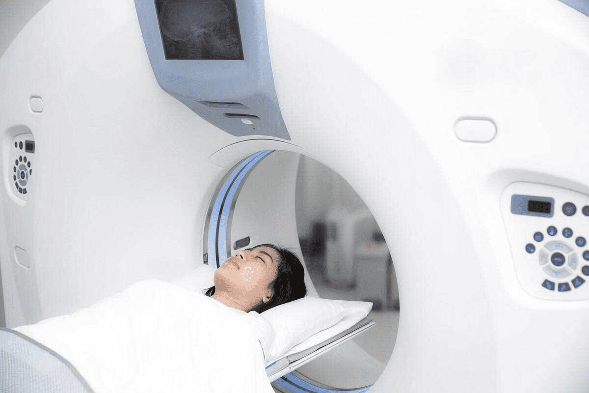
CT and MRI have changed how we see head and neck vasculature. These scans show the complex relationships between vessels, muscles, and other structures.
The vertebral artery runs through the foramina transversaria of cervical vertebrae. Knowing these relationships helps in interpreting scans and planning surgeries.
“The use of cross-sectional imaging has significantly improved the diagnosis and treatment of vascular diseases in the head and neck.”
— Expert Opinion
Imaging Correlations: CT, MRI, and Angiography
Different imaging methods help us see the blood vessels in the head and neck. CT angiography shows arterial anatomy well. MRI angiography looks at both arteries and veins without radiation.
- CT angiography is great for checking atherosclerosis.
- MRI is good for looking at vascular malformations and tumors.
- Conventional angiography is the best for some procedures.
By using labeled anatomy and imaging, healthcare professionals can improve diagnosis and treatment for vascular conditions in the head and neck.
Flowchart of Head and Neck Arterial Supply
Knowing how arteries supply the head and neck is key for treating vascular issues. The network of arteries in this area is complex. It starts at the aortic arch and branches into arteries for different parts.
From Aortic Arch to Terminal Branches
The journey starts at the aortic arch. Here, the common carotid and vertebral arteries begin. The common carotid splits into internal and external carotid arteries. Each supplies different areas.
The internal carotid artery mainly feeds the brain. The external carotid branches to the face and neck. The vertebral arteries join to form the basilar artery, vital for the brain’s back side.
Branching Patterns and Variations
Arteries in the head and neck branch differently in everyone. Knowing these differences is key for correct diagnosis and treatment.
| Artery | Origin | Supply Region |
| Common Carotid | Aortic Arch | Neck and Head |
| Internal Carotid | Common Carotid | Brain |
| External Carotid | Common Carotid | Face and Neck |
| Vertebral | Subclavian | Posterior Brain |
Clinical Decision-Making Based on Vascular Anatomy
Doctors need to know the arterial supply flowchart for vascular treatments. The anatomy can affect surgery planning and results.
For example, knowing the external carotid’s branches helps in procedures like embolization for malformations.
Understanding the head and neck arterial supply flowchart helps doctors. It leads to better patient care.
Venous Drainage: Complementing the Arterial System
The venous drainage system of the head and neck is vital. It works with the arterial system to ensure blood flows well. This is key for good circulation and health.
Internal and External Jugular Veins
The internal and external jugular veins are important for draining blood. The internal jugular vein collects blood from the brain, face, and neck. It starts from the sigmoid sinus and goes down to join the subclavian vein.
The external jugular vein drains blood from the face, scalp, and neck. It lies just under the sternocleidomastoid muscle and merges with the subclavian vein. Both veins are essential for blood return from the head and neck.
Dural Venous Sinuses
The dural venous sinuses are channels between the dura mater layers. They get blood from the brain’s veins and cerebrospinal fluid. The superior sagittal sinus runs along the brain’s midline, draining into the confluence of sinuses.
These sinuses are vital for brain venous drainage. They are closely tied to the brain’s blood circulation.
Venous Anastomoses and Clinical Significance
Venous anastomoses connect different venous channels. They provide backup paths for blood flow. In the head and neck, they are key for dealing with venous blockages.
Knowing about venous anastomoses is vital for doctors. It helps in managing conditions like venous thrombosis. These structures are critical for keeping the brain’s blood flowing and overall health.
Conclusion: Mastering Head and Neck Vascular Anatomy
Understanding the head and neck’s vascular anatomy is key for good clinical training and surgery. The arteries in this area are vital for blood flow to the brain, face, and neck. Knowing them well is critical for diagnosing and treating vascular diseases.
We’ve looked at the main arteries like the common carotid, internal carotid, external carotid, and vertebral arteries. We’ve also seen how important they are in treating cerebrovascular disease and stroke.
Knowing the head and neck’s vascular anatomy helps doctors make better decisions during surgery. It’s a basic part of understanding vascular diseases. As we keep improving in training and technology, knowing this anatomy is more important than ever for good patient care.
FAQ
What are the main arteries that supply blood to the head and neck?
The main arteries for the head and neck are the common carotid arteries and the vertebral arteries. The common carotid arteries split into the internal and external carotid arteries. These arteries supply different parts of the head and neck.
What is the significance of understanding the carotid bifurcation?
Knowing the carotid bifurcation is key for medical training and surgery. It’s a vital spot for carotid artery disease and stroke. The carotid bifurcation is where the common carotid artery splits into the internal and external carotid arteries.
What are the four segments of the internal carotid artery?
The internal carotid artery has four segments: cervical, petrous, cavernous, and cerebral. Each segment has unique features and supplies different brain areas.
What is the role of the vertebral arteries in the posterior circulation?
The vertebral arteries are vital for the posterior circulation. They supply blood to the brainstem, cerebellum, and posterior inferior cerebellar arteries. They merge to form the basilar artery, which continues to supply the posterior circulation.
What is the Circle of Willis, and what is its clinical importance?
The Circle of Willis is a key network in the brain. It provides backup blood flow to the brain. Knowing the Circle of Willis is vital for treating brain diseases.
How do the external carotid arteries supply the face and neck?
The external carotid arteries supply the face and neck through their branches. These include the superior thyroid, facial, maxillary, occipital, and posterior auricular arteries. They provide blood to various face and neck areas.
What is the thyrocervical trunk, and what is its clinical relevance?
The thyrocervical trunk is a subclavian artery branch. It supplies blood to the thyroid gland, neck, and shoulders. It’s important in thyroid procedures for its blood supply to the thyroid gland.
What is the importance of understanding the venous drainage of the head and neck?
Knowing the venous drainage of the head and neck is key for diagnosing and treating conditions. The internal and external jugular veins, dural venous sinuses, and venous anastomoses are critical for draining blood.
How do imaging modalities like CT, MRI, and angiography help in understanding head and neck vascular anatomy?
Imaging like CT, MRI, and angiography gives valuable insights into head and neck vascular anatomy. They help identify landmarks, diagnose diseases, and plan surgeries.
Why is mastering head and neck vascular anatomy important for clinical practice and surgical training?
Mastering head and neck vascular anatomy is essential for medical practice and training. It lays the groundwork for understanding vascular diseases. It helps healthcare professionals diagnose and treat conditions effectively and safely.
References
- StatPearls. (2023). Anatomy, Head and Neck: Carotid Arteries. In StatPearls https://www.ncbi.nlm.nih.gov/books/NBK545238/
- Jones, O. (2023, February). Major arteries of the head and neck. TeachMeAnatomy. https://teachmeanatomy.info/neck/vessels/arterial-supply/
- Foran, P., & Co-authors. (2023). Clinical basis for the knowledge of anatomy of the carotid artery: A review article. Yenagoa Medical Journal, 5(2), 24-29. https://yenagoamedicaljournal.net/clinical-basis-for-the-knowledge-of-anatomy-of-the-carotid-artery-a-review-article/


