Last Updated on November 27, 2025 by Bilal Hasdemir
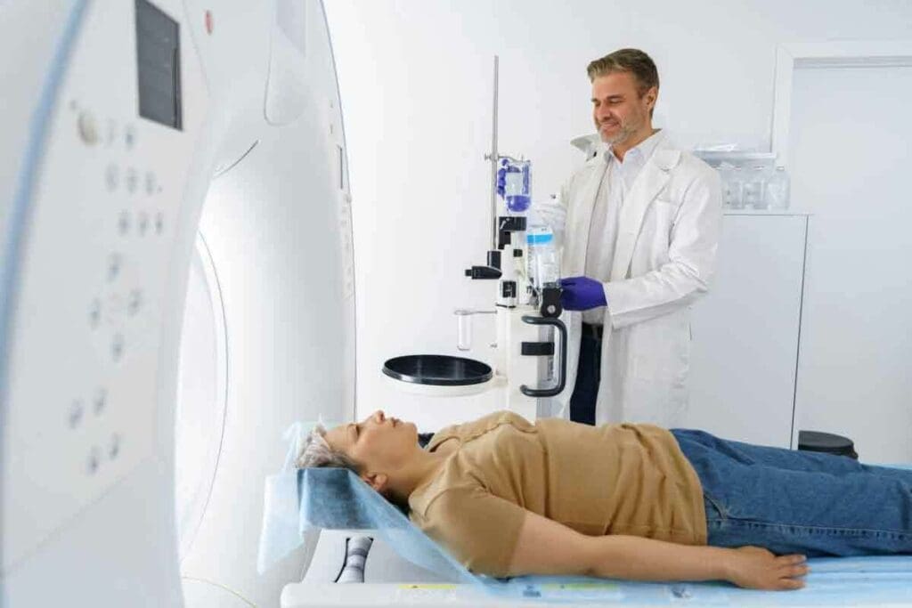
At Liv Hospital, we use top-notch bone scan machines to get clear bone scan pictures. These pictures help us spot and figure out different bone problems. A bone scan uses a special radioactive tracer to find bone damage or disease. It’s done in a hospital’s nuclear medicine unit.
These bone scan images give us important info about bone health. They help doctors diagnose and treat issues like fractures, tumors, and infections well.
Key Takeaways
- Bone scan pictures help detect bone-related conditions early.
- The procedure involves using a radioactive tracer.
- It’s performed in a nuclear medicine unit.
- Detailed images aid in diagnosis and treatment.
- Conditions like fractures, tumors, and infections can be identified.
The Fundamentals of Nuclear Bone Scanning
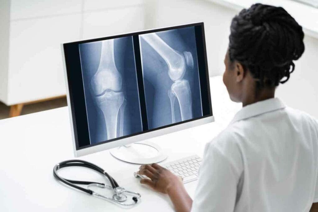
Nuclear bone scanning uses radiotracers to show bone activity. It’s a key tool in medicine, helping doctors see bone health clearly.
This method uses small amounts of radioactive tracers. They build up in bones that are very active. This helps find issues like fractures, infections, and cancers.
How Radiotracers Illuminate Bone Metabolism
Radiotracers are key in nuclear bone scanning. They show where bones are most active. A nuclear bone scan machine then takes pictures of these areas.
Using radiotracers has many benefits. It’s very good at finding bone problems. It can scan the whole skeleton at once. And it’s safe, with low radiation.
The Role of Technetium-99m in Bone Imaging
Technetium-99m is the top choice for bone scanning. It’s short-lived and emits the right kind of radiation. It sticks to bone where it’s most active.
Technetium-99m has many pluses. It gives clear images of bone activity. It’s safe, leaving the body quickly. And it spots many bone issues, from small fractures to cancer.
Knowing how Technetium-99m works helps doctors use it better. This improves care for patients.
Bone Scan Pictures: What They Reveal About Skeletal Health
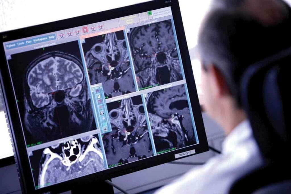
Bone scan pictures give us a peek into our skeletal health. They show details that are key for diagnosis and treatment. These images are more than just pictures; they hold important info about bone activity.
Healthcare experts can spot different skeletal system issues by looking at these images.
Decoding the Visual Language of Bone Scans
The language of bone scans is complex. “Hot spots” on the image mean high bone activity. This could be due to fractures, infections, or tumors. “Cold spots” show low bone activity, which might mean other issues.
A leading expert says,
“Bone scans are invaluable in detecting skeletal abnormalities, providing a complete view of bone health that other imaging might miss.”
It’s key to understand bone scan pictures for accurate diagnosis and treatment planning.
Distinguishing Normal vs. Abnormal Bone Activity
Telling normal from abnormal bone activity is vital. Normal bone activity looks even on the scan. Abnormal activity shows up as hot or cold spots. For example, a fracture might show up as a hot spot because of bone repair.
Bone scan pictures can spot many conditions, like cancer, infections, and metabolic bone diseases. They help doctors find problems early, track disease, and check treatment success. As we learn more about bone scan technology, it’s clear these images are a strong tool for skeletal health care.
Inside the Bone Scan Machine: Components and Technology
Bone scan machines are complex devices. They have key parts and technology that work together. These machines, also called gamma cameras, detect signals from radiotracers in the body.
Gamma Camera Systems and Detection Mechanisms
The gamma camera is the heart of a bone scan machine. It captures radiation patterns and turns them into images. This uses scintillation detectors to change gamma rays into light. Then, photomultiplier tubes make these light signals strong enough to see.
To learn more about bone scans, check out RadiologyInfo.org. It’s a great place for detailed info on radiologic procedures.
Patient Table Design and Imaging Configurations
The patient table is key for good images. Modern machines have tables that move smoothly. This lets them scan the whole body in one go.
| Feature | Description | Benefit |
| Gamma Camera System | Captures gamma rays emitted by radiotracers | Detailed imaging of bone activity |
| Patient Table Design | Smooth and precise movement under the gamma camera | Comprehensive whole-body scanning |
| Scintillation Detectors | Convert gamma rays into visible light | Accurate detection of radiotracer signals |
Modern bone scan machines use advanced technology. They give high-quality images. These images help doctors diagnose and monitor skeletal conditions.
Key Fact #1: Bone Scan Images Detect Fractures Invisible to X-rays
We use bone scan images to find fractures that X-rays can’t see. This helps us diagnose and treat early. It’s key for stress fractures or injuries that X-rays miss.
Bone scan technology is very sensitive. It spots small changes in bone activity. This is great for catching stress fractures and early injuries, so we can act fast.
Stress Fractures and Early Injury Detection
Stress fractures are hard to spot with regular imaging. Bone scan images show them by highlighting active bone areas.
- Early detection of stress fractures
- Identification of bone stress before it becomes a full fracture
- Monitoring of bone health in athletes or individuals with high-risk activities
Comparative Sensitivity with Other Imaging Methods
Bone scan images are better than X-rays for finding fractures early. X-rays might not catch them until they’re more obvious.
| Imaging Method | Sensitivity for Fracture Detection |
| X-ray | Moderate |
| Bone Scan | High |
| CT Scan | High |
Using bone scan images shows how important advanced tools are in medicine. They help us give better diagnoses and treatment plans to our patients.
Key Fact #2: How “Hot Spots” and “Cold Spots” Guide Diagnosis
In bone scan imaging, “hot spots” and “cold spots” are key. They help us find the right diagnosis and treatment. These spots show us where the bone is active or not, helping us spot different bone problems.
Interpreting Areas of Increased Tracer Uptake
“Hot spots” on a bone scan mean the tracer is more concentrated. This usually means the bone is working hard, like when there’s a fracture, infection, or tumor. We look at how bright the spot is, where it is, and how it looks to figure out what’s going on.
For example, a “hot spot” in a bone that bears weight might mean a stress fracture. But if there are many “hot spots” all over, it could be a sign of cancer spreading to the bones. By looking at these patterns, we can start to guess what might be wrong and what to do next.
Understanding Regions of Decreased Bone Activity
“Cold spots” show up when there’s less tracer than expected. This can mean things like bone death, cysts, or areas where the bone isn’t working right. It’s just as important to understand “cold spots” as “hot spots” because they both tell us about bone health.
The table below shows the main differences between “hot spots” and “cold spots” and what they might mean for your health.
| Feature | Hot Spots | Cold Spots |
| Tracer Uptake | Increased | Decreased |
| Typical Causes | Fractures, infections, tumors | Avascular necrosis, bone cysts, suppressed bone activity |
| Clinical Implication | Indicates heightened bone metabolism | Suggests reduced or absent bone activity |
By looking at both “hot spots” and “cold spots” on bone scans, we get a full picture of a patient’s bones. This info is key for making the right diagnosis, planning treatment, and keeping an eye on how the disease is doing or how well treatment is working.
Key Fact #3: Bone Scans Excel at Cancer Detection and Monitoring
Bone scans are key in finding cancer that has spread to bones. They use special machines to spot unusual bone activity. This can mean cancer cells are present.
Identifying Metastatic Bone Disease
Metastatic bone disease happens when cancer spreads to bones from other parts. Bone scans are great at finding this, mainly in cancers like prostate, breast, and lung.
The machine uses a tiny bit of radioactive material to show bone activity. Cancer changes bone structure, so it takes up more of the material. This shows up as “hot spots” on the scan.
Tracking Treatment Response in Skeletal Malignancies
Bone scans also help see how well treatment is working. By comparing scans, we can tell if treatment is working or if the disease is getting worse.
This info is key for changing treatment plans. It helps make sure patients get the best care. Watching how bone metastases change over time is very important.
Comparison of Imaging Modalities for Cancer Detection
| Imaging Modality | Sensitivity for Bone Metastases | Use in Cancer Detection |
| Bone Scan | High | Excellent for detecting bone metastases |
| CT Scan | Moderate | Good for assessing bone structure and soft tissue |
| MRI | High | Excellent for soft tissue evaluation and marrow involvement |
| PET Scan | Very High | Excellent for detecting metabolically active cancer cells |
In summary, bone scans are very important for finding and tracking cancer. They help doctors make better decisions and improve patient care.
Key Fact #4: Whole Body Bone Scan Machines Provide Comprehensive Assessment
Whole body bone scan machines have changed nuclear medicine a lot. They let doctors see the whole skeleton at once. This helps find problems in bones all over the body, making diagnosis and treatment better.
We use these machines to check the skeleton fully. They help find bone disorders and track disease progress.
Full Skeletal Imaging Capabilities and Benefits
Whole body bone scan machines can see the whole skeleton in one go. This means they can spot many problems, like fractures, infections, and tumors. These issues might not show up on regular X-rays or local bone scans.
The good things about whole body bone scanning are:
- They check the whole skeleton at once.
- They find many problems in one scan.
- They help doctors make more accurate diagnoses.
- They help track how diseases are doing and how treatments work.
A study in the Journal of Nuclear Medicine found whole body bone scans are very good at finding bone metastases in cancer patients. They found over 90% of cases (1).
| Imaging Modality | Sensitivity | Specificity |
| Whole Body Bone Scan | 95% | 80% |
| Localized Bone Scan | 80% | 70% |
| X-ray | 60% | 90% |
Protocol Optimization for Different Clinical Questions
To get the most from whole body bone scans, we need to tailor the imaging. This means adjusting things like the amount of radiotracer, how fast the scan is, and how the images are taken.
For example, if we think a patient might have bone metastases, we might use more radiotracer. But for kids, we use less to keep radiation low while keeping image quality good.
“Optimizing imaging protocols is key to getting top-notch whole body bone scans. These scans give doctors the info they need to help patients.”
Nuclear Medicine Expert
By fine-tuning whole body bone scan protocols, we help patients get the right diagnosis and treatment. This leads to better health outcomes for everyone.
Key Fact #5: Bone Scans Use Minimal Radiation for Maximum Diagnostic Value
Bone scans are great because they give a lot of information without using much radiation. This makes them a good choice for many patients.
Radiation Exposure Levels and Safety Considerations
Bone scans use about as much radiation as a regular X-ray. They expose patients to around 6.3 millisieverts (mSv). This is much less than many other tests.
Every year, we all get about 2.4 mSv of background radiation. We do our best to keep radiation low. This includes using the least amount of radioactive tracer needed and optimizing our scans.
Risk-Benefit Analysis Across Patient Populations
It’s important to weigh the benefits of bone scans against the risks of radiation. For most people, the benefits are much greater. This is true for diagnosing serious conditions like cancer or osteoporosis.
But, for pregnant women or young kids, the decision is more complex. In these cases, we might choose other imaging options if they’re available.
We always look for ways to improve our tests. We want to give our patients the best information possible while keeping radiation low. This is our top priority in diagnostic imaging.
Key Fact #6: Modern Bone Scintigraphy Machines Feature Advanced Integration
Modern bone scintigraphy machines have changed a lot. They now use new technologies to help doctors make better diagnoses. This is thanks to the use of hybrid imaging and digital tools.
Hybrid Imaging Technologies
Hybrid imaging, like SPECT/CT, is a big step forward. It mixes SPECT’s functional info with CT’s detailed images. This gives doctors a clearer view of bone health and problems.
- Enhanced Diagnostic Accuracy: SPECT/CT combines two types of images. This makes diagnoses more precise, helping doctors spot issues better.
- Better Localization of Lesions: The mix of SPECT and CT images helps pinpoint where problems are. This is key for planning treatments.
Digital Enhancement and Quantitative Analysis Tools
Modern machines also have digital tools for better analysis. These tools help doctors get more out of bone scans. They can use this data to make treatment plans.
The digital tools offer several benefits:
- Improved Image Quality: New algorithms make bone scan images clearer. This makes it easier to spot small issues.
- Quantitative Assessment: These tools give numbers on bone activity. This helps doctors track how diseases progress and how well treatments work.
These updates show how bone scintigraphy is using the latest tech to help patients. As we keep improving, we’ll see even better ways to diagnose and treat diseases.
Key Fact #7: The Patient Experience During Bone Scan Procedures
It’s important to know what patients go through during bone scan procedures. This includes everything from getting ready to aftercare. We aim to make this experience as smooth as possible.
Preparation and Radiotracer Administration
Before a bone scan, patients get instructions on how to prepare. They might be told to avoid certain foods or meds. A healthcare pro then gives them a radioactive tracer through an IV.
This tracer goes to areas of the bone that are most active. This helps create detailed images for the scan.
Getting the radiotracer is a key step. It’s what makes the scan images clear. Patients then wait a few hours for the tracer to spread through their bones.
The Scanning Process and Timeline
After the tracer spreads, patients lie on a table that moves into a gamma camera. The scan itself takes about 30 minutes to an hour. The camera picks up the radiation from the tracer, making detailed bone images.
For more details on the scanning, patients can check NHS guidelines. They offer a lot of info on medical imaging.
Post-Procedure Care and Results Interpretation
After the scan, patients can go back to their usual activities. The tracer leaves the body in 24-48 hours. Drinking lots of water helps get rid of it faster.
The scan results are checked by a radiologist or nuclear medicine expert. They look for any unusual bone activity. This could mean anything from fractures to cancer.
| Stage | Description | Timeline |
| Preparation | Patients receive instructions and undergo radiotracer administration. | Variable, typically 1-2 hours before scanning |
| Scanning | The patient is scanned using a gamma camera. | 30 minutes to 1 hour |
| Post-Procedure | Patients resume normal activities; radiotracer is eliminated. | 24-48 hours |
Understanding the patient experience helps us support them better. We want to make sure they get the best care during and after the test.
Conclusion: The Enduring Value of Bone Scan Imaging in Modern Medicine
We’ve looked into bone scan imaging and its role in health. It’s key for spotting and tracking bone issues. This technology keeps giving value in today’s medicine, helping doctors make accurate diagnoses and treatments.
Bone scans are a top tool for checking bone health and diseases. They help doctors decide on the best treatments, leading to better patient results. They play a big part in top-notch healthcare, helping patients worldwide.
Thanks to new bone scan machines and methods, doctors can give more precise diagnoses and treatments. As this technology gets better, it will keep helping patients get the best care possible.
FAQ
Reference
What is a bone scan, and how does it work?
A bone scan is a test that uses a tiny bit of radioactive material. This material, called a radiotracer, helps doctors see the bones. It shows where the bones are most active, helping find problems.
What is Technetium-99m, and why is it used in bone scans?
Technetium-99m is a special radiotracer used in bone scans. It shows where the bones are most active. This helps doctors find issues. It’s safe because it doesn’t stay in the body for long.
What do “hot spots” and “cold spots” on bone scan images indicate?
“Hot spots” mean the bones are very active. “Cold spots” mean they’re not. These help doctors figure out what’s wrong and how to fix it.
How do bone scan machines work, and what are their components?
Bone scan machines use special cameras to find the radiotracer. They have a camera, a table for the patient, and settings for clear images.
What are the benefits of whole body bone scan machines?
Whole body bone scan machines check the whole skeleton. They help doctors find problems in bones. This gives a full view of the skeleton, helping with diagnosis and treatment.
How much radiation exposure is involved in a bone scan, and is it safe?
Bone scans use very little radiation. This makes them safe and useful for finding problems. Doctors make sure the benefits are worth the small risk.
What is the patient experience like during a bone scan procedure?
Getting ready for a bone scan includes a small amount of radioactive material. The scan itself is quick. After, doctors explain the results. Our team makes sure patients are comfortable and informed.
How do bone scans detect fractures that are invisible to X-rays?
Bone scans find fractures that X-rays can’t by showing where bones are most active. This helps doctors spot stress fractures and early injuries.
Can bone scans be used to monitor cancer treatment response?
Yes, bone scans can track how cancer treatment is working. They show changes in bone activity. This helps doctors adjust treatment plans.
What are the advantages of modern bone scintigraphy machines?
Modern machines have advanced features like SPECT/CT and digital tools. These improve accuracy and give more detailed bone health information.
How do bone scan images compare to other imaging methods in detecting bone abnormalities?
Bone scan images are very good at finding bone problems. They are often better than X-rays. This helps doctors diagnose and treat bone issues effectively.
RadiologyInfo.org – Bone Scan https://www.radiologyinfo.org/en/info/bone-scan
Explains using a gamma camera and radiotracer technetium-99m for skeletal imaging. Normal scans show symmetric, even tracer uptake, while abnormal scans reveal hot or cold spots indicating fractures, tumors, or infection.


