Last Updated on November 27, 2025 by Bilal Hasdemir

We use bone scintigraphy, or a bone scan, to find different bone problems, including cancer and arthritis. It works by using a radioactive isotope to show where bones are acting strangely.
Many people wonder what bone scan pictures of cancer look like. These images highlight areas where abnormal activity occurs in the bones, helping doctors see possible cancer spread or damage.
Recent studies have shown that radionuclide bone scanning is very effective at finding bone issues. It spots areas where bones are not working right. This helps doctors diagnose and keep track of diseases like cancer and arthritis.
Key Takeaways
- A bone scan is a tool to find cancer, arthritis, and other bone issues.
- It uses a radioactive isotope to see where bones are acting strangely.
- Bone scintigraphy is great at finding osteoblastic lesions in metastatic cancers.
- Bone scans can show changes that might mean arthritis or other bone problems.
- Radionuclide bone scanning is a key tool in medical care.
Understanding Bone Scan Technology

Picture background
Bone scans use radioactive isotopes to show abnormal bone activity. They are key in finding bone problems like cancer and arthritis. This technology is both interesting and complex.
How Bone Scans Work
A small amount of radioactive material is injected into the blood. This material, often a radioactive isotope, goes to bones with unusual activity. A special camera picks up the radiation, making bone images.
A leading nuclear medicine expert says, “Bone scintigraphy is very good at finding bone metastases and other skeletal issues.” This is because the radioactive isotope shows areas with more bone activity.
Radioactive Isotopes in Bone Scanning
Technetium-99m (Tc-99m) is the most used isotope in bone scans. It has a short half-life, making it safe and quick for imaging. Tc-99m helps see bone metabolism, spotting abnormal bone activity.
“The use of radioactive isotopes in bone scanning has revolutionized the field of nuclear medicine, enabling the early detection of bone-related diseases.”
-Nuclear Medicine Specialist
The Procedure: What to Expect
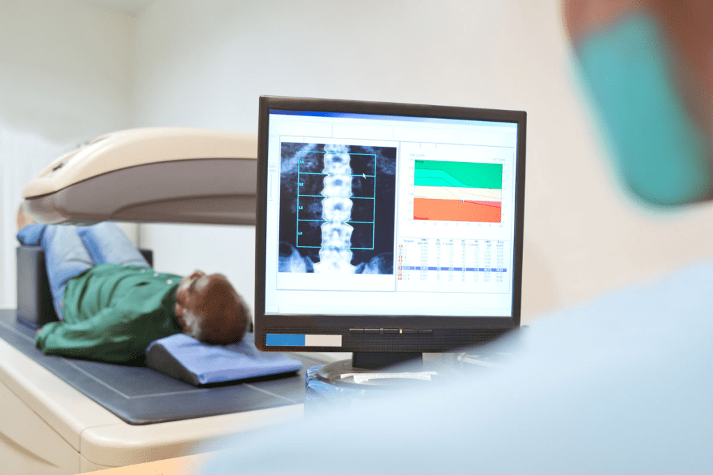
A small amount of a radioactive isotope is injected into a vein. The patient waits a few hours for it to spread. Then, they lie on a table, and the gamma camera takes skeletal images.
The whole process takes a few hours. It’s painless, and the radioactive material leaves the body in a day or two.
| Diagnostic Method | Sensitivity for Bone Metastases | Use of Radioactive Isotopes |
| Bone Scan | High | Yes |
| X-ray | Low to Moderate | No |
| CT Scan | Moderate to High | No |
| PET/CT Scan | High | Yes |
Knowing how bone scans work shows their importance in diagnosing and tracking bone issues. They use radioactive isotopes to give vital information for treatment plans.
The Science Behind Bone Scintigraphy

Bone scintigraphy, or radionuclide bone scanning, is key in diagnosing bone diseases. This diagnostic tool is vital for spotting bone issues like cancer, arthritis, and metabolic bone diseases.
Principles of Radionuclide Bone Scanning
Radionuclide bone scanning uses tiny amounts of radioactive tracers, like Technetium-99m methylene diphosphonate (Tc-99m MDP). These tracers are injected into the blood. They gather in areas of high bone activity, making them visible during scans.
The idea is that bones with disease will take up more of the tracer.
How Abnormal Bone Activity Appears
Abnormal bone activity shows up as “hot spots” on the scan. These spots can mean fractures, infections, or cancer. Bone scintigraphy is great at finding osteoblastic lesions, which are linked to some cancers that make bones grow faster.
Types of Bone Scintigraphy Machines
There are various bone scintigraphy machines, each with unique features. The gamma camera is the most common. It’s used for both planar and SPECT imaging. SPECT imaging gives detailed three-dimensional views of bone activity.
The right machine choice depends on the diagnostic needs and the level of detail required.
Bone Scan Pictures of Cancer: Visual Interpretation
Bone scan pictures of cancer offer key insights into the disease’s presence and spread. These images are vital for diagnosing and staging cancer. They also help track how well treatments are working.
Identifying Cancerous Lesions on Scans
Cancerous lesions on bone scans show up as “hot spots” where the radioactive tracer uptake is high. These hot spots point to abnormal bone activity. This can mean different things, including cancer.
Healthcare experts look for specific patterns and features to spot cancerous lesions. They check for:
- Intensity of uptake: Cancerous lesions usually have very high tracer uptake.
- Location: Some cancers spread to specific areas, like the spine or pelvis.
- Distribution: Seeing many hot spots in a pattern can suggest metastatic disease.
Differentiating Primary vs. Metastatic Bone Cancer
Differentiating primary bone cancer from metastatic bone cancer is key to the right treatment. Primary bone cancers start in the bone itself. Metastatic bone cancer comes from cancer cells spreading to the bone from another place.
Bone scans help tell these two apart by looking at the pattern and spread of lesions:
- Primary bone cancer usually has one main lesion.
- Metastatic bone cancer has many lesions in different spots.
Common Patterns and “Hot Spots” in Bone Metastases
Bone metastases often show specific patterns on bone scans. These patterns include:
- Multiple random hot spots
- Solitary hot spots in typical locations (e.g., spine, pelvis)
- Diffuse uptake in the axial skeleton
Knowing these patterns is key for accurate bone scan interpretation and effective cancer care.
Types of Cancer Detectable Through Bone Scans
Bone scans are key in finding cancer in bones. They can spot different cancers, both those that start in bones and those that spread to bones from elsewhere.
Primary Bone Cancers
Primary bone cancers start in the bones. Osteosarcoma, Ewing’s sarcoma, and chondrosarcoma are examples. These cancers are rare but can grow fast and need quick treatment.
Metastatic Cancers That Spread to Bone
Bone scans often find cancers that have spread to bones. Breast cancer, prostate cancer, and lung cancer are common culprits. They help see how far cancer has spread, which is key for treatment plans.
Limitations in Cancer Detection
Bone scans are great for many bone cancers, but have limits. They might miss some cancers, like certain multiple myeloma types. They can also show false positives, showing activity that’s not cancer.
Knowing these limits helps doctors understand bone scan results better. It also tells them when more tests are needed.
Accuracy and Sensitivity of Bone Scans for Cancer
It’s key to know how accurate bone scans are for cancer. They help find cancer, like when it spreads to bones. But, how well they work can change based on a few things.
Detection Rates for Different Cancer Types
Research shows bone scans catch different cancers at different rates. For example, prostate and breast cancer often spread to bones. These cancers are usually found well by bone scans. The scans are 85% to 93% accurate in spotting bone metastases from these cancers.
“The sensitivity of bone scans in detecting metastatic bone disease is very high for osteoblastic lesions,” studies say. Osteoblastic lesions are tumors that cause bones grow more. This makes them easier to spot on bone scans.
Osteoblastic vs. Osteolytic Lesions
Bone metastases can be either osteoblastic (bone-forming) or osteolytic (bone-destroying). Osteoblastic lesions show up well on bone scans because they take up more of the radioactive tracer. Osteolytic lesions might not show up as much, which can make them harder to find.
Factors Affecting Scan Accuracy
Many things can affect how accurate bone scans are. These include the cancer type, where and how big the metastases are, and if they are osteoblastic or osteolytic. Also, the quality of the imaging equipment and the doctor’s skill can play a part.
We need to think about these factors when we look at bone scan results. This helps make sure we diagnose and treat patients correctly. Knowing what bone scans can and can’t do helps doctors make better choices for their patients.
Bone Scans and Arthritis: What They Reveal
An arthritis diagnosis usually doesn’t use bone scans. Yet, these scans can show bone activity changes that hint at arthritis. We’ll look at how bone scans relate to arthritis, what they show, and their limits.
Inflammatory Markers in Arthritis
Bone scans can spot inflammatory markers linked to arthritis. These markers show bone activity increase, hinting at arthritis. The presence of these markers doesn’t confirm arthritis, but they’re key in diagnosis.
Active arthritis can change bone metabolism, leading to more radioactive tracer uptake. This shows up on scans, giving clues about the condition’s extent and severity.
Differentiating Arthritis from Cancer on Bone Scans
Telling arthritis from cancer on bone scans is tough because they share signs. Both can show “hot spots” on scans. Yet, some signs can help tell them apart.
- Cancer spots are usually more focused, while arthritis is more spread out.
- The spot’s location can also give clues; arthritis has specific patterns.
- It’s vital to match scan results with patient symptoms and history for accurate diagnosis.
Why Bone Scans Aren’t Typically Used for Arthritis Diagnosis
Despite showing inflammatory markers, bone scans aren’t main tools for arthritis diagnosis. The main reasons are:
- They’re not specific; scans can’t tell different causes apart well.
- There are direct and specific tests for arthritis, like clinical exams and lab tests.
- They involve radiation, making them less ideal for routine use in non-life-threatening conditions like arthritis.
In summary, bone scans can hint at arthritis but aren’t the main diagnostic tool. They’re more of a supporting role, adding extra info when needed.
Comparing Bone Scans to Other Diagnostic Methods
It’s important to compare bone scans with other imaging methods. This helps us see their strengths and weaknesses. Each imaging technique has its own benefits and is best for certain conditions.
Bone Scans vs. X-rays and CT Scans
X-rays and CT scans show bone structures well. But, they’re not the same as bone scans. Bone scans look at bone activity, which is useful for finding early changes not seen on X-rays or CT scans.
| Imaging Technique | Primary Use | Advantages | Limitations |
| Bone Scan | Detecting bone metabolism changes | Sensitive to early bone changes, whole-body imaging | Less specific, radiation exposure |
| X-ray | Bone structure visualization | Quick, widely available, low cost | Limited soft tissue detail, radiation exposure |
| CT Scan | Detailed bone and soft tissue imaging | High detail, fast scanning | Higher radiation dose, cost |
Bone Scans vs. MRI
MRI focuses on soft tissues and some bone marrow issues. It’s safer because it doesn’t use radiation. But, MRI is pricier and not as common as bone scans.
Key differences between Bone Scans and MRI:
- Bone scans are more sensitive to bone metabolism changes.
- MRI provides better soft tissue detail.
- Bone scans involve radiation, while MRI does not.
Bone Scans vs. PET/CT Scans
PET/CT scans mix PET’s metabolic info with CT’s anatomy. They give a detailed look at bone and tissue. Though they’re very accurate, they cost more and use more radiation than bone scans.
In summary, bone scans are special in the world of imaging. Knowing how they compare to X-rays, CT scans, MRI, and PET/CT scans helps doctors choose the best test for patients.
Other Bone Conditions Revealed by Bone Scans
Bone scans are great at finding many bone problems, like fractures and diseases. They help doctors see and treat different bone issues, not just cancer.
Fractures and Trauma
Bone scans are super at finding fractures, even when X-rays can’t. They’re great for spotting stress fractures or occult fractures that X-rays miss.
“Bone scans are often used in the evaluation of stress fractures, mainly in athletes or those who are very active,” a study says.
Infections and Osteomyelitis
It’s hard to find infections like osteomyelitis with just X-rays. But bone scans can spot areas where the bone is more active, showing infection.
- Increased uptake on bone scans can indicate the presence of osteomyelitis.
- Bone scans can help tell apart bone infection from other issues.
- They’re really helpful when the infection is deep or in people with certain health problems.
Metabolic Bone Diseases
Metabolic bone diseases, like Paget’s disease or osteoporosis, can be checked with bone scans. These diseases cause specific changes in bone activity that scans can spot.
Paget’s disease shows a distinctive pattern of intense uptake in affected bones. This makes it easy to diagnose and track the disease.
Bone scans are key in helping doctors care for patients. They help create treatment plans and keep track of how the disease is doing.
Conclusion: The Value and Limitations of Bone Scans
We’ve looked at how bone scans help diagnose and manage bone issues like cancer and arthritis. It’s key to know their worth for better use in healthcare.
Bone scans are a great tool for spotting bone activity and finding abnormal bone metabolism. But, we must also know their limits, like how well they work in different situations.
Research has shown both the good and bad sides of bone scans. They’re useful for finding some cancers and bone problems. So, bone scans are a big part of diagnosing, giving a special view on bone health.
In the end, bone scans are very useful when used right, knowing their strengths and weaknesses. This helps doctors make better choices, improving care for patients.
FAQ
What is a bone scan, and how does it work?
A bone scan, or bone scintigraphy, uses a tiny amount of radioactive material. It’s injected into your bloodstream to see where bones are most active. This helps us spot any problems.
Does a bone scan show arthritis?
A bone scan might show signs of inflammation, like in arthritis. But it’s not the go-to test for diagnosing arthritis. It can’t always tell the difference between arthritis and other issues, like cancer.
What cancers can a bone scan detect?
Bone scans can find cancers that start in the bones or spread there. This includes cancers from places like the breast, prostate, and lungs.
Can a bone scan detect bone cancer?
Yes, bone scans can spot bone cancer by highlighting abnormal bone activity. But, they might not always tell the difference between bone cancer and cancer that has spread.
How accurate are bone scans in detecting cancer?
The accuracy of bone scans for cancer depends on several things. These include the cancer type, where the tumor is, and how sensitive the scan is. They work best for certain cancers, like prostate cancer.
What is the difference between osteoblastic and osteolytic lesions on a bone scan?
Osteoblastic lesions show up as active bone areas on a scan, meaning bone is forming. Osteolytic lesions show up as less active, meaning bone is being destroyed. Different cancers cause different types of lesions.
Can a bone scan be used to diagnose fractures or trauma?
Yes, bone scans can find fractures or trauma, even if X-rays can’t. They show where bone activity is high, indicating healing.
How does a bone scan compare to other diagnostic imaging methods like X-rays, CT scans, and MRI?
Bone scans are good at showing bone activity changes. X-rays and CT scans give detailed bone structure views. MRI is better for soft tissue and some bone lesions.
What is radionuclide bone scanning, and how does it work?
Radionuclide bone scanning uses a radioactive isotope to see bone activity. The isotope is injected and goes to active bone areas, helping us find issues.
What does a bone scan reveal about bone conditions?
A bone scan can show many bone conditions, like cancer, fractures, infections, and metabolic diseases. It gives us important info on bone activity, helping us diagnose and manage these conditions.
References:
- Even-Sapir, E., Metser, U., Mishani, E., Lerman, H., & Leibovitch, I. (2006). Bone scintigraphy in the detection of metastatic bone disease: Review of the literature. Seminars in Nuclear Medicine, 36(2), 76-85. https://pubmed.ncbi.nlm.nih.gov/16443156/
- Jadvar, H. (2015). Flow-sensitive nuclear medicine imaging of bone metastases: Current status and future directions. PET Clinics, 10(2), 161-176. https://pubmed.ncbi.nlm.nih.gov/26210191/
- Buvat, I., Dogan, A. U., Hoetjes, N. J., & Xu, J. (2020). Role of radionuclide imaging in diagnosis and monitoring of arthritis: An update. Seminars in Nuclear Medicine, 50(2), 127-143. https://pubmed.ncbi.nlm.nih.gov/32087230/


