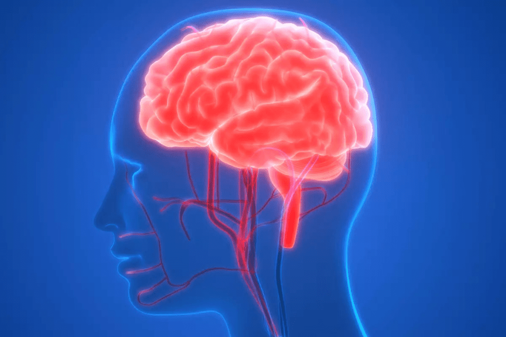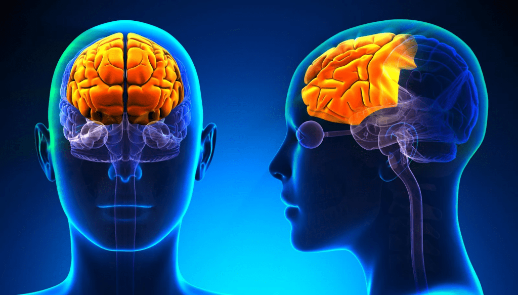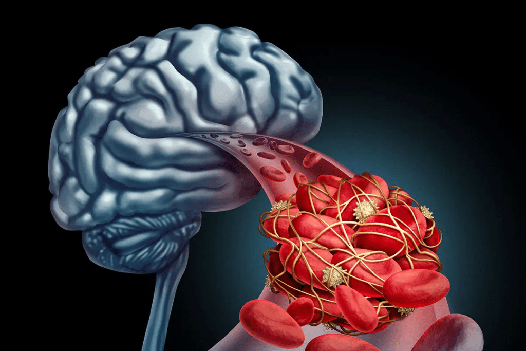
Did you know that problems with cerebral circulation can lead to neurological disorders? These issues affect millions of people around the world. It’s important to check blood flow to the brain to diagnose and treat these conditions. We will look at the different tests used to measure brain blood flow. We’ll see how they are used in medical practice and why they matter.
Key Takeaways
- Understanding cerebral circulation is vital for neurological health.
- Diagnostic tests for brain blood flow are key for diagnosis.
- There are many tests, each with its own use.
- Checking blood flow to the brain helps understand brain function.
- These tests are essential for managing neurological conditions.
The Critical Role of Brain Blood Flow in Neurological Health
Keeping cerebral blood flow at the right level is key for brain health. The brain needs a lot of blood to work well. We’ll look at how blood flow helps keep the brain healthy.
How Cerebral Circulation Works
Cerebral circulation is a complex process. It involves many blood vessels working together to give the brain what it needs. The brain gets about 15% of the heart’s blood, showing how much it needs.
Autoregulation helps the brain keep a steady blood flow. It does this by changing the size of blood vessels. When blood pressure goes down, vessels get bigger to let more blood in. When it goes up, they get smaller to keep blood flow steady.
Why Adequate Blood Flow Is Essential for Brain Function
Good brain circulation is vital for brain function. The brain needs a steady supply of oxygen and glucose. Without it, brain problems or damage can happen.
| Condition | Effect on Cerebral Blood Flow | Neurological Impact |
| Stroke | Reduced or blocked blood flow | Cognitive and motor deficits |
| Alzheimer’s Disease | Decreased blood flow | Memory loss and cognitive decline |
| Traumatic Brain Injury | Variable effects on blood flow | Cognitive, emotional, and behavioral changes |
Knowing how cerebral circulation affects brain health is important. It helps doctors diagnose and treat brain problems. Tests help check blood flow and guide treatment.
Overview of Diagnostic Tests for Brain Blood Flow

Understanding brain perfusion is key. Several tests are used to check it. These tests help diagnose and manage neurological issues, giving insights into brain function.
Categories of Brain Perfusion Tests
Brain perfusion tests fall into main categories. They vary in how they work and what they show. Here are the main types:
- Imaging Techniques: Computed Tomography (CT) Perfusion, Magnetic Resonance Imaging (MRI) Perfusion, and Positron Emission Tomography (PET) are examples. Each gives unique views into brain blood flow and metabolism.
- Nuclear Medicine Tests: Single-Photon Emission Computed Tomography (SPECT) is another type. It shows brain function and blood flow.
- Ultrasonography: Transcranial Doppler Ultrasonography is used to check blood flow in brain arteries.
When Brain Blood Flow Testing Is Recommended
Brain blood flow tests are needed in many cases. Here are some examples:
- Stroke and Cerebrovascular Diseases: They help see how much damage there is and what treatment to choose.
- Dementia and Cognitive Disorders: They help figure out what kind of dementia someone has and check blood flow.
- Epilepsy: They help find where seizures start and understand blood flow during and between seizures.
- Traumatic Brain Injury: They check how brain circulation is affected by injury.
These tests are very important in neurology. They help doctors make the best decisions for their patients. Knowing about these tests helps us see how they help manage brain conditions.
Computed Tomography (CT) Perfusion Scan
CT perfusion imaging is key in checking how blood flows in the brain. It helps spot areas where blood flow is low. This is very important for treating strokes and other brain problems.
How CT Perfusion Measures Cerebral Blood Flow
CT perfusion scans track how blood moves through the brain’s vessels. They use quick CT scans after giving iodinated contrast. This creates maps of blood flow and volume in the brain.
These maps show how well the brain’s blood is flowing. Doctors use them to find areas that might be saved during a stroke.
Clinical Applications of CT Perfusion
CT perfusion is very useful in treating strokes. It helps doctors:
- See how big the stroke area is
- Decide if clot-busting drugs are right
- Check if brain tissue can be saved
It’s also used for other brain problems, like when blood vessels narrow after a brain bleed.
Advantages and Limitations
CT perfusion is fast and easy to get. It gives detailed info on blood flow. But, it has downsides too.
It uses radiation and iodinated contrast. These can be bad for some patients.
| Advantages | Limitations |
| Rapid acquisition time | Radiation exposure |
| Wide availability | Need for iodinated contrast |
| Quantitative CBF information | Potential for artifacts |
Even as we get better at brain imaging, CT perfusion is a big help. It’s key for understanding and treating brain blood flow issues.
Magnetic Resonance Imaging (MRI) Brain Perfusion

MRI has greatly improved how we check brain blood flow and its health effects. It’s a non-invasive way to see how blood moves in the brain. This helps doctors understand brain health better.
Perfusion Techniques
MRI uses special methods and contrast agents to look at brain blood flow. Dynamic Susceptibility Contrast (DSC) MRItracks a contrast agent through the brain. It’s a key method used today.
Arterial Spin Labeling (ASL) is another method. It uses blood as a natural tracer. It’s great for those who can’t have contrast agents.
Clinical Applications in Neurology
MRI brain perfusion helps in many neurology areas. It’s used for stroke, brain tumors, and diseases like Alzheimer’s. It helps doctors see if brain tissue is alive and decide on treatments.
In stroke cases, MRI shows where blood flow is low. This helps doctors see how much damage there is. It also helps decide if clot-busting drugs should be used.
Benefits and Limitations of MRI Perfusion
MRI perfusion is non-invasive and doesn’t use harmful radiation. It also shows soft tissues well. But, it needs special equipment and experts. Some people can’t have it because of MRI or contrast agent issues.
Even with its challenges, MRI brain perfusion is very useful. It gives doctors important information about brain blood flow. This helps in making treatment plans.
Single-Photon Emission Computed Tomography (SPECT) Brain Imaging
SPECT brain imaging is a key tool in neurological diagnostics. It helps evaluate brain perfusion. This method uses nuclear medicine to show brain function and blood flow, helping diagnose and manage neurological conditions.
SPECT Procedure and Technology
SPECT imaging uses a small radioactive tracer injected into the blood. A SPECT camera then detects gamma rays from the tracer. This creates detailed 3D images of brain activity and blood flow.
Advanced SPECT cameras provide high-resolution images. This helps doctors see brain function clearly. The procedure is done in a nuclear medicine department or a specialized imaging center.
Clinical Uses of SPECT in Neurology
SPECT brain imaging is used for many neurological conditions. It helps diagnose and monitor:
- Alzheimer’s disease and other dementias
- Stroke and cerebral vasculature disorders
- Epilepsy, for finding seizure foci
- Traumatic brain injuries
- Certain psychiatric disorders
It helps doctors create personalized treatment plans. This improves care for neurological patients.
Advantages and Disadvantages
SPECT imaging has many benefits. It provides functional brain information and is relatively available. But, it has drawbacks like radiation exposure and lower resolution than MRI.
We consider these factors when deciding if SPECT imaging is right for our patients. We balance the benefits against the risks.
| Aspect | Description | Clinical Relevance |
| Procedure | Injection of radioactive tracer, followed by SPECT scanning | Diagnostic imaging for neurological conditions |
| Technology | Advanced SPECT cameras for high-resolution imaging | Precise evaluation of brain function and blood flow |
| Clinical Uses | Diagnosis and monitoring of neurological disorders | Tailoring treatment plans for improved patient outcomes |
| Advantages | Provides functional brain information, relatively accessible | Valuable for diagnosing and managing neurological conditions |
| Disadvantages | Involves radiation exposure, lower spatial resolution | Considerations for patient selection and risk assessment |
Positron Emission Tomography (PET) Brain Scan
Positron Emission Tomography (PET) brain scans give us a peek into the brain’s activity. They help find and treat many brain problems. By using tiny amounts of radioactive tracers, PET scans show how the brain works. This helps doctors diagnose and treat brain disorders better.
Measuring Brain Metabolism and Blood Flow
PET scans track brain metabolism and blood flow by finding the radiation from tracers. The most common tracer is fluorodeoxyglucose (FDG). It shows how active brain cells are, helping doctors spot problems.
We use PET scans to check how well the brain gets blood and uses energy. This is key for diagnosing diseases like Alzheimer’s. In Alzheimer’s, some brain areas don’t use energy right.
Clinical Applications in Neurology
PET brain scans help in many ways in neurology. They are used to:
- Diagnose and track neurodegenerative diseases like Alzheimer’s and Parkinson’s.
- Check brain tumors and see how they react to treatment.
- Look at seizure disorders and find where seizures start.
- Study inflammatory and infectious brain diseases.
PET scans give us detailed info on brain function and blood flow. They work with MRI and CT scans to give a full picture of the brain.
Limitations and Considerations
Even though PET scans are very useful, they have some downsides. These include:
| Limitation | Description |
| Radiation Exposure | PET scans use a little radiation, which might worry some patients. |
| Cost and Availability | PET scans cost more and are not everywhere, making them hard to get. |
| Image Resolution | PET scans are great for function but not as clear as MRI or CT scans. |
Despite these issues, PET brain scans are very helpful in neurology. They give important info for diagnosis and treatment. We keep working to make PET scans even better for doctors and patients.
Transcranial Doppler Ultrasonography
Transcranial Doppler ultrasonography is a non-invasive tool for checking brain blood flow. It helps us see how fast blood moves through brain vessels. This gives us important info on brain circulation.
Principles and Technique
This method uses the Doppler effect. It changes sound wave frequency when hitting moving objects, like red blood cells. We use an ultrasound device to send sound waves through the skull.
These sound waves bounce off blood cells and come back to us. This lets us figure out blood flow speed.
To do this, we look for special spots in the skull. These spots are thinner, letting sound waves pass better. The most used spots are the temporal, transorbital, and suboccipital windows.
Clinical Applications
Transcranial Doppler is key in neurology and neurosurgery. It lets us watch brain blood flow live, which is vital during surgeries or in critical care. It also helps spot problems like vasospasm after bleeding in the brain.
It’s also used to check if the brain’s blood flow has stopped. Plus, it helps see if treatments are working to improve blood flow.
It’s also used for sickle cell disease. It predicts stroke risk by measuring blood flow speed in the brain.
Advantages and Limitations
One big plus is it’s non-invasive, making it safe and easy to do again. It gives us live info, which is super helpful in urgent situations. But, it can be less effective if the skull is too thick.
Even with its limits, it’s a key tool for checking brain blood flow. We keep working to make it better with new tech.
Brain Blood Flow Assessment in Stroke
In stroke cases, checking how well the brain gets blood is key. This helps doctors decide the best treatment. Imaging tests are vital for this.
Acute Stroke Evaluation
When someone has a stroke, quick blood flow checks are needed. We use CT perfusion and MRI perfusion to see how much brain is at risk. These tests help find brain areas that might be saved.
These methods also tell us if it’s an ischemic or hemorrhagic stroke. Knowing this helps us choose the right treatment.
Perfusion-Diffusion Mismatch
The perfusion-diffusion mismatch is important in stroke care. It shows the difference between brain areas with low blood flow and those already damaged.
Finding this mismatch is key. It shows us which brain areas might be saved with quick action. We use special imaging to spot this and guide treatment.
| Imaging Technique | Clinical Use in Stroke | Advantages |
| CT Perfusion | Assesses cerebral blood flow and identifies ischemic areas | Quick, widely available, sensitive to acute hemorrhage |
| MRI Perfusion | Evaluates cerebral perfusion and diffusion characteristics | High sensitivity for ischemic stroke, detailed tissue characterization |
Monitoring Treatment Response
After starting stroke treatment, we keep an eye on how the patient is doing. We use imaging to see if the treatment is working.
By watching how blood flow changes, we can tweak the treatment plan. This helps improve the patient’s chances of recovery.
Brain Perfusion Testing in Dementia and Cognitive Disorders
Brain perfusion testing is key in diagnosing and managing dementia and cognitive disorders. It helps doctors understand why cognitive decline happens. By checking blood flow to the brain, they get important insights.
We use different tests to look at brain perfusion. Each test gives unique info about brain circulation and its effect on thinking. These tests help us tell apart different types of dementia and find ways to help.
Alzheimer’s Disease Patterns
In Alzheimer’s, brain perfusion tests show specific patterns of reduced blood flow. Studies show that certain brain areas, like the temporal and parietal lobes, have less blood flow as thinking skills decline. We use SPECT or PET scans to see these patterns and help confirm Alzheimer’s diagnosis.
The table below shows what Alzheimer’s looks like on different brain perfusion tests:
| Imaging Modality | Typical Findings in Alzheimer’s Disease |
| SPECT | Reduced perfusion in temporal and parietal lobes |
| PET | Hypometabolism in posterior cingulate and temporoparietal regions |
| CT Perfusion | Decreased cerebral blood flow in affected areas |
Vascular Dementia Assessment
Vascular dementia, the second most common dementia, is linked to poor blood flow to the brain. Brain perfusion tests help spot areas with less blood flow due to blood vessel problems. These tests help us see how much damage there is and how it affects thinking.
Key features of vascular dementia on brain perfusion tests include:
- Multiple areas of reduced perfusion corresponding to vascular territories
- Perfusion deficits often seen in areas surrounding infarcts
- Variable patterns depending on the underlying vascular pathology
Other Neurodegenerative Conditions
Brain perfusion testing is also useful for other brain diseases that affect thinking. For example, in frontotemporal dementia, we see changes in the frontal and anterior temporal regions. These tests help us diagnose and track how the disease is progressing.
By combining clinical checks with advanced brain perfusion tests, we can make more accurate diagnoses. This helps us create better treatment plans for patients with dementia and other cognitive disorders.
Epilepsy and Brain Blood Flow Imaging
Managing epilepsy has changed thanks to brain blood flow imaging. We use advanced imaging to check how blood flows in the brain. This is key for diagnosing and treating epilepsy well.
Ictal vs. Interictal Studies
Imaging the brain during seizures and between them gives us important clues. Ictal studies show how blood flow changes during a seizure. Interictal studies show the brain’s normal blood flow.
By comparing these, we can find where seizures start and how severe they are.
Localizing Seizure Focus
Finding where seizures start is vital for surgery. Imaging like SPECT and PET helps find seizure areas. This is key for planning surgery.
By looking at blood flow patterns, doctors can pinpoint seizure start zones. This is essential for surgery planning.
Presurgical Evaluation
Before surgery, we thoroughly check brain function and structure. Brain blood flow imaging is key here. It tells us about brain region function.
We use this info to plan surgery carefully. This makes sure the surgery is safe and works well.
Advanced Applications of Brain Blood Flow Testing
Brain blood flow testing is now used in new ways. It helps in studying psychiatric disorders, traumatic brain injuries, and neurodevelopmental disorders. This technology is giving us new insights into these complex conditions.
Psychiatric Disorder Assessment
Functional brain scans are being used more to study psychiatric disorders. They help researchers understand the causes of conditions like depression and schizophrenia by looking at brain blood flow.
Key Findings:
- Altered brain perfusion patterns in psychiatric disorders
- Potential biomarkers for diagnosis and treatment monitoring
- Insights into the neural circuits involved in psychiatric conditions
Traumatic Brain Injury Evaluation
Traumatic brain injury (TBI) can cause big problems with thinking and function. Brain blood flow testing helps figure out how severe TBI is and how well someone is recovering.
| Parameter | Normal Value | TBI Value |
| Cerebral Blood Flow (CBF) | 50-60 ml/100g/min | <30 ml/100g/min |
| Cerebral Blood Volume (CBV) | 4-6 ml/100g | >6 ml/100g |
Neurodevelopmental Disorders
Neurodevelopmental disorders, like autism spectrum disorder (ASD), show changes in brain blood flow. Functional brain scans offer important info on the brain’s workings in these conditions.
Research Implications:
- Understanding the role of brain perfusion in neurodevelopmental disorders
- Identifying possible treatment targets
- Creating early diagnostic markers
As we dive deeper into brain blood flow testing, we learn more about brain function and various conditions. This knowledge helps us find better ways to diagnose and treat these issues.
Emerging Technologies in Brain Blood Flow Assessment
New medical technologies are changing how we check brain blood flow. The field of neurology is growing fast. Now, we can diagnose and treat brain issues better than before.
Functional Near-Infrared Spectroscopy (fNIRS)
Functional Near-Infrared Spectroscopy (fNIRS) is a new way to watch brain activity without hurting it. It looks at blood oxygen levels to see how the brain works. This tool is great for checking brain function right away, helping doctors and researchers a lot.
fNIRS is easy to use, small, and not too expensive. It’s good for many uses, like watching brain activity in patients or studying kids’ brains.
Advanced MRI Techniques
New MRI methods like arterial spin labeling (ASL) and blood oxygen level-dependent (BOLD) imaging help us see brain blood flow better. They give detailed info on blood flow and oxygen levels. This helps doctors diagnose and keep track of brain conditions more accurately.
These MRI methods are special because they make clear images without needing special dyes. This is good for people who can’t have certain dyes.
AI and Machine Learning Applications
Artificial Intelligence (AI) and Machine Learning (ML) are making a big difference in brain blood flow checks. AI can spot things in images that humans might miss. This means we can find problems sooner and treat them better.
Machine Learning helps make image analysis better too. By learning from lots of data, it makes tests more accurate. This leads to better care for patients.
Practical Considerations When Undergoing Brain Blood Flow Tests
When you’re getting ready for brain blood flow tests, there are a few things to keep in mind. These steps help make sure you get accurate results and have a smooth experience. We know that getting tested can make you feel nervous.
Preparation for Testing
Getting ready is important for getting good results from brain tests. You might need to avoid certain foods, medicines, or activities beforehand. For example, some nuclear brain scans ask you to fast or stop taking certain medicines.
It’s key to listen to what your doctor or the testing place tells you. This might mean getting there early to fill out papers, taking off jewelry, or following special diet rules.
Radiation Exposure Concerns
Some brain tests, like perfusion brain tests, might worry you about radiation. Tests like CT perfusion scans do involve some radiation.
Even though the risk from one test is usually small, it’s good to talk to your doctor about it. They can explain the benefits and risks. They might also suggest tests without radiation, like MRI perfusion, for your situation.
| Test Type | Radiation Exposure | Typical Use |
| CT Perfusion Scan | Yes | Acute stroke evaluation, tumor assessment |
| MRI Perfusion | No | Brain tumor evaluation, stroke assessment |
| SPECT Scan | Yes | Epilepsy, dementia, and stroke evaluation |
Insurance Coverage and Costs
It’s also important to know about the costs and what your insurance covers. Prices can change based on the test, where you go, and more.
Check with your insurance to see what they cover and what you might have to pay for. Some tests need approval before they can be done. Knowing this can help you avoid surprises.
If you don’t have insurance or it doesn’t cover much, many places offer help. They might have programs or advice to help with the costs of tests.
Conclusion
Understanding brain blood flow is key to diagnosing and managing neurological conditions. We’ve looked at different tests like CT perfusion scans, MRI brain perfusion, SPECT, and PET scans. Each test gives unique insights into how blood flows through the brain.
By checking brain blood flow, doctors can spot areas that don’t get enough blood. This helps them make better treatment plans. It also improves how well patients do.
Keeping the brain healthy needs a full plan, including good blood flow. With advanced tests, doctors can give care that fits each patient. This helps improve brain health and overall well-being.
FAQ
What is a brain perfusion scan, and how does it measure blood flow to the brain?
A brain perfusion scan is a test that checks blood flow to the brain. It uses different methods like CT perfusion, MRI perfusion, SPECT, and PET scans. These tests help find out where blood flow is low and diagnose brain problems.
What is the difference between a CT perfusion scan and an MRI perfusion scan?
CT perfusion scans use X-rays and dye to measure blood flow. MRI perfusion scans use magnetic fields and dye for the same purpose. MRI is better at seeing certain brain tissues and can show more about brain metabolism.
How does transcranial Doppler ultrasonography work, and what does it measure?
Transcranial Doppler ultrasonography is a non-invasive test. It uses sound waves to check blood flow in brain arteries. It helps find problems like narrowed or blocked arteries.
What is the role of brain blood flow assessment in diagnosing and managing stroke?
Assessing brain blood flow is key in diagnosing and treating stroke. Tests like CT perfusion, MRI perfusion, and transcranial Doppler ultrasonography show where blood flow is low. They help doctors decide how to treat the stroke.
Can brain blood flow testing help diagnose dementia and cognitive disorders?
Yes, brain blood flow tests can help find dementia and cognitive disorders. Tests like SPECT and PET scans show where blood flow is low. This helps doctors diagnose conditions like Alzheimer’s disease and vascular dementia.
How does brain blood flow imaging contribute to the evaluation and management of epilepsy?
Brain blood flow imaging, like SPECT and PET scans, helps find where seizures start. It also checks brain function in people with epilepsy. This info is useful for planning surgery and treatment.
Are there any emerging technologies in brain blood flow assessment?
Yes, new technologies like functional near-infrared spectroscopy (fNIRS), advanced MRI, and AI are being developed. They aim to better understand and measure cerebral blood flow.
What are the practical considerations when undergoing brain blood flow tests?
When getting brain blood flow tests, consider preparation, radiation concerns, and insurance and costs. Talk to your doctor about these to make informed choices.
How can I improve blood flow to my brain?
To improve brain blood flow, try lifestyle changes like exercise, a healthy diet, stress management, and enough sleep. Some supplements and medications might help too. Always talk to a doctor before trying anything new.
What are the benefits of enhanced cerebral blood flow?
Boosting cerebral blood flow can improve thinking, memory, and brain health. It may also lower the risk of brain diseases and enhance life quality.
References
- Weinstein, H. C., Hijdra, A., van Royen, E. A., & Derix, M. M. (1989). Determination of cerebral blood flow by SPECT: A valuable tool in the investigation of dementia? Clinical Neurology and Neurosurgery, 91(1), 13-19. https://pubmed.ncbi.nlm.nih.gov/2538276/
- Amen, D. G., et al. (2021). A new way forward: How brain SPECT imaging can improve diagnosis and treatment of neurological disorders. Frontiers in Psychiatry, 12, Article 715315. https://www.frontiersin.org/articles/10.3389/fpsyt.2021.715315/full
- Seeley, M. C., et al. (2025). Novel brain SPECT imaging unravels abnormal cerebral perfusion in patients with postural orthostatic tachycardia syndrome and cognitive dysfunction. Scientific Reports, 15, Article 87748. https://www.nature.com/articles/s41598-025-87748-4
- Roberts, J. D. (2013). Cerebral perfusion (SPECT) studies. Australian Family Physician, 42(3), 140“142. https://www.racgp.org.au/afp/2013/march/spect-studies


