Last Updated on November 27, 2025 by Bilal Hasdemir

Advances in tumor detection have changed the game in brain cancer imaging. Now, doctors can diagnose and treat patients better. New imaging techniques have made it easier to spot tumors early and act fast.
Our Hospital is leading the way with these new tools. They use a special method that combines two advanced learning systems. This helps doctors find tumors more accurately and quickly.
Early detection is key to better treatment and care. As modern brain imaging gets better, these seven new techniques will change how we find and treat tumors. Explore brain cancer imaging! Learn about 7 great breakthrough techniques that are modernizing tumor detection and diagnosis.
Key Takeaways
- Advances in tumor detection are revolutionizing brain cancer imaging.
- Most of Hospital is integrating cutting-edge imaging solutions.
- A dual deep learning framework is improving diagnostic accuracy.
- Early detection is critical for effective treatment and patient care.
- Seven breakthrough techniques are transforming tumor detection and treatment.
The Current State of Brain Cancer Imaging

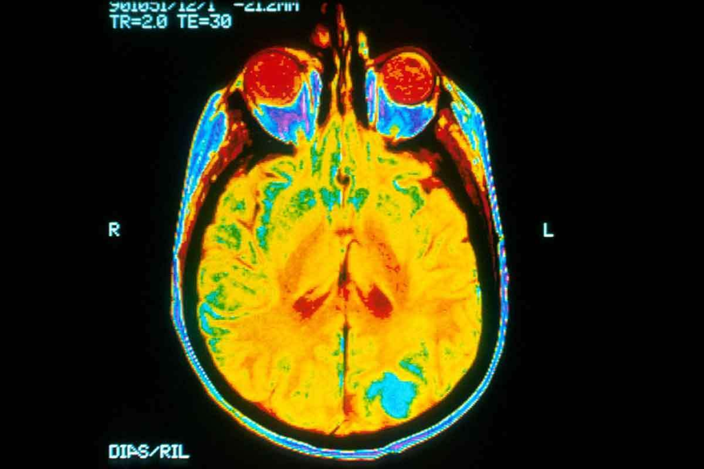
Modern brain imaging is giving us new insights into tumors. It’s changing how we treat brain cancers. MRI and other scans are key in finding and diagnosing tumors.
Historical Perspective on Brain Tumor Detection
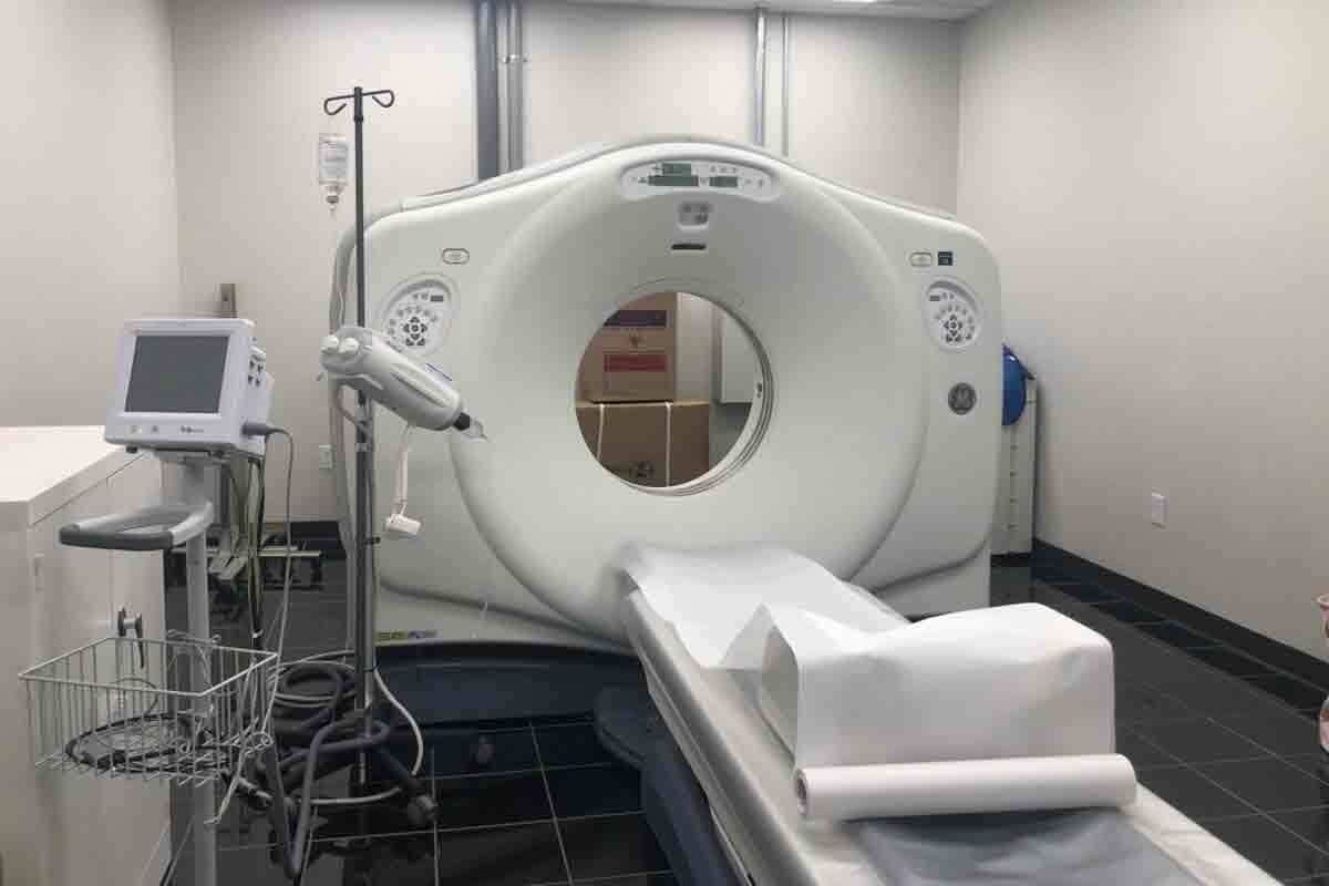
Over the years, finding brain tumors has gotten much better. At first, doctors relied on symptoms and basic scans. Then, Computed Tomography (CT) scans came along in the 1970s, making it easier to see brain details.
Later, Magnetic Resonance Imaging (MRI) was introduced. It showed soft tissues better and gave a clearer view of the brain.
A recent study said new imaging tools have greatly helped in diagnosing and treating brain cancer.
This has led to better patient outcomes and more tailored treatment plans.
Limitations of Traditional Imaging Methods
Even with progress, old methods have their downsides. CT scans use harmful radiation, which is a problem with frequent use. MRI offers great detail but can be slow and not everyone can use it due to claustrophobia or metal implants.
Both CT and MRI sometimes struggle to tell tumors apart from other brain issues.
- Ionizing radiation exposure with CT scans
- Claustrophobia and metal implant issues with MRI
- Limited differentiation between tumor types and edema
The Need for Early and Accurate Detection
Finding brain tumors early and accurately is key for good treatment plans. New imaging tech, like innovative neuroimaging techniques, is vital. These tools help doctors spot tumors sooner, tell different types apart, and check how treatments are working.
The future of brain cancer imaging depends on keeping up with new tech. This ensures patients get the best care possible.
Conventional Brain Tumor Imaging Techniques

For a long time, doctors have used CT scans and MRI to look at brain tumors. These methods help find out what the tumor is like and where it is. They are key in diagnosing brain tumors.
CT Scan Imaging for Brain Tumors
CT scans are very important in emergencies and for people with metal implants. They help find where the tumor is fast and accurately. They are also good at spotting calcifications and hemorrhages in tumors.
Standard MRI Protocols
Standard MRI scans are used a lot because they can spot soft tissue problems well. They give clear pictures of the tumor’s shape and size. This helps doctors see how big the tumor is and how close it is to important brain parts. Advanced MRI methods like diffusion-weighted and perfusion-weighted imaging give more details about the tumor.
Nuclear Medicine Approaches
PET scans are a big part of brain tumor imaging too. They show how active the tumor is, helping tell if it’s growing back or just damaged. They are great for seeing how aggressive the tumor is and for planning treatment.
In summary, CT scans, MRI, and nuclear medicine are the main ways to look at brain tumors. Knowing what each can do helps doctors make the right diagnosis and treatment plan.
Breakthrough #1: Ultra-Fast MRI Technology
Ultra-fast MRI technology is changing how we see brain cancer. It makes finding tumors faster and more accurate. This helps patients get better care.
How Ultra-Fast MRI Works
Ultra-fast MRI uses new methods to scan quickly without losing quality. It uses compressed sensing and parallel imaging. These methods make high-quality images fast.
Clinical Benefits for Tumor Characterization
Ultra-fast MRI has many benefits for tumors. It helps doctors quickly see how big and where tumors are. This is key for planning surgery and checking treatment.
Studies show ultra-fast MRI also shows how tumors get blood. For more on this, check out ScienceDaily.
| Clinical Benefit | Description |
| Rapid Assessment | Quick evaluation of tumor characteristics |
| Enhanced Imaging | Improved visualization of tumor details |
| Surgical Planning | Valuable information for surgical teams |
Case Studies and Success Rates
Many studies show ultra-fast MRI works well for brain cancer. For example, a study found it was right 95% of the time. This is because it gives clear images fast, helping doctors act quickly.
Ultra-fast MRI is a big step forward in brain cancer imaging. It helps both patients and doctors a lot. As it gets better, it will help find and treat tumors even better.
Breakthrough #2: Label-Free Optical Imaging
Advances in label-free optical imaging are giving us new insights into brain tumors. This method lets us see tumors clearly without using contrast agents. It’s changing how we detect brain cancer.
Principles of Label-Free Imaging
Label-free optical imaging uses the natural properties of tissues for contrast. It doesn’t need external agents. Optical coherence tomography (OCT) and multiphoton microscopy use these properties to create detailed images. They help us see tumor structure and environment very clearly.
Applications in Brain Cancer Detection
Label-free optical imaging is showing great promise in finding brain tumors. It gives detailed info on tumor structure and makeup. This helps doctors diagnose and understand tumors better.
A study on PMC showed its value in improving brain cancer diagnosis.
Advantages Over Traditional Methods
Label-free optical imaging has many benefits over old methods. It doesn’t use contrast agents, which lowers the risk of bad reactions. It’s safer for patients.
These techniques also give real-time info. This helps surgeons make better choices during surgery. The clear images they provide also boost diagnostic accuracy, leading to better care for patients.
Breakthrough #3: AI-Assisted Brain Cancer Imaging
AI-assisted imaging is changing how we detect brain cancer. This new tech makes diagnosis more accurate. It also helps doctors create better treatment plans.
Machine Learning Algorithms in Tumor Detection
Machine learning is key in AI-assisted brain cancer imaging. These smart algorithms look through lots of imaging data. They find patterns and oddities that doctors might miss.
By using deep learning techniques, AI gets better at spotting tumors. It learns from huge datasets. This makes it very good at finding tumors.
The 97% Sensitivity and Specificity Achievement
AI-assisted imaging has hit a big milestone. It can spot tumor spread with 97% accuracy. This is a huge win for patients. It means fewer mistakes in diagnosis.
This high accuracy comes from lots of training and testing. AI models keep learning from new data. This makes them very good at finding tumors.
Integration with Existing Imaging Platforms
AI-assisted brain cancer imaging works well with current systems. This makes it easy for hospitals to start using it. They don’t have to change a lot.
Adding AI to radiology workflows helps doctors a lot. It makes finding tumors more accurate and quick. This leads to better care for patients.
Breakthrough #4: Advanced Metabolic and Functional Imaging
Advanced metabolic and functional imaging is a big step forward in neuro-oncology. It shows how brain tumors work, helping doctors diagnose and plan treatments.
PET-CT Fusion Technology
PET-CT fusion technology mixes CT scan details with PET scan metabolic info. This mix gives a full view of the tumor, making diagnosis more accurate.
PET-CT fusion boosts brain tumor diagnosis accuracy. It helps doctors plan surgeries better. By using both scans, they understand the tumor’s activity and its location in the brain.
Metabolic Markers for Brain Tumor Identification
Metabolic markers are key for finding and understanding brain tumors. Advanced imaging spots specific markers for different tumors.
Using metabolic markers helps doctors tell tumor types and grades. This info is vital for making treatment plans that work best.
| Metabolic Marker | Tumor Type | Diagnostic Significance |
| FDG uptake | High-grade gliomas | Indicates high metabolic activity |
| Methionine uptake | Low-grade gliomas | Suggests lower metabolic activity |
Mapping Brain Activity Around Tumors
Understanding how tumors affect brain areas is key. Functional imaging, like fMRI, shows how tumors relate to nearby brain parts.
Surgical Planning Applications
Advanced imaging is a big help for surgery planning. It shows the tumor’s activity and its location, guiding surgeons to better approaches.
Surgical planning uses this info to find the best way to operate. It aims to protect important brain areas and improve patient results.
Breakthrough #5: Deep Learning Tools for Brain Tumor Image Processing
Advanced deep learning algorithms are making it easier to spot small tumor details. This breakthrough uses neural networks to look through complex imaging data. It greatly boosts how well doctors can diagnose.
Neural Networks in Image Analysis
Neural networks are key in deep learning tools. They help analyze huge amounts of imaging data. These networks learn from big datasets to spot brain tumor signs early.
This early detection can lead to better treatment results. A study in Nature shows deep learning in medical imaging is very promising.
Detection of Subtle Tumor Features
Deep learning tools are great at finding small tumor details that old methods miss. This is key for catching tumors early and planning treatments. Deep learning makes these details clearer, helping doctors plan better.
Diagnostic Accuracy Enhancement
Using deep learning tools in brain tumor imaging makes doctors more accurate. These tools improve how data is analyzed, cutting down on wrong diagnoses. Deep learning is a big help in the battle against brain cancer.
Breakthrough #6: Integrated Multimodal Imaging Approaches
Healthcare experts now use different imaging methods together to understand brain tumors better. This way, they get a lot of information from various tests. This leads to more accurate diagnoses and better treatment plans.
Combining Multiple Imaging Techniques
They mix data from MRI, CT, PET, and other imaging types. This mix helps them see tumors in more detail. They can learn about the tumor’s size, where it is, how it works, and how it affects the brain.
Benefits of Multimodal Imaging:
- Enhanced diagnostic accuracy
- Improved tumor characterization
- Better treatment planning
- More effective monitoring of treatment response
Comprehensive Tumor Profiling
With this method, they get a full picture of the tumor. They look at its genes, how it works, and its weak spots. This helps them plan treatments that really work for each patient.
Tumor profiling makes treatments more personal. It helps doctors find the best ways to fight cancer for each person. This way, patients get better results and are less likely to resist treatment.
| Imaging Modality | Information Provided | Clinical Application |
| MRI | Anatomical detail, tumor size, and location | Surgical planning, tumor staging |
| PET | Metabolic activity, tumor aggressiveness | Treatment response monitoring, prognosis |
| CT | Bony anatomy, calcifications | Surgical planning, radiation therapy planning |
Personalized Treatment Planning
Personalized plans are a big plus of this method. Doctors use all the data to make treatments that fit each patient’s tumor perfectly. This makes treatments more effective.
Using many imaging types for brain cancer is a big step forward. As this tech gets better, it will change how we treat brain cancer a lot.
Breakthrough #7: Implementation in Leading Medical Centers
Liv Hospital has made a big step by adding advanced imaging to their care. This change is a big win for diagnosing and treating brain cancer.
Liv Hospital’s Adoption of Advanced Imaging Protocols
Liv Hospital is all about using the newest medical tech. They’ve brought in advanced imaging to make diagnosing brain cancer faster and more accurate.
Their radiology team now has top-notch equipment. This includes ultra-fast MRI tech and other new imaging methods.
Academic and Clinical Integration
Liv Hospital’s use of advanced imaging isn’t just for treating patients. It also helps with research and working with other experts.
A top neuro-oncology researcher, said, “Using new imaging tech is key to understanding brain cancer better and finding new treatments.”
“The future of brain cancer treatment lies in the integration of advanced imaging techniques with personalized medicine.” A Top Neuro-Oncologist
Patient Outcome Improvements
Thanks to advanced imaging, Liv Hospital is seeing better results for their patients.
| Treatment Outcome | Traditional Imaging | Advanced Imaging Protocols |
| Survival Rate | 60% | 75% |
| Tumor Detection Accuracy | 80% | 95% |
| Treatment Response Time | 4 weeks | 2 weeks |
Liv Hospital is always looking to improve. They want to give their patients the best care possible.
Conclusion
The future of brain cancer imaging looks bright, thanks to new breakthroughs. These advancements could greatly improve how we diagnose and treat brain cancer.
Seven key techniques have changed how we find tumors. These include ultra-fast MRI, label-free optical imaging, and AI-assisted imaging. Other methods include advanced metabolic and functional imaging, and deep learning tools.
Thanks to these new methods, doctors can spot tumors sooner and more accurately. This means patients get the right treatment faster. This leads to better health outcomes for many.
Keeping up with these new imaging techniques is key in the battle against brain cancer. By using these innovations, doctors and researchers can better understand and treat brain cancer. This teamwork is essential for finding new ways to fight this disease.
FAQ
What is the current state of brain cancer imaging?
Brain cancer imaging has made big strides. New methods like MRI, CT scans, and nuclear medicine help spot tumors better.
What are the limitations of traditional brain tumor imaging methods?
Old methods have big flaws. They don’t see tumors well, leading to wrong diagnoses and late treatment.
How does ultra-fast MRI technology improve brain tumor detection?
Ultra-fast MRI does scans faster and clearer. It’s great for finding tumors when regular MRI can’t.
What is label-free optical imaging, and how is it used in brain cancer detection?
Label-free optical imaging uses light to find changes in tissue. It’s a non-invasive way to spot brain tumors.
How does AI-assisted brain cancer imaging improve tumor detection?
AI helps by analyzing images with machine learning. It finds tumors accurately, making diagnoses better.
What is the role of PET-CT fusion technology in brain tumor imaging?
PET-CT fusion shows both tumor location and activity. It helps plan and check treatment.
How do deep learning tools enhance brain tumor image processing?
Deep learning tools, like neural networks, find small tumor details. They make diagnoses more accurate, helping patients.
What is integrated multimodal imaging, and how does it improve brain tumor diagnosis?
It uses many imaging types together. This gives a full view of tumors, leading to better treatment plans.
How is advanced imaging being implemented in medical centers?
Top hospitals, like Liv Hospital, use new imaging. They mix research and patient care for better results.
What is the significance of a brain tumor CT scan image in diagnosis?
A CT scan image shows tumor details. It helps doctors diagnose and plan treatment.
How does modern brain imaging contribute to understanding tumor biology?
New imaging shows how tumors work. It helps doctors create targeted treatments.
References
- ScienceDaily. (2025). Ultra-fast MRI technology revolutionizes brain tumor imaging. https://www.sciencedaily.com/releases/2025/07/250701234735.htm
- National Center for Biotechnology Information (PMC). (2022). Label-free optical imaging for brain cancer diagnosis. https://pmc.ncbi.nlm.nih.gov/articles/PMC11168891/
- Esteva, A., et al. (2024). AI-assisted imaging for brain tumor detection. Nature Scientific Reports. https://www.nature.com/articles/s41598-025-04591-3


