Last Updated on November 27, 2025 by Bilal Hasdemir

When you notice symptoms that might mean brain cancer, finding it early and accurately is key. At Liv Hospital, we use top-notch imaging to spot brain tumors well and care for you fully.
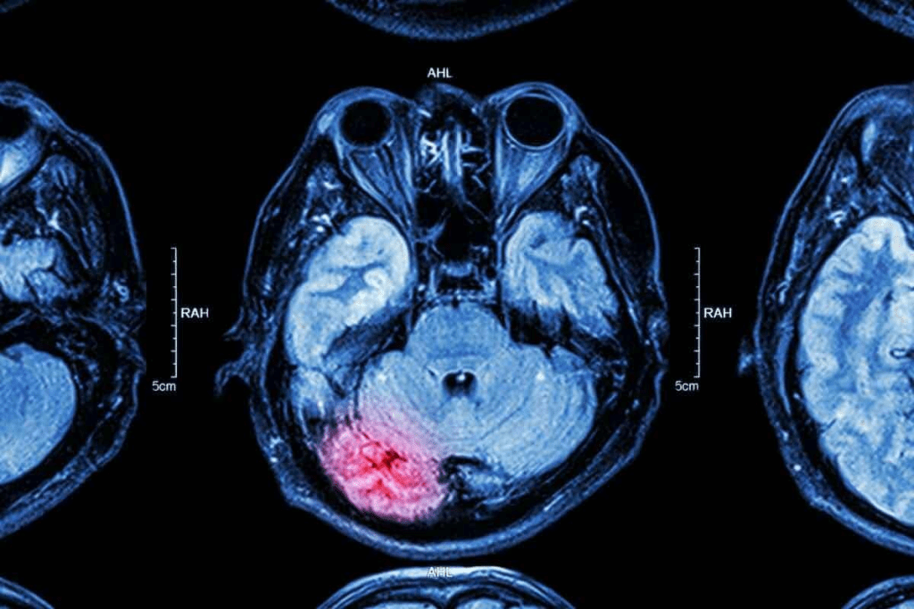
CT scans and MRI are key in brain cancer screening. CT scans use X-rays to find body issues. But MRI scans give more detail and are better at finding brain tumors. How does a brain cancer scan work? This essential guide compares CT vs MRI and explains the accuracy of modern tumor detection.
Key Takeaways
- Advanced imaging techniques are vital for good brain cancer screening.
- CT scans and MRI scans are the main ways to find brain tumors.
- MRI scans are better because they show more detail and are more sensitive.
- Finding cancer early is important for good care and management.
- Liv Hospital uses the newest imaging tech for the best patient care.
Understanding Brain Cancer and the Need for Imaging

It’s key to know about brain cancer to see why imaging is needed. Brain tumors can really hurt someone’s life, so finding them early is very important.
Common Symptoms That Warrant Brain Cancer Screening
Brain tumor symptoms can be different for everyone. They might include headaches, seizures, and changes in how you think. These signs can mean other things ,too. But if they keep happening or get worse, you need to get checked for brain cancer screening.
- Headaches that are persistent or severe
- Seizures or convulsions
- Cognitive changes, such as memory loss or confusion
Risk Factors for Brain Tumors
Some things can make you more likely to get brain tumors. These include genes you might have and being exposed to radiation.
| Risk Factor | Description |
| Genetic Predisposition | Family history of brain tumors or certain genetic conditions |
| Radiation Exposure | Previous exposure to ionizing radiation, such as from radiation therapy |
We stress how vital it is to know these risk factors and symptoms. This helps find brain tumors early. By spotting the signs and risks, people can get medical help fast. This might help them get better sooner.
The Science Behind Brain Cancer Scans

Brain cancer scans use advanced imaging to find and show tumors. These techniques are key for spotting abnormal tissue and seeing how big tumors are.
How Medical Imaging Detects Abnormal Tissue
CT and MRI scans find abnormal tissue in different ways. CT scans use X-rays to make detailed brain images, showing where things are off. MRI uses magnetic fields and radio waves for clear images of soft tissues, helping spot brain tumors.
Experts say MRI is better than CT for finding some brain tumors, like small ones or those in hard-to-see spots.
Contrast agents make tumors easier to see during scans. These substances, like gadolinium for MRI or iodine for CT, are given to patients before scanning. They highlight abnormal tissue, helping doctors see tumors more clearly.
This clear view helps doctors make accurate diagnoses and plan treatments. Medical experts say contrast agents make imaging better, helping doctors see tumors more precisely.
Using these advanced methods and contrast agents, we can give more accurate diagnoses. This helps us create better treatment plans for brain cancer patients.
Types of Brain Cancer Scan Procedures
Diagnosing brain cancer uses different imaging techniques. Each has its own benefits and uses. At our institution, we use a variety of methods to give accurate and detailed assessments to our patients.
Primary Diagnostic Imaging Methods
The main imaging methods for brain cancer are CT (Computed Tomography) scans and MRI (Magnetic Resonance Imaging). These are key to finding and understanding brain tumors.
CT scans use X-rays to show detailed images of the brain. They help spot tumors and other issues. MRI, with its strong magnetic field and radio waves, gives detailed images of brain tissue. It’s great for seeing tumors clearly because it shows soft tissues well.
Supplementary Testing Procedures
Along with main imaging, we use extra tests to help diagnose and plan treatment. These include functional MRI (fMRI) to check brain function, diffusion tensor imaging (DTI) to see white matter tracts, and magnetic resonance spectroscopy (MRS) to study tumor metabolism.
We also use PET (Positron Emission Tomography) scans. They show how active tumors are. This helps tell tumor types and check how well treatments work.
By mixing main imaging with extra tests, we get a full picture of the tumor. This helps plan better treatments and improves patient results.
CT Scans for Brain Tumor Detection
CT scans are key in finding brain tumors. They are fast and easy to get. This makes them great for quick checks, like in emergencies.
How CT Technology Works
CT scans use X-rays to make detailed pictures of the brain. An X-ray machine moves around the head. It takes many pictures to make a full view of the brain.
This helps us see the brain’s layout and find any problems, like tumors.
Can a CT Scan Show a Brain Tumor?
Yes, CT scans can spot brain tumors. They work well for big tumors and those that change the brain’s shape a lot. But, they’re not as good as MRI scans for finding small tumors.
They’re best used in urgent cases, like when someone has a stroke or bleeding in the brain.
Limitations of CT in Brain Cancer Detection
Even though CT scans are helpful, they have some downsides. They might not show small tumors or those in hard-to-reach places as clearly as MRI scans. Also, they use radiation, which is a concern, mainly for kids or those needing many scans.
| Imaging Modality | Detection Capability | Radiation Exposure |
| CT Scan | Good for larger tumors and emergency situations | Yes |
| MRI Scan | Excellent for detailed imaging, including smaller tumors | No |
In short, CT scans are good for initial checks of brain tumors, mainly in urgent cases. But, they can’t do everything. We also use MRI scans for a closer look at tumors.
MRI Scans: The Gold Standard for Brain Cancer Screening
MRI scans are the top choice for finding brain tumors. They show brain tissue in great detail. This helps us give our patients accurate diagnoses and treatment plans.
How MRI Technology Visualizes Brain Tissue
MRI scans use magnetic fields and radio waves to make detailed brain images. This tech lets us see brain structures and any tumors. The clear images help our team understand tumor size, location, and type.
Types of MRI Sequences Used for Brain Tumors
There are various MRI sequences for brain tumors. These include T1-weighted, T2-weighted, FLAIR, and diffusion-weighted imaging. Each sequence gives different information about the tumor. By using many sequences, we get a full picture of the tumor for treatment planning.
Can MRI Detect Brain Tumors That CT Misses?
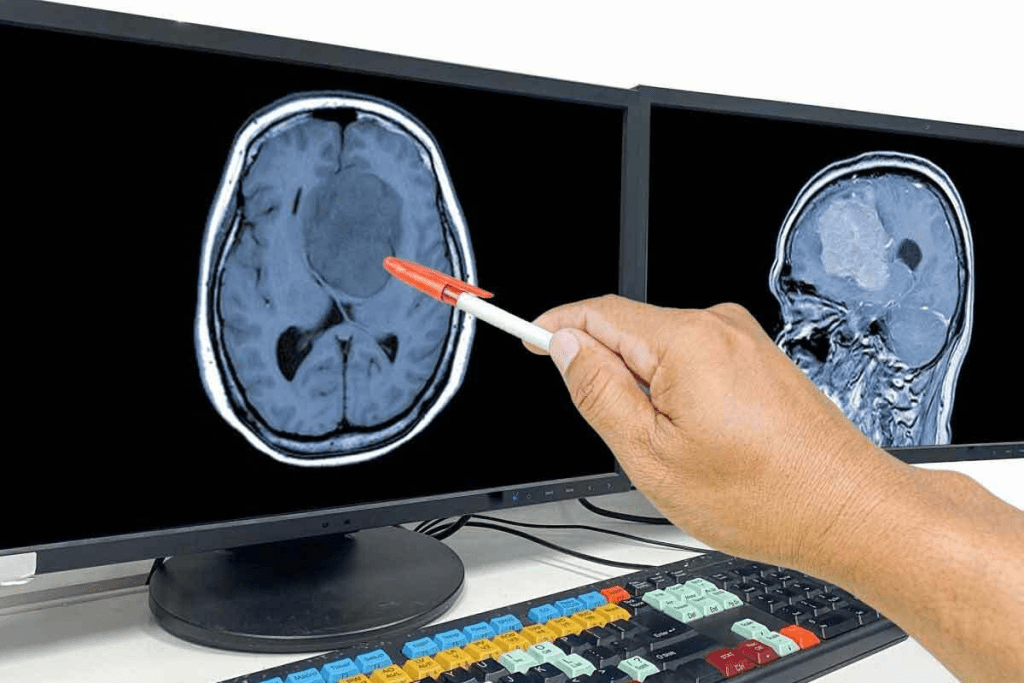
Yes, MRI scans can spot tumors that CT scans might miss. MRI’s better soft-tissue contrast helps see small or hard-to-find tumors. It also shows how tumors relate to nearby brain areas, key for surgery and radiation therapy.
In short, MRI scans are key to finding and treating brain tumors. They offer detailed and sensitive results. We keep using MRI technology to give our patients the best care.
CT vs MRI: Comparing Brain Tumor Detection Capabilities
It’s key to know the differences between CT and MRI scans for finding brain tumors. These tools help us understand the size, location, and presence of tumors in the brain.
Sensitivity and Specificity Differences
MRI scans are better at finding brain tumors than CT scans. MRI shows soft tissues like the brain in detail. MRI’s high sensitivity helps spot tumor boundaries and details.
CT scans, though, are faster and easier to get. They’re good for quick checks in emergencies. But, they might not show as much detail as an MRI for some tumors.
When CT is Preferred Over MRI
CT scans are better in some cases. They’re fast, which is important in emergencies. They’re also used when MRI isn’t safe due to metal implants or other reasons.
CT scans are chosen when quick imaging is needed or an MRI isn’t available. The choice between CT and MRI depends on the patient’s needs.
Cost and Accessibility Considerations
Cost and where to get them matter when picking CT or MRI. CT scans are cheaper and easier to find. They’re good for first checks or in places with limited resources.
But MRI’s better detail and accuracy make it better for detailed plans and treatments. We consider these points to give our patients the best care.
The Complete Brain Cancer Scan Process
We help our patients through every step of the brain cancer scan process. We make sure they are well-prepared and informed. This helps reduce anxiety and uncertainty, letting patients focus on their diagnosis and treatment.
Before Your Scan: Preparation Guidelines
Getting ready for a Brain Cancer Scan is important. Patients should follow specific guidelines to get accurate and useful images. These guidelines include:
- Removing any metal objects, such as jewelry or glasses, before the scan
- Avoiding certain foods or medications that could interfere with the scan
- Arriving early to complete any necessary paperwork
During the Procedure: What Happens
During the scan, patients lie on a table that slides into the machine. The scanning process is typically painless, but some might feel uncomfortable due to the space or staying very quiet for a long time.
Our medical team is there to make sure you’re comfortable and safe. We use the latest technology to get detailed brain images. These images help us diagnose and monitor brain cancer.
After Your Scan: Next Steps
After the scan, our team reviews the images and gives a detailed report to your healthcare provider. The next steps will depend on the findings of the scan. This might include more tests, talking to a specialist, or starting a treatment plan.
We know waiting for results can be tough. We’re here to support you. Your healthcare team will explain the next steps and answer any questions you have.
Advanced Brain Tumor Imaging Technologies
We are seeing big changes in how we find and treat brain tumors. New imaging tools are helping us do a better job.
Functional MRI and Diffusion Tensor Imaging
Functional MRI (fMRI) and Diffusion Tensor Imaging (DTI) are key tools in finding brain tumors. fMRI shows brain activity by looking at blood flow changes. This helps us see how tumors affect the brain.
DTI maps white matter tracts, which is vital for surgery planning. It helps keep brain function safe.
These tools are very important in brain cancer care. They give us detailed views of tumors and their surroundings. This helps doctors plan surgeries and treatments better.
Spectroscopy and Perfusion Studies
Magnetic Resonance Spectroscopy (MRS) and Perfusion-weighted Imaging (PWI) give us more info on brain tumors. MRS tells us about tumor metabolism, helping us tell tumor types apart. PWI shows how well blood flows to the tumor, which tells us about its growth and treatment response.
These methods add to what we see with regular scans. They help doctors make better diagnoses and track treatment progress.
Emerging Technologies in Brain Tumor Imaging
New brain tumor imaging methods are being developed. Advanced diffusion techniques like diffusion kurtosis imaging give us even more tumor details. Arterial spin labeling is another method that doesn’t need contrast agents.
These new tools are likely to make diagnosing and treating brain tumors even better. As research keeps going, we’ll see these advances become a big part of treating brain tumors.
Interpreting Brain Scan Results
Understanding brain scan results is key to diagnosing and treating brain tumors. It’s vital to accurately interpret these scans. This helps identify the presence, size, and type of tumors, guiding treatment choices.
How Radiologists Identify Brain Tumors
Radiologists analyze brain scans for abnormalities that might show tumors. They look at the size, shape, and location of any suspicious areas. They also check how these areas interact with the surrounding brain tissue.
When scanning, radiologists consider the patient’s medical history and symptoms. This detailed approach ensures accurate diagnoses.
Common Findings and What They Mean
Brain scan results can show various findings, from benign cysts to malignant tumors. Radiologists must carefully evaluate these findings to understand their clinical significance.
| Finding | Possible Interpretation |
| Enhancing mass | Potential tumor, possibly malignant |
| Cystic lesion | Could be benign or a tumor with cystic components |
| Edema surrounding a lesion | Indicates tumor infiltration or compression of surrounding brain tissue |
The Role of AI in Improving Detection Accuracy
Artificial intelligence (AI) is being used more to help radiologists with brain scans. AI algorithms can spot patterns that humans might miss, boosting detection accuracy.
By studying large datasets of brain scans, AI learns to recognize tumor characteristics. This helps radiologists make more accurate diagnoses and plan effective treatments.
Detection Accuracy: Can Brain Tumors Be Missed?
Brain tumor detection is complex, influenced by many factors. These include the tumor’s characteristics and the imaging methods used. Knowing these factors helps both doctors and patients make better choices about diagnosis and treatment.
Factors Affecting Detection Sensitivity
Several factors affect how well brain tumors are detected. Tumor size and location are key; smaller or harder-to-reach tumors are tougher to spot. The type of imaging used also matters. MRI is better than CT scans for finding soft tissue tumors, like brain tumors.
Other important factors include the quality of imaging equipment and the radiologist’s expertise. Better technology and skilled professionals can greatly improve detection rates.
False Positives and False Negatives
Brain tumor detection can lead to two types of errors: false positives and false negatives. A false positive means a test says there’s a tumor when there isn’t. This can cause unnecessary worry, more tests, and treatments.
A false negative is when a test misses a real tumor. This can delay treatment, which might harm the patient’s outcome.
When Additional Testing Is Needed
Because of these errors, more tests are often needed to confirm a diagnosis. This might include different imaging methods or tests like functional MRI or PET scans. We also look at the patient’s symptoms and medical history to make sure the diagnosis is right.
It’s vital to understand the limits of brain tumor detection for good patient care. By knowing what can affect detection, we can improve the diagnostic process and make better decisions.
From Scan to Diagnosis: Tests for Brain Cancer
Getting a brain cancer diagnosis involves several steps. It starts with imaging tests and might end with a biopsy. We’re here to help you through each step with care and knowledge.
The Role of Biopsy in Confirming Brain Cancer
A biopsy is a key step. It involves taking a small tissue sample from the tumor. This helps confirm if it’s cancer and what type it is.
The biopsy process typically involves:
- Preparation: This includes stopping certain medications and discussing any concerns or allergies.
- Procedure: The biopsy is usually performed under general anesthesia or local anesthesia with sedation, using imaging guidance to ensure accuracy.
- Recovery: Post-procedure care includes monitoring for any complications and managing pain.
As noted by the
American Cancer Society, “A biopsy is the only way to know for sure if you have a brain tumor and what type it is.”
How to Get Checked for a Brain Tumor
If you have symptoms like headaches, seizures, or changes in thinking, see a doctor. The first steps are a detailed medical history and a neurological exam.
| Diagnostic Step | Description |
| Medical History | Reviewing your medical history to identify any risk factors or previous conditions. |
| Neurological Examination | Assessing neurological function, including cognitive status, motor skills, and sensory responses. |
| Imaging Tests | Utilizing MRI or CT scans to visualize the brain and identify any abnormalities. |
Staging and Grading Brain Tumors
After confirming a diagnosis, we stage and grade the tumor. Staging shows how far the tumor has spread. Grading tells us how aggressive the tumor is.
We’re committed to accurate diagnoses and care. Our team creates a treatment plan tailored to your tumor’s specifics.
Conclusion: Advances in Brain Cancer Detection
Imaging technologies have greatly improved how we find and treat brain cancer. MRI technology is now key for spotting brain tumors. It helps us see tumors clearly and track how well treatments work.
We are always looking for new ways to help our patients. New brain cancer scan technologies, like MRI, are very important. They help us find cancer early and manage it better. This means we can give our patients better care and save more lives.
The future of finding brain cancer looks bright with new imaging tech. We’re committed to keeping up with these advances. This way, we can make sure our patients get the best care possible.
FAQ
What is the primary purpose of a brain cancer scan?
Brain cancer scans aim to find and diagnose brain tumors. They use technologies like CT and MRI scans for this purpose.
Can a CT scan detect a brain tumor?
Yes, CT scans can spot brain tumors. But MRI scans give more detailed images. CT scans are often used first.
Why is MRI considered the gold standard for brain cancer screening?
MRI is top because it shows brain tissue details better. It can find tumors that CT scans might miss.
What is the difference between CT and MRI scans in terms of sensitivity and specificity for brain tumor detection?
MRI scans are more sensitive and specific for brain tumors. This is true, even for certain tumor types.
How do I prepare for a brain cancer scan?
Preparing varies by scan type. Usually, remove metal objects. Some scans need contrast agents.
What happens during a brain cancer scan?
The patient lies on a table that slides into the scanner. The scanner then takes images of the brain.
How are brain scan results interpreted?
Radiologists look at images for abnormal tissue or tumors. They use their knowledge to spot issues.
Can brain tumors be missed during a scan?
Yes, tumors can be missed. This happens if the scan isn’t sensitive enough or if the tumor is small or hard to see.
What is the role of biopsy in confirming brain cancer?
A biopsy takes a tumor sample. It’s the best way to confirm if a tumor is cancerous.
How do I get checked for a brain tumor?
See a healthcare professional for a check. They’ll check your symptoms and choose tests like CT or MRI scans.
What are the latest advancements in brain tumor imaging technologies?
New tech includes functional MRI and diffusion tensor imaging. These help in more precise diagnosis and treatment planning.
Can an X-ray show a brain tumor?
No, X-rays can’t detect brain tumors. They’re not good for soft tissue in the brain.
How does MRI technology visualize brain tissue?
MRI uses a strong magnetic field and radio waves. It creates detailed images of brain tissue, spotting tumors and abnormalities.
What are the factors that affect detection sensitivity for brain tumors?
Detection sensitivity depends on the imaging tech, tumor size, and location, and the radiologist’s skill.
References
- Whelan, H. T., et al. (1988). Comparison of CT and MRI brain tumor imaging using a stereotactic external reference frame. American Journal of Neuroradiology, 9(3), 551-559. https://pubmed.ncbi.nlm.nih.gov/3242530/
- Javed, M. Y., et al. (2025). Detection and classification of brain tumors using a hybrid stacking classifier and multimodal brain tumor segmentation framework. Scientific Reports, 15, Article 18979. https://www.nature.com/articles/s41598-025-18979-8


