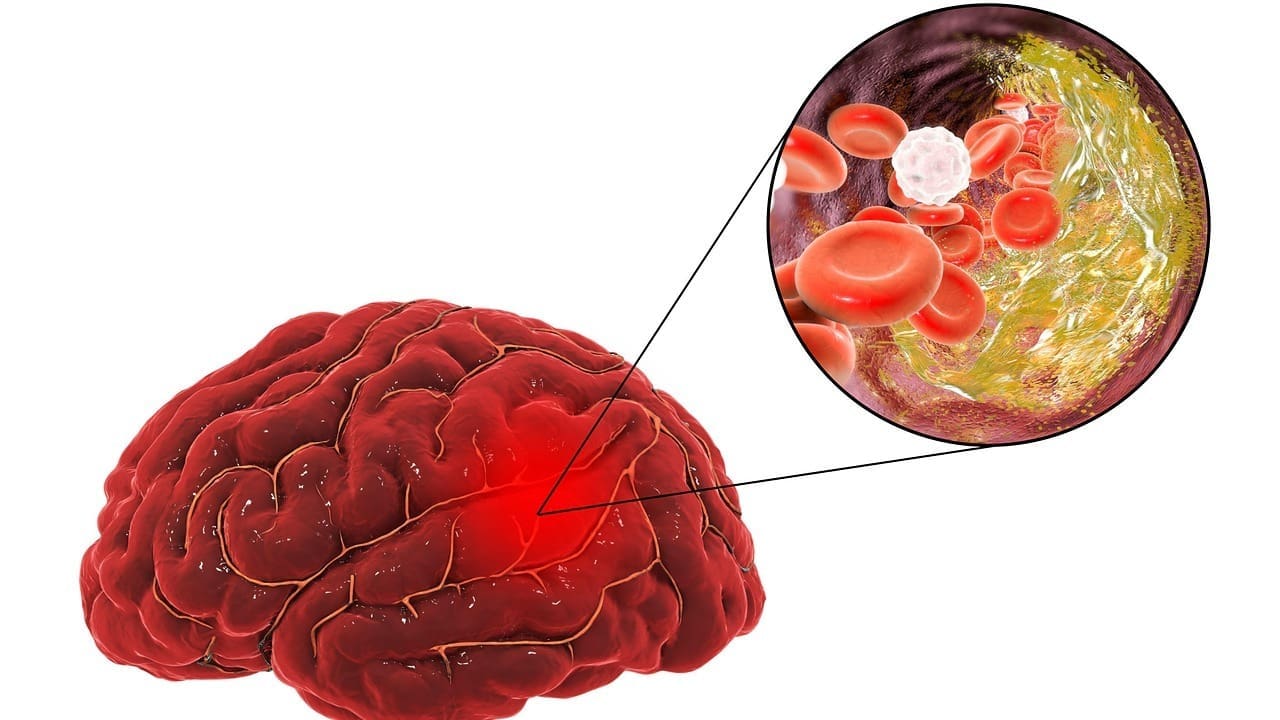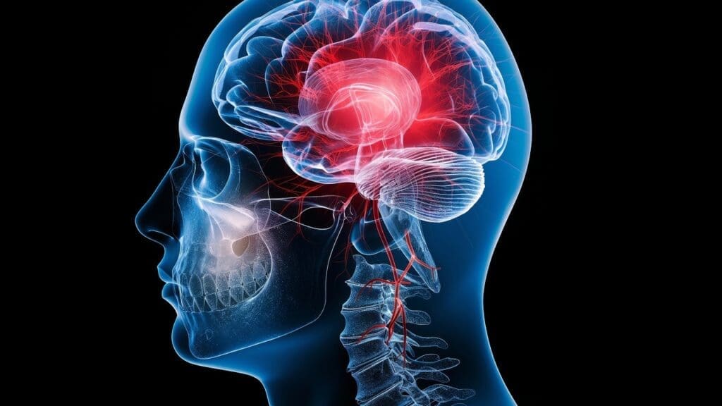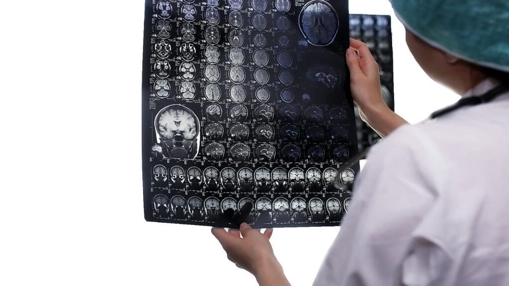
We lead in medical innovation with top-notch high-resolution imaging services. These services help doctors diagnose and plan treatments accurately. Our use of advanced technology lets us see the brain’s structure and function clearly.
We use seven main techniques: Magnetic Resonance Imaging (MRI), functional MRI (fMRI), Computed Tomography (CT), Positron Emission Tomography (PET), Single Photon Emission Computed Tomography (SPECT), Electroencephalography (EEG), and Diffusion Tensor Imaging (DTI). Each method gives us special information about brain function and dysfunction. This helps our doctors create specific treatment plans.
Key Takeaways
- Advanced brain mapping techniques for precise diagnosis
- Seven key imaging modalities for a full understanding
- High-resolution imaging for detailed views
- Targeted treatment plans based on imaging insights
- Combination of imaging modalities for better patient results
Understanding Brain Imaging Detailed H in Modern Medicine
The field of neuroimaging has grown a lot in recent years. This growth is thanks to new technologies and a better understanding of the brain. We are now getting unprecedented insights into the complexities of the human brain.
The Evolution of Neuroimaging Technologies
Neuroimaging technologies have changed a lot over the past decades. We’ve moved from early CT scans to today’s functional magnetic resonance imaging (fMRI). Each new technology has helped us understand the brain better. “The development of high-resolution brain imaging techniques has been a game-changer in neurology,” experts say.
Why High-Resolution Brain Imaging Matters
High-resolution brain imaging is key for diagnosing and treating neurological disorders. It gives detailed images of brain structures. This helps doctors spot problems that lower resolution images might miss.
Impact on Diagnosis and Treatment Planning
The effect of high-resolution brain imaging on diagnosis and treatment planning is huge. It lets doctors see the brain in great detail. This helps them make accurate diagnoses and plan treatments that work better. Patients get better care, leading to better results.
Magnetic Resonance Imaging (MRI): Structural Analysis with High Spatial Detail
Magnetic Resonance Imaging (MRI) has changed neuroimaging a lot. It gives us detailed pictures of the brain. This is key for finding and treating brain problems.
Principles and Physics Behind MRI Technology
MRI works on nuclear magnetic resonance. It uses strong magnetic fields and radio waves to show brain images. The science behind it is about aligning hydrogen nuclei in the body to make detailed pictures.
T1, T2, and FLAIR Sequences in Brain Visualization
Sequences like T1, T2, and FLAIR are important for brain pictures. T1 images show clear anatomy. T2 images spot changes in tissue water. FLAIR is great for finding lesions. We use these to see the brain fully.
Clinical Applications in Neurological Disorders
MRI is key for diagnosing and treating brain issues. It helps us see problems like stroke, multiple sclerosis, and tumors. MRI’s clear images help us make the right diagnosis and treatment plans.
Functional MRI (fMRI): Detecting Real-Time Brain Activity
Functional MRI (fMRI) has changed neuroscience by showing brain activity in real-time. It helps us see how brain parts work together. We use fMRI for surgeries and to study how we think and act.
Blood Oxygen Level Dependent (BOLD) Signal
The BOLD signal is key to fMRI. It uses blood oxygen changes in the brain to show activity. When brain areas work hard, they need more oxygen, which changes blood flow.
This change is what fMRI detects. It lets us see which brain parts are active during tasks.
Mapping Cognitive Functions and Neural Networks
fMRI helps us map brain functions and networks clearly. By looking at the BOLD signal, we find out which brain areas are active during tasks. This is important for understanding how the brain works and how areas talk to each other.
We’ve used it to study complex brain processes like memory and decision-making.
Applications in Neurosurgery and Cognitive Research
fMRI has many uses in neurosurgery and cognitive research. In neurosurgery, it helps map brain areas important for functions like language and movement. This helps surgeons plan safer surgeries.
In cognitive research, fMRI lets us study how the brain works. It gives us insights into brain and mental health issues. For example, fMRI studies have helped us understand Alzheimer’s and depression.
Computed Tomography (CT): Vital Tool for Rapid Brain Assessment
CT scans are key in emergency brain care, giving quick and detailed images. Our emergency department uses top CT scanners for fast and accurate diagnoses.
Creating Cross-Sectional Brain Images
CT scans use X-rays to make detailed brain images. They work by moving an X-ray source and detectors around the patient. This captures data for images.
Key aspects of CT imaging include:
- Rapid acquisition time, vital in emergencies
- High-resolution images for precise diagnosis
- Ability to see many tissues and structures
Advantages in Trauma and Acute Conditions
For trauma or sudden brain issues, CT scans are essential. They quickly show hemorrhages, fractures, and other urgent injuries.
The benefits of CT in these situations include:
| Benefit | Description |
|---|---|
| Speed | Rapid scanning time is vital in emergencies |
| Accuracy | High-resolution images help in exact diagnosis |
| Accessibility | CT scanners are common in hospitals |
Balancing Diagnostic Value and Radiation Exposure
CT scans are very useful but also expose to radiation. We balance their benefits with risks, mainly for young patients.
To cut down radiation, we follow the ALARA principle. We make scan protocols to get good images with the least dose.
Positron Emission Tomography (PET): Tracking Metabolic Processes and Disease Activity
PET imaging is a key tool in modern neurology. It gives a unique look into the brain’s metabolic functions and disease states. PET scans use radiopharmaceuticals to show detailed metabolic activity in brain tissues. This helps in diagnosing and planning treatments for various neurological conditions.
Radiopharmaceuticals and Glucose Metabolism
At the core of PET imaging are radiopharmaceuticals. These substances emit positrons and help visualize metabolic processes. The most used one is FDG (Fluorodeoxyglucose), which builds up in areas with high glucose metabolism. This lets doctors see the metabolic activity of brain tumors and track changes in glucose metabolism linked to neurological diseases.
Applications in Dementia and Brain Tumors
PET imaging is vital in managing dementia and brain tumors. In dementia, PET scans spot areas with low glucose metabolism. This helps diagnose Alzheimer’s disease and other dementias. For brain tumors, PET imaging checks how aggressive the tumor is and how well it responds to treatment.
| Condition | PET Imaging Application | Clinical Benefit |
|---|---|---|
| Dementia | Assessing glucose metabolism | Aids in early diagnosis and monitoring disease progression |
| Brain Tumors | Evaluating tumor metabolism | Helps in assessing tumor aggressiveness and treatment response |
Integration with Other Imaging Modalities
PET works best when combined with other imaging like MRI and CT. This mix boosts diagnostic accuracy and gives a full view of brain anatomy and function. It’s super useful in complex cases where detailed metabolic and anatomical info is needed for treatment planning.
By mixing PET’s metabolic data with MRI or CT’s anatomical details, doctors can craft better treatment plans for patients with neurological disorders.
Single Photon Emission Computed Tomography (SPECT): Revealing Cerebral Blood Flow Patterns
Single Photon Emission Computed Tomography (SPECT) is key in checking how blood flows to the brain. At our place, we use SPECT to see how the brain works and to help diagnose and treat brain problems.
Technical Principles and Radiotracer Use
SPECT uses tiny amounts of radioactive tracers, or radiotracers, injected into the blood. These tracers send out gamma rays that the SPECT camera catches. This lets us make detailed, 3D pictures of blood flow in the brain.
The type of radiotracer used is very important. Different tracers work better for different brain studies. For example, Technetium-99m HMPAO is often used because it sticks well to brain tissue and stays stable.
Clinical Value in Cerebrovascular Disorders
SPECT is very useful in studying and treating brain blood flow problems. It helps doctors figure out if someone has had a stroke or a temporary blockage in the brain.
It also helps check if treatments are working to improve blood flow. This is really helpful for people with moyamoya disease or other brain blood flow issues.
Comparative Advantages to Other Functional Imaging Methods
Compared to other brain imaging methods like PET, SPECT has some benefits. While PET is super sensitive to brain activity, SPECT is more common and cheaper. This makes SPECT a good choice for many brain studies.
| Imaging Modality | Cerebral Blood Flow Assessment | Availability | Cost |
|---|---|---|---|
| SPECT | High | Wide | Moderate |
| PET | Very High | Limited | High |
| fMRI | Indirect | Moderate | High |
In summary, SPECT is a great tool for looking at brain blood flow. It’s useful for diagnosing and treating brain blood flow problems. Its role in brain imaging shows how important it is today.
Electroencephalography (EEG): Measuring Electrical Signals for Neurological Assessment
We use Electroencephalography (EEG) to capture the brain’s electrical signals. This helps us diagnose and manage neurological conditions. EEG is non-invasive, recording electrical activity from the scalp’s surface. It gives us important information about brain function.
Recording and Interpreting Brain Wave Patterns
EEG records different brain waves like delta, theta, alpha, beta, and gamma. Each type shows different brain states. By looking at these patterns, we can spot problems and diagnose conditions like epilepsy and sleep disorders.
Diagnostic Applications in Epilepsy and Consciousness Disorders
EEG is key in diagnosing and managing epilepsy. It can spot seizure activity and understand patterns. It also helps with assessing consciousness disorders, like coma and vegetative state, by checking brain activity levels.
Advanced Quantitative Analysis Techniques
Advanced EEG analysis, like spectral analysis and source localization, gives us more detailed brain activity info. These methods help us measure brain function changes and pinpoint abnormal areas. This improves our ability to diagnose and treat neurological conditions.
Using EEG and advanced analysis, we can make diagnoses more accurate. This leads to better treatment plans for patients with neurological disorders.
Diffusion Tensor Imaging (DTI): Mapping White Matter Connectivity Networks
Diffusion Tensor Imaging (DTI) has changed neuroimaging by giving us detailed views of white matter connections. This advanced imaging lets us see the detailed paths of white matter tracts in the brain. It shows us how the brain’s structure works.
Principles of Diffusion-Weighted Imaging
DTI uses the principles of diffusion-weighted imaging to measure water movement in the brain. It looks at how water moves differently in white matter tracts. This helps us understand the complex structure of white matter networks.
“The ability to non-invasively map white matter tracts has significant implications for both clinical diagnosis and research,” as noted by leading neuroimaging experts. This ability helps us understand neurological conditions better and plan surgeries more precisely.
Fiber Tracking and Structural Connectivity Analysis
Fiber tracking, or tractography, is a key part of DTI. It helps us follow the paths of white matter tracts. By looking at the diffusion tensor data, we can see how these tracts connect. This is key for understanding how our brains work and how they are affected by diseases.
- DTI enables the detailed mapping of white matter tracts.
- It provides insights into the structural integrity of neural pathways.
- Fiber tracking facilitates the analysis of complex neural networks.
Clinical Applications in White Matter Disorders and Surgical Planning
DTI has many uses in clinical settings, mainly for diagnosing and treating white matter disorders. It helps doctors understand conditions like multiple sclerosis and leukodystrophy. It also helps neurosurgeons plan surgeries by showing them the brain’s complex anatomy and avoiding important tracts.
As we learn more about DTI, we can better diagnose and treat complex brain conditions. The insights from DTI are very important for both doctors and researchers. They help us find new ways to treat diseases.
Recent Advances in Brain Imaging Detailed H: Pushing Resolution Boundaries
The field of brain imaging is changing fast. New technologies are improving how we see the brain. We’re seeing big steps forward in high-field MRI, multiband imaging, and using artificial intelligence to analyze images.
High-Field MRI and Multiband Imaging Techniques
High-field MRI is a key tool for better brain images. It uses stronger magnetic fields for clearer views of brain structures. Multiband imaging makes MRI scans faster and more comfortable for patients.
A leading reseacher says, “High-field MRI could change brain imaging by showing more detail than ever before.”
“The development of high-field MRI represents a significant leap forward in our ability to visualize the brain in detail.”
Multimodal Integration for Comprehensive Brain Assessment
Multimodal integration combines data from various imaging methods. This gives a deeper look at brain structure and function. By mixing MRI, PET, and other scans, we understand neurological issues better.
| Imaging Modality | Primary Use | Benefits |
|---|---|---|
| MRI | Structural Analysis | High-resolution images of brain structures |
| PET | Metabolic Activity | Insights into brain metabolism and function |
| fMRI | Functional Analysis | Real-time brain activity monitoring |
Artificial Intelligence in Image Analysis and Interpretation
Artificial intelligence (AI) is making brain imaging better. AI finds patterns and oddities in images, making diagnoses more accurate.
Machine Learning Applications in Neuroimaging
Machine learning, a part of AI, is creating predictive models from neuroimaging data. These models help forecast patient outcomes and guide treatments.
Future Directions in Automated Diagnosis
The future of brain imaging is bright with AI and advanced imaging. As AI grows, we’ll see better automated diagnoses and personalized care.
Conclusion: The Transformative Impact of Advanced Brain Imaging
Advanced brain imaging has changed the game in neuroscience. It lets us diagnose and plan treatments with great accuracy. At Liv Hospital, we use the newest brain imaging tech to give our patients top-notch care.
By using tools like functional MRI and diffusion tensor imaging, doctors get a full picture of the brain. This helps us create treatment plans that really work. Our goal is to improve patients’ lives and quality of care, thanks to advanced brain mapping.
We’re always looking to improve brain imaging at Liv Hospital. We’re committed to giving international patients the best healthcare. Our team works hard to make sure each patient gets the best care possible, using the latest in brain imaging and more.
What is brain imaging detailed H, and how does it contribute to neurological diagnosis?
Brain imaging detailed H uses MRI, fMRI, CT, PET, SPECT, EEG, and DTI. These methods show the brain’s structure and function in detail. This helps doctors make accurate diagnoses and plan treatments.
What are the benefits of using high-resolution brain imaging techniques?
High-resolution brain imaging gives a deep look into brain function and problems. It helps doctors create specific treatment plans. This leads to better patient results.
How does MRI contribute to the diagnosis of neurological disorders?
MRI shows the brain’s structure clearly. It helps doctors spot and track conditions like stroke, tumors, and neurodegenerative diseases.
What is the role of fMRI in neurosurgery and cognitive research?
fMRI maps brain functions and networks. It guides surgeries and studies how the brain works and behaves.
How does CT scanning help in emergency neurological care?
CT scans quickly check for brain injuries and conditions. They help doctors make fast and accurate diagnoses in emergencies.
What is the significance of PET imaging in dementia and brain tumors?
PET imaging looks at brain metabolism and disease activity. It helps doctors see how brain tumors work and track neurological diseases like dementia.
How does SPECT imaging contribute to the diagnosis of cerebrovascular disorders?
SPECT imaging checks blood flow in the brain. It helps doctors diagnose and treat cerebrovascular disorders.
What is the role of EEG in diagnosing and managing neurological disorders?
EEG measures brain electrical activity. It’s key for diagnosing and managing conditions like epilepsy and consciousness disorders.
How does DTI contribute to the understanding of white matter connectivity networks?
DTI maps white matter networks. It helps doctors check white matter health and plan surgeries.
What are the latest advances in brain imaging detailed H?
New advances include high-field MRI and multiband imaging. There’s also multimodal integration and AI in image analysis. These advancements improve resolution and diagnosis.
How do advanced brain imaging techniques improve patient outcomes?
Advanced imaging lets doctors understand brain function and problems fully. This leads to better diagnosis and treatment plans. It improves patient results.


