
The human body’s complex neural network is partly controlled by the 12 cranial nerves. These nerves connect the brain to the head, neck, face, and vital organs.brainstem anatomy cranial nervesArteries of the Head: 7 Key Facts and Anatomy
It’s important to understand the origins and functions of these nerves. They are linked to the brainstem. The brainstem, made up of the midbrain, pons, and medulla oblongata, controls key functions. These include vision, hearing, taste, smell, facial movement, and the regulation of autonomic organs.
Knowing the anatomical origins and functions of the 12 cranial nerves is key. It helps clinicians diagnose and manage neurological problems well.
Key Takeaways
- The 12 cranial nerves are critical for controlling various bodily functions.
- Ten pairs of these nerves originate directly from the brainstem.
- Understanding the origins and functions of cranial nerves is vital for neurological diagnosis.
- The brainstem plays a key role in controlling essential bodily functions.
- Knowledge of cranial nerves is essential for effective neurological pathology management.
The Cranial Nervous System: An Overview

The cranial nervous system is key in our anatomy. It handles sensory and motor functions of the head and neck. This is vital for our neurological health.
Definition and Significance of Cranial Nerves
Cranial nerves are 12 nerves from the brain and brainstem. They control eye movements and send sensory info from the face and head. They also manage important functions like swallowing and heart rate.
The cranial nervous system is essential for our body’s actions. It controls both voluntary and involuntary actions. Knowing about cranial nerves helps in diagnosing and treating neurological issues.
General Organization of the Peripheral Nervous System
The peripheral nervous system (PNS) has two parts: the somatic and autonomic nervous systems. The PNS connects the CNS to the rest of the body. Cranial nerves are key in this system, carrying sensory and motor signals.
- The somatic nervous system handles voluntary actions, like movements and sensations.
- The autonomic nervous system controls involuntary actions, like heart rate and digestion.
Cranial nerves link the CNS to the body’s peripheral parts, mainly in the head and neck.
In summary, the cranial nervous system is complex and vital. It controls many functions we need every day.
Brainstem Anatomy and Cranial Nerves: The Fundamental Connection

Knowing the brainstem’s anatomy helps us understand cranial nerves better. The brainstem links the cerebrum to the spinal cord. It controls many automatic body functions.
The Three Regions of the Brainstem
The brainstem has three main parts: the midbrain, pons, and medulla oblongata. Each part has its own structures and roles. Together, they help cranial nerves control the body.
- The midbrain is at the top. It handles hearing and vision.
- The pons is below the midbrain. It helps with sleep and staying awake.
- The medulla oblongata is at the bottom. It connects to the spinal cord. It controls things like breathing and heart rate.
Cranial Nerve Nuclei within the Brainstem
Cranial nerve nuclei are groups of neurons in the brainstem. They are linked to cranial nerves. These nuclei handle sensory input, motor control, and autonomic functions.
The brainstem is home to many cranial nerve nuclei. Knowing where they are and what they do is key for treating nerve disorders.
Classification of Cranial Nerves
It’s important to know how cranial nerves are classified. They have different roles in our body. There are mainly three types: sensory, motor, and mixed nerves.
Sensory Cranial Nerves
Sensory cranial nerves send information from our body to the brain. They help us understand the world around us.
The main sensory nerves are the Olfactory nerve (CN I), Optic nerve (CN II), and Vestibulocochlear nerve (CN VIII). They deal with smell, vision, and balance/hearing.
Motor Cranial Nerves
Motor cranial nerves control our muscles. They send signals from the brain to muscles, helping us move.
Examples include the Oculomotor nerve (CN III), Trochlear nerve (CN IV), Abducens nerve (CN VI), and Hypoglossal nerve (CN XII). They help with eye movements, facial expressions, and tongue movements.
Mixed Cranial Nerves
Mixed nerves have both sensory and motor fibers. They are important for activities like swallowing and tasting.
The Trigeminal nerve (CN V), Facial nerve (CN VII), Glossopharyngeal nerve (CN IX), and Vagus nerve (CN X) are examples. They can send sensory info and control muscles.
Type of Cranial Nerve | Examples | Primary Functions |
Sensory | Olfactory (CN I), Optic (CN II), Vestibulocochlear (CN VIII) | Smell, Vision, Balance/Hearing |
Motor | Oculomotor (CN III), Trochlear (CN IV), Abducens (CN VI), Hypoglossal (CN XII) | Eye movements, Facial expressions, Tongue movements |
Mixed | Trigeminal (CN V), Facial (CN VII), Glossopharyngeal (CN IX), Vagus (CN X) | Facial sensation, Taste, Swallowing, Various bodily functions |
Cranial Nerves Originating from the Cerebrum
The cerebrum is the source of the olfactory and optic nerves. These nerves are key for our sense of smell and sight. They help us understand and react to what’s around us.
Olfactory Nerve (CN I): Structure and Function
The olfactory nerve carries smell information from our nose to our brain. It starts in the nasal cavity and ends in the olfactory bulb. There, it’s processed.
This nerve is special because it connects our brain directly to the outside world. This makes it more susceptible to damage from injuries or infections.
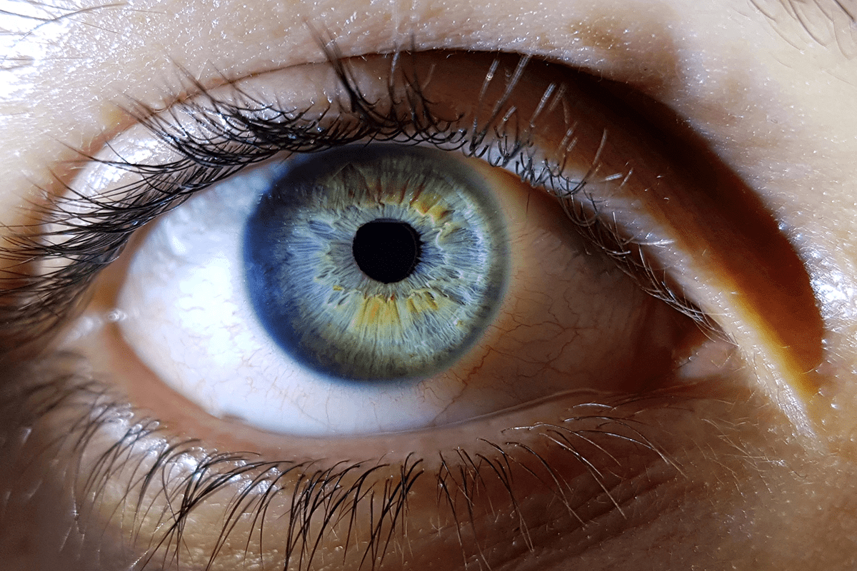
Optic Nerve (CN II): Structure and Function
The optic nerve is vital for our vision. It sends visual information from the retina to the brain. The optic nerve is made of axons from ganglion cells in the retina.
It carries visual signals to the brain for interpretation. Damage to the optic nerve can cause various vision problems, depending on the damage’s extent and location.
Midbrain Cranial Nerves
The midbrain is a key part of the brainstem. It is where two important cranial nerves start. These nerves help control eye movements and functions. We will look at the oculomotor and trochlear nerves, where they come from, and what they do.
Oculomotor Nerve (CN III): Origin and Function
The oculomotor nerve, or cranial nerve III, controls most eye movements. It helps rotate the eyeball, narrow the pupil, and keep the eyelid open. It starts from the oculomotor nucleus in the midbrain. The oculomotor nerve controls several extraocular muscles, including the medial rectus, superior rectus, inferior rectus, and inferior oblique muscles.
- Controls eyeball rotation
- Regulates pupil constriction
- Manages eyelid opening
Trochlear Nerve (CN IV): The Unique Dorsal Emergence
The trochlear nerve is the fourth cranial nerve. It is special because it comes out from the back of the brainstem. It starts from the trochlear nucleus in the midbrain. This muscle is key for rotating the eyeball, mainly when looking down.
The trochlear nerve’s role is vital for eye movements. Damage to it can make it hard to look down or move the eye in a certain way.
In summary, the midbrain is where the oculomotor and trochlear nerves start. Knowing about them helps in diagnosing and treating eye movement problems.
Pons and Pontomedullary Junction Cranial Nerves
We will explore the cranial nerves that arise from the pons and pontomedullary junction. These nerves are very important. The pons is a key part of the brainstem. It connects different parts of the brain and helps with many important functions.
The cranial nerves from the pons control facial expressions and send sensory information. They also help regulate the body’s functions. Let’s look at these nerves in more detail.
Trigeminal Nerve (CN V): The Three Branches
The trigeminal nerve is very complex. It handles both sensory and motor functions. It has three main branches: the ophthalmic, maxillary, and mandibular nerves.
- The ophthalmic branch is sensory, sending info from the eye and nearby areas.
- The maxillary branch is sensory, sending info from the mid-face.
- The mandibular branch has both sensory and motor fibers. It controls muscles for chewing.
The trigeminal nerve’s diverse functions make it key for facial sensation and motor control.
Abducens Nerve (CN VI): Eye Movement Control
The abducens nerve controls the lateral rectus muscle. This muscle is for moving the eye outward. Damage to this nerve can cause eye movement problems.
“The abducens nerve’s role in controlling eye movement shows how complex vision coordination is.”
Facial Nerve (CN VII): Facial Expression and Taste
The facial nerve has many roles. It controls facial expressions, sends taste info from the tongue, and controls some neck muscles.
Damage to the facial nerve can cause facial paralysis and taste issues. This shows how important it is for both motor and sensory functions.
Vestibulocochlear Nerve (CN VIII): Balance and Hearing
The vestibulocochlear nerve handles sound and balance info. It has two parts: the cochlear nerve for hearing and the vestibular nerve for balance.
Dysfunction of the vestibulocochlear nerve can cause hearing loss, vertigo, and balance problems. This can greatly affect quality of life.
In conclusion, the cranial nerves from the pons and pontomedullary junction are vital. They help with facial expressions, eye movements, hearing, and balance. Understanding these nerves is key for diagnosing and treating neurological disorders.
Medulla Oblongata Cranial Nerves
The medulla oblongata is a key part of the brainstem. It gives rise to four important cranial nerves. These nerves help control swallowing, talking, and tongue movement.
Glossopharyngeal Nerve (CN IX): Throat and Tongue
The glossopharyngeal nerve, or cranial nerve IX, is vital for swallowing and taste. It sends signals from the back third of the tongue. It also helps the stylopharyngeus muscle in swallowing.
Damage to this nerve can cause trouble swallowing and losing taste on the back tongue. Doctors check the gag reflex and taste to assess this nerve.
Vagus Nerve (CN X): The Wanderer
The vagus nerve, or cranial nerve X, is very complex and wide-reaching. It helps many organs in the chest and belly. It’s key in the parasympathetic nervous system.
This nerve controls the larynx muscles, heart rate, and digestion. Damage can cause voice problems, swallowing issues, and other autonomic problems.
Accessory Nerve (CN XI): Head and Shoulder Movement
The accessory nerve, or cranial nerve XI, is special because it has both cranial and spinal roots. It helps the sternocleidomastoid and trapezius muscles for head and shoulder movements.
Problems with this nerve can weaken or paralyze these muscles. This makes it hard to rotate the head or lift the shoulder. Doctors test the strength of these muscles.
Hypoglossal Nerve (CN XII): Tongue Control
The hypoglossal nerve, or cranial nerve XII, controls tongue movements. It works with all the tongue’s muscles.
Damage to this nerve can weaken or paralyze the tongue. This makes speaking and swallowing hard. Doctors check tongue strength and movement.
Cranial Nerve | Function | Clinical Significance |
Glossopharyngeal (IX) | Swallowing, taste sensation | Dysphagia, loss of taste |
Vagus (X) | Parasympathetic functions, vocalization | Vocal cord paralysis, autonomic dysfunction |
Accessory (XI) | Head and shoulder movement | Weakness in sternocleidomastoid and trapezius muscles |
Hypoglossal (XII) | Tongue movement | Dysarthria, dysphagia |
In conclusion, the cranial nerves from the medulla oblongata are very important. They control essential functions. Knowing about them helps in diagnosing and treating neurological problems.
Comprehensive Map of Cranial Nerves on the Brainstem
The network of cranial nerves on the brainstem is complex and interesting. It needs a detailed visual guide and mnemonic aids for better understanding. Knowing the origins and functions of these nerves is key for medical professionals and students.
Visual Guide to Cranial Nerve Origins
A visual guide is essential for grasping the anatomy of cranial nerves on the brainstem. Looking at a detailed map helps see the exact locations and functions of each nerve. The brainstem, made up of the midbrain, pons, and medulla oblongata, is where 10 of the 12 cranial nerves start.
Cranial nerves III and IV start from the midbrain. The oculomotor nerve (III) controls eye movements. The trochlear nerve (IV) is the thinnest and also controls eye movement. The pons is where cranial nerves V, VI, VII, and VIII begin. These nerves handle facial sensation, eye movement, facial expression, and hearing and balance.
Mnemonic Devices for Remembering Cranial Nerves
Mnemonic devices help remember the order and names of cranial nerves. A well-known mnemonic is: “On Old Olympus’ Towering Top, A Finn And German Viewed Some Hops.” Each word starts with the first letter of the 12 cranial nerves in order.
“The key to understanding cranial nerves lies not just in memorizing their names, but in visualizing their origins and functions on the brainstem.”
Using a detailed visual guide and mnemonic devices helps learners understand cranial nerves better. This approach improves retention and recall. It’s very useful for both medical education and clinical practice.
Clinical Assessment of Cranial Nerve Function
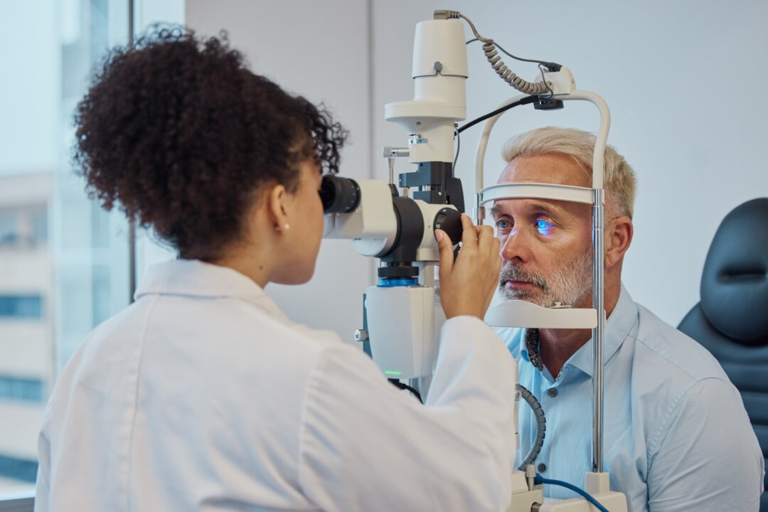
Evaluating cranial nerve function is key for diagnosing neurological disorders. We will look at standard examination techniques and how they work together. This highlights their role in clinical practice.
Standard Examination Techniques
Standardized techniques are vital for accurately checking cranial nerve function. These methods involve a detailed check of each nerve, using specific tests. For example, the olfactory nerve (CN I) is tested by smelling different odors.
Other nerves are checked in different ways. The oculomotor (CN III), trochlear (CN IV), and abducens (CN VI) nerves are tested by looking at eye movements. The trigeminal nerve (CN V) is checked through facial sensation and jaw clenching tests. The facial nerve (CN VII) is tested by looking at facial expressions and taste.
Integrated Approach to Cranial Nerve Testing
An integrated approach combines individual nerve tests into a full neurological exam. This helps spot patterns of nerve problems that might point to specific conditions. For instance, a patient with double vision might need tests on CN III, CN IV, and CN VI, along with other nerves.
This approach also looks at how nerves work together with the rest of the nervous system. It lets doctors diagnose complex conditions and create specific treatment plans.
By mixing standard tests with an integrated approach, doctors can do a complete check of cranial nerve function. This detailed assessment is essential for making accurate diagnoses and effective treatment plans for patients with neurological issues.
Common Cranial Nerve Disorders and Pathologies
Cranial nerve disorders can really affect someone’s life. It’s important to know what causes them and how they show up. These issues can come from things like blood vessel problems, injuries, infections, or tumors.
The way these disorders show up can change a lot. It depends on which nerves are affected and why.
Cranial Nerve Palsies and Their Presentations
Cranial nerve palsies make the muscles controlled by the nerve weak or paralyzed. For example, a problem with the third nerve can cause droopy eyelids, double vision, and big pupils. A neurologist says, “Understanding the nerves and how they work is key to diagnosing these palsies.”
“The clinical presentation of cranial nerve palsies can be quite varied, making a detailed neurological examination critical for diagnosis.”
The sixth nerve palsy can cause double vision because it can’t move the eye sideways. Bell’s palsy, affecting the seventh nerve, leads to weakness or paralysis on one side of the face.
Neurodegenerative and Inflammatory Conditions
Diseases like Alzheimer’s and Parkinson’s can harm cranial nerves. This can lead to memory loss, trouble moving, and problems with automatic functions. Inflammatory diseases, like multiple sclerosis, can also affect nerves, causing vision problems, double vision, and facial pain.
Handling these conditions needs a team effort. Doctors, eye specialists, and physical therapists work together. They might use medicine, therapy, or surgery to help.
Learning more about cranial nerve disorders is important. Early diagnosis and treatment can make a big difference. By spotting the signs early, doctors can give better care.
Conclusion
Learning about the 12 cranial nerves is key to understanding their roles in our body. We’ve looked at where they come from, what they do, and why they’re important. This gives us a full picture of cranial nerves.
The cranial nerves start in the brainstem and help control many body functions. They handle things like feeling sensations, moving muscles, and keeping our body’s systems running smoothly. Knowing about these nerves helps doctors diagnose and treat problems related to them.
To wrap it up, we’ve covered the 12 cranial nerves in detail. We’ve seen how they’re vital for our health and how they help our body work right. By grasping their complex roles, we can better understand how our body functions.
FAQ
What are the 12 cranial nerves and their functions?
The 12 cranial nerves start from the brain. They include the olfactory and optic nerves. Also, the oculomotor, trochlear, and trigeminal nerves are part of this group.
Other nerves like the abducens, facial, and vestibulocochlear nerves are also included. The glossopharyngeal, vagus, accessory, and hypoglossal nerves round out the list. These nerves help us smell, see, move our eyes, and more.
What is the brainstem and its relation to cranial nerves?
The brainstem is key and includes the midbrain, pons, and medulla oblongata. It’s where most cranial nerves start or end. This makes it essential for controlling our body’s functions.
How are cranial nerves classified?
Cranial nerves are divided into three types. Sensory nerves send information to the brain. Motor nerves control our movements. Mixed nerves do both.
What is the significance of understanding cranial nerves?
Knowing about cranial nerves helps doctors diagnose and treat neurological issues. It’s vital for assessing and treating patients with nerve problems.
What are some common cranial nerve disorders?
Common issues include nerve palsies and conditions like multiple sclerosis. Meningitis is another example. These affect how our nerves work.
How are cranial nerves tested clinically?
Doctors use specific tests to check cranial nerves. They look at sensory and motor functions, and reflexes. Each nerve has its own test.
What is the role of the cranial nerves in controlling eye movement?
The oculomotor, trochlear, and abducens nerves control our eye movements. The oculomotor nerve works with several eye muscles. The trochlear and abducens nerves control the superior oblique and lateral rectus muscles, respectively.
How do cranial nerves relate to the peripheral nervous system?
Cranial nerves are part of the peripheral nervous system, along with spinal nerves. They help transmit information between the brain and the body.
What are some mnemonic devices for remembering cranial nerves?
Mnemonics like “On Old Olympus’ Towering Top, A Finn And German Viewed Some Hops” can help remember the 12 cranial nerves.
Where can I find a detailed map of cranial nerves on the brainstem?
Detailed maps of cranial nerves can be found in neuroanatomy resources. Textbooks and online atlases provide visual guides to the nerves’ origins and paths.
References
Government Health Resource. Pituitary Gland: Alternative Names and Functions. Retrieved from https://histology.siu.edu/erg/pituit.htm
.


