
Last Updated on October 31, 2025 by Saadet Demir
Discovering a lump in the neck can be alarming. But, modern imaging technology offers reassurance. Neck cancer affects the head and neck, impacting speech and swallowing.
Early detection is key for effective treatment. CT scans play a significant role in showing detailed images of internal structures. We will look into the effectiveness of CT scans in detecting neck cancer, their accuracy, and what you need to know about the process.
Can a CT scan detect neck cancer? Yes, it is a primary tool for staging and identifying tumors in the throat, lymph nodes, and surrounding structures.
Neck cancer includes many types, such as salivary gland cancer and thyroid cancer. It’s important to know the risk factors and the need for early detection. This knowledge helps in understanding and treating neck cancer effectively.
Neck cancers vary, each with its own challenges. Salivary gland cancer affects the glands that make saliva. Carotid body tumors are rare growths near the carotid artery. Thyroid cancer involves the thyroid gland, and lymphoma is a cancer of the lymphatic system. Squamous cell carcinoma is a common type that can arise in various parts of the neck.
The symptoms of neck cancer can vary. Common signs include lumps or swelling, sore throat, and difficulty swallowing. Risk factors like tobacco use, alcohol consumption, and Human Papillomavirus (HPV) infection play a big role in developing neck cancer.
Risk Factor | Description | Impact on Neck Cancer |
Tobacco Use | Includes smoking and chewing tobacco | Increases risk of developing neck cancer |
Alcohol Consumption | Excessive alcohol intake | Heightens risk, specially with tobacco use |
HPV Infection | Certain strains of the Human Papillomavirus | Linked to oropharyngeal cancers |
Early detection of neck cancer is key. It improves treatment outcomes and survival rates. Advanced CT technology helps in accurate diagnosis.
Being aware of risk factors and symptoms is vital. This awareness helps in seeking medical attention early. Understanding neck cancer types and their implications is essential for timely action.
CT scans are key in finding neck cancer. They give us clear pictures of the neck area. We can see bones, soft tissues, blood vessels, and lymph nodes.
CT imaging uses X-rays and computers to make detailed images. It lets us see inside the neck well. This helps us spot problems that might mean cancer.
The machine moves around the body, taking pictures from all sides. Then, a computer puts these images together. This makes detailed pictures of the neck’s inside.
CT scans can be with or without contrast. Contrast material makes some areas stand out more. This makes it easier to see tumors and other issues. We often use contrast for neck cancer scans because it shows soft tissues and blood vessels better.
CT scans can show many things in the neck. This includes lymph nodes, salivary glands, thyroid gland, and soft tissues. This is important for spotting abnormal growths and changes that might mean cancer.
Some important things CT scans can spot include:
By showing us these details, CT scans help us understand how big the cancer is. This helps us plan the best treatment.
It’s important to know how well CT scans work in finding neck cancer. They are a key tool in diagnosing and understanding the extent of neck cancer. But, their ability to find cancer can depend on several things.
Research shows CT scans can spot neck cancer tumors in 68 to 83 out of 100 cases. This is known as sensitivity. It shows how often the scan finds the tumor correctly.
CT scans are also good at ruling out neck cancer, with a specificity of 69% to 93%. Specificity is about how well the scan says who doesn’t have cancer. A higher specificity means fewer false alarms.
When it comes to finding cancer that has come back, CT scans are less effective. They have a sensitivity of about 63% and a specificity of 80%. Finding cancer again is hard because it can change after treatment. CT scans are helpful but not perfect in these cases.
Understanding these numbers is key when looking at CT scan results. CT scans are good but not 100% accurate in finding neck cancer. How well they work can depend on the tumor’s size, where it is, and the scan’s quality.
Talking to a healthcare provider about CT scan results is important. They can help explain what the results mean and what to do next.
Advanced CT technologies are key in improving neck cancer detection. Techniques like iterative reconstruction and metal artifact reduction have boosted CT scan quality.
Iterative reconstruction has changed CT imaging. It makes images clearer and reduces noise. This method helps us make more precise diagnoses by improving image quality.
“The use of iterative reconstruction in CT scans has been a game-changer in diagnostic imaging,” says a leading radiologist. “It has enabled us to provide more accurate diagnoses and develop more effective treatment plans.”
Metal artifact reduction is another big step in CT technology. It reduces distortions from metal objects, like dental fillings or implants. This makes images clearer, leading to more accurate assessments.
Together, iterative reconstruction and metal artifact reduction greatly improve diagnostic precision. They help us detect neck cancer more effectively. This means patients get accurate diagnoses and timely treatment.
When a patient gets a CT scan for a neck lump, these technologies help us understand it better. They tell us if it’s a benign cyst or a malignant tumor. This clarity is very important.
As we go forward, these advanced CT technologies will keep improving our ability to diagnose and treat neck cancer. By using these innovations, we can give better care to our patients and improve outcomes in the fight against neck cancer.
In neck cancer diagnosis, MRI and CT scans are both key. They each have their own strengths. The choice between them depends on the tumor’s type and the patient’s needs.
Both MRI and CT scans are good at finding neck cancer. MRI is as good as CT scans for some neck cancer details. But, the right choice depends on the situation.
MRI is great for seeing soft tissue details, which is important for cancer spread. CT scans are quicker and easier to get, perfect for urgent cases.
MRI is better at showing soft tissue details. This is key for seeing how far cancer has spread. It helps in planning treatment.
MRI is safer because it doesn’t use ionizing radiation. This is good for patients needing many scans or who are sensitive to radiation. CT scans use radiation, but doses are kept low.
MRI is better when you need to see soft tissue details clearly. It’s also safer for avoiding radiation, like in kids or for many scans.
In summary, MRI and CT scans both help in neck cancer care. The choice depends on the situation, patient needs, and tumor details. Knowing each modality’s strengths helps doctors make the best decisions for patients.
PET-CT is a top choice for diagnosing neck cancer. It combines Positron Emission Tomography (PET) and Computed Tomography (CT) in one scan. This makes it a powerful tool for both functional and anatomical imaging.
PET-CT scans use PET’s metabolic info and CT’s detailed images. This mix lets doctors spot tumors and see how active they are. It gives a clear picture of the tumor’s size and how it’s growing.
Key benefits of PET-CT include:
PET-CT is great at finding cancer in lymph nodes. It’s very accurate, with sensitivity rates up to 96.4% and specificity around 86.4%. This is key for planning treatment.
The high accuracy of PET-CT in detecting lymph node involvement is critical for treatment planning. It helps doctors know how far the cancer has spread. This info guides decisions on surgery, radiation, or other treatments.
PET-CT is very effective but can be pricey and hard to find. It’s not as common as CT or MRI machines. But, for many, its benefits in diagnosis and staging are worth it.
Access to PET-CT varies by region and healthcare system. Yet, for neck cancer patients, it’s a valuable tool. It can greatly influence their treatment plan.
Endoscopy is key in finding neck cancer. It lets doctors see the throat and larynx up close. They can spot any odd spots or growths.
Endoscopy lets doctors see inside the throat and larynx. This direct visualization helps find growths or odd spots that other tests miss. Doctors use a special camera to look closely at the inside.
The high-resolution images from modern endoscopes help doctors see everything clearly. This is very useful for finding cancers or growths early on.
Endoscopy is great for taking tissue samples. Doctors can take samples from any odd spots. Then, these samples are checked to see if there are cancer cells.
The biopsy capability of endoscopy is key for a sure diagnosis. By studying the samples, doctors can figure out the cancer type and stage. This helps plan the best treatment.
Endoscopy works well with CT scans to fully understand neck cancer. CT scans show how big the cancer is and if it’s spread. But endoscopy lets doctors see and take samples directly.
Using both endoscopy and imaging studies gives a full picture of the cancer. This helps doctors stage the cancer accurately and plan the best treatment. The complementary use of endoscopy and imaging studies makes diagnosis and treatment more precise.
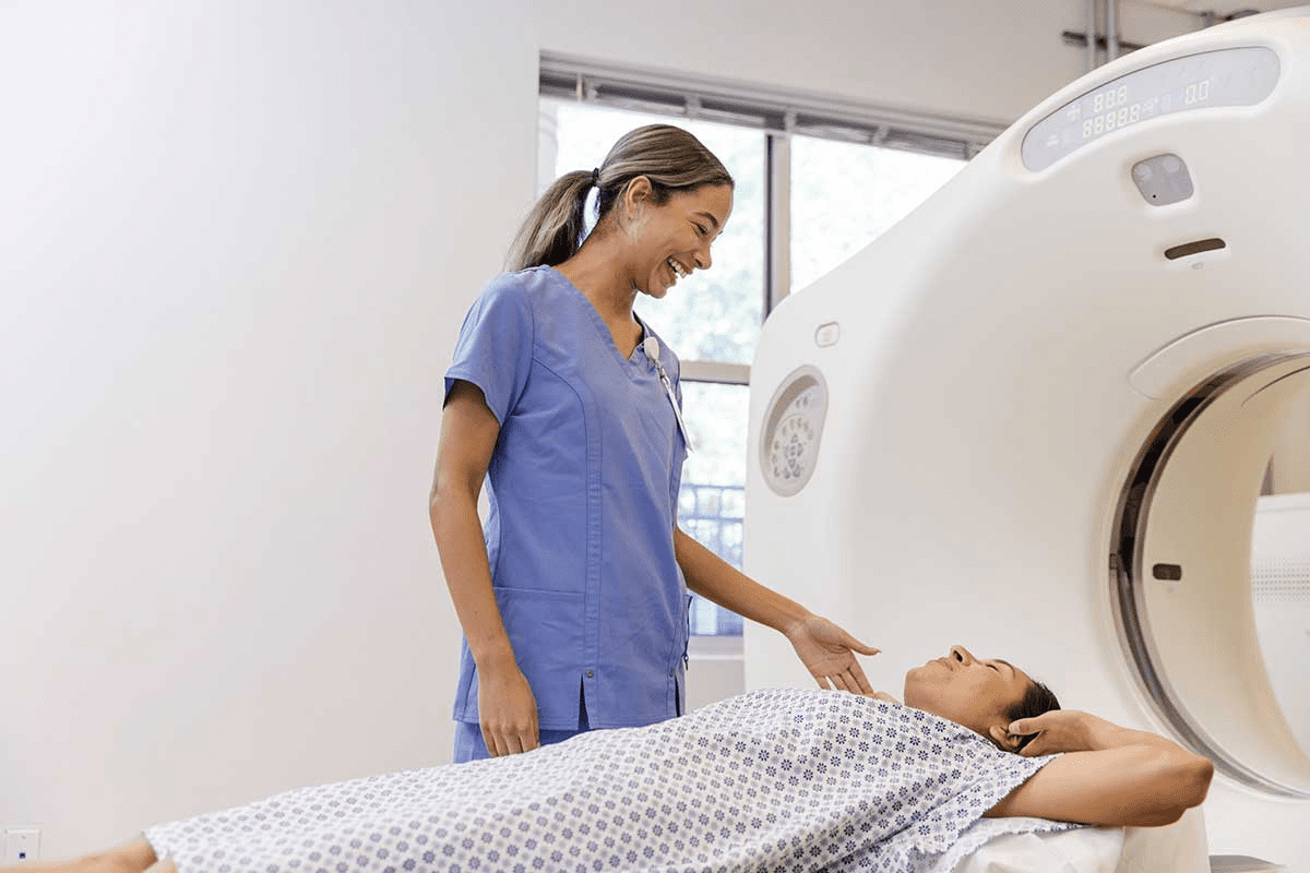
If you’re getting a neck CT scan, you might wonder what it’s like. A neck CT scan is a test that lets doctors see inside your neck. It helps find problems or cancer in your lymph nodes. Getting ready right is key for a good scan.
To get ready for your neck CT scan, you’ll need to follow some steps. These steps include:
Also, arriving early is a good idea. This lets you fill out paperwork and talk to the medical team about any worries.
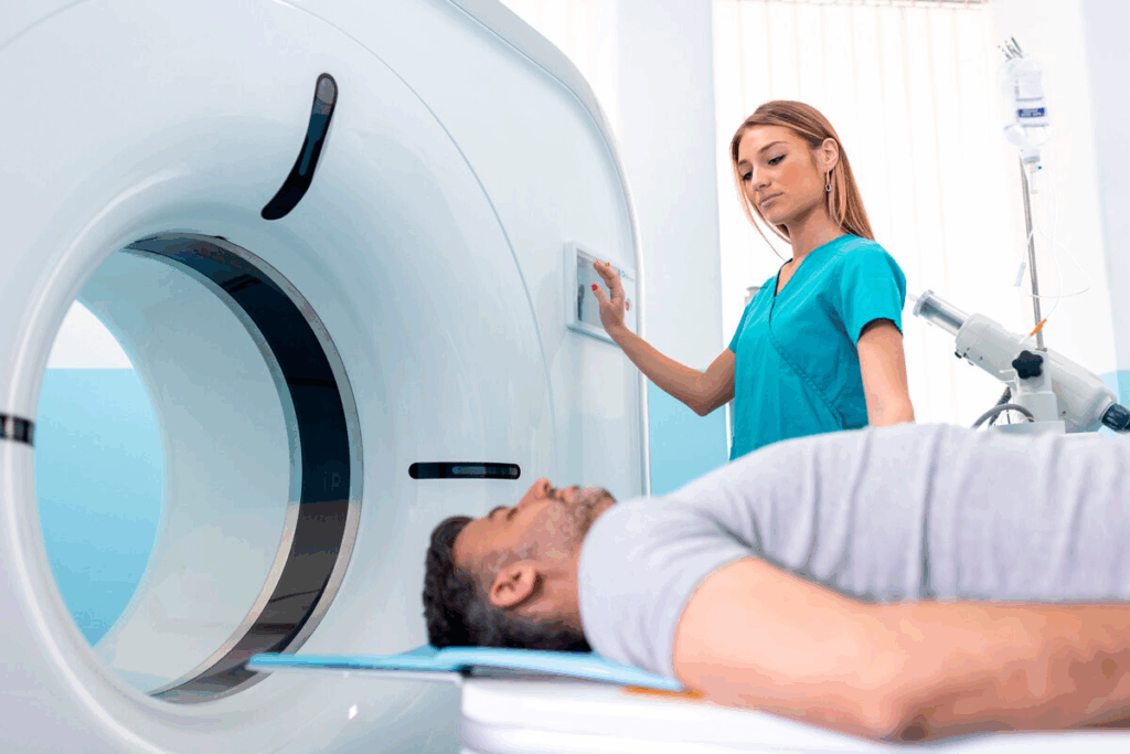
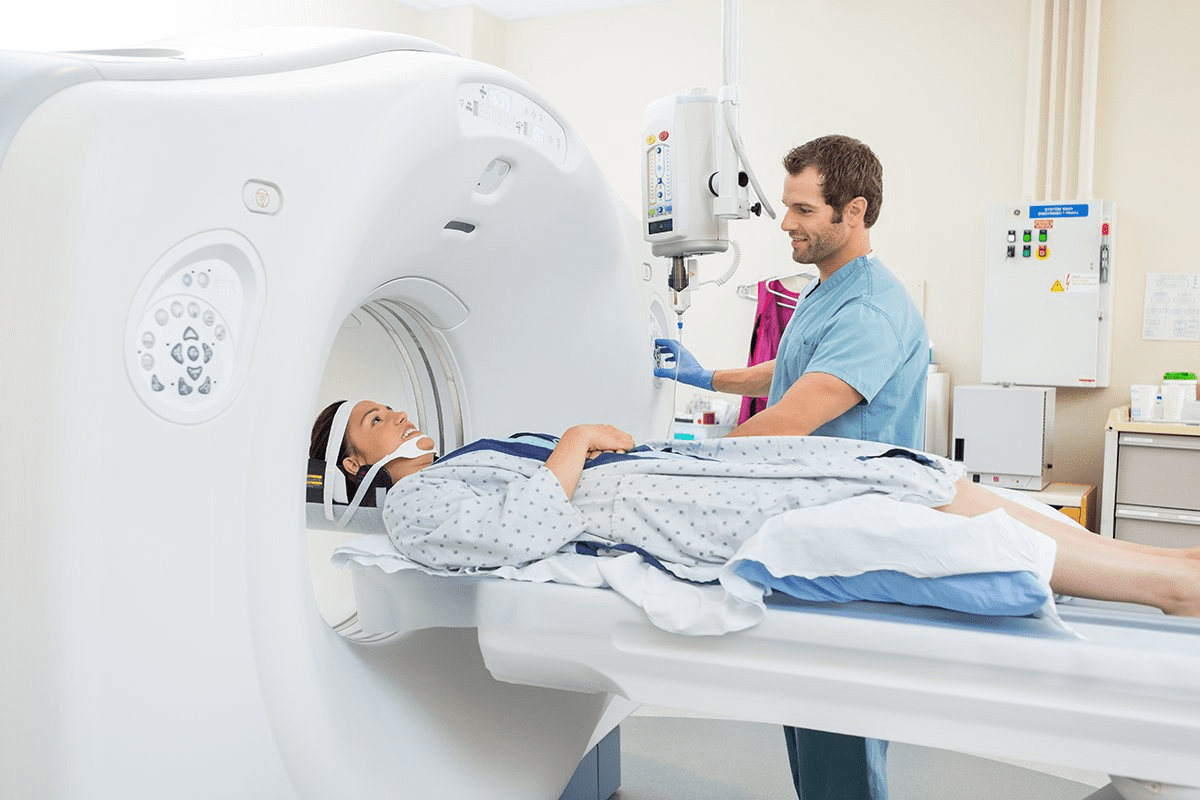
The CT scan itself is quick and easy. You’ll lie on a table that moves into a big, doughnut-shaped machine. The machine will move around you, taking X-ray images from different angles. If contrast is used, it goes through an IV in your arm.
You’ll need to stay very quiet and might have to hold your breath sometimes. The whole thing usually takes just a few minutes.
Even though CT scans are safe, there are some possible side effects and risks. These include:
Talking to your doctor or the team doing the scan is important. They can answer your questions and make you feel more at ease.
Finding out why a neck lump appears can be tricky. It needs a careful plan to figure it out. When someone notices a lump in their neck, a detailed first check is key to decide what to do next.
The first step is a full medical history and physical check. We look at the lump’s size, where it is, and if it hurts. If it might be cancer, seeing a specialist is usually the next move.
For neck cancer, scans are very important to find and understand the disease. A CT scan of the neck is often first because it shows the neck’s details well.
A CT scan can spot the main tumor, check lymph nodes, and find any spread. Sometimes, MRI or PET-CT scans are needed too to see how far the disease has spread.
After the scans, we look at the results to plan the next steps. If it’s neck cancer, we talk about treatment options. This could be surgery, radiation, or chemo.
“Accurate diagnosis and staging are key for a good treatment plan for neck cancer patients.”
Neck Cancer Treatment Guidelines
We team up with many experts to give full care. This is from finding out the diagnosis to treatment and aftercare.
Understanding the role of CT scans in neck lumps and lymph nodes is key. CT scans give us detailed images. These images help us spot signs that might mean cancer.
CT scans help us tell if a lump is harmless or might be cancer. We look at size, shape, and density. A big or irregular lymph node might suggest cancer.
Knowing if a lump is benign or malignant is very important. It helps us give the right care. Some lumps, like those from infections, can grow big. But, certain CT scan signs can point to cancer.
Cancerous lymph nodes show certain signs on CT scans. These include:
Characteristic | Benign | Malignant |
Size | Typically <1 cm | Often >1 cm |
Shape | Oval or Round | Irregular |
Necrosis | Absent | Present |
Grouping | Scattered | Clustered |
If a CT scan shows suspicious findings, we need to investigate more. This might include PET-CT or MRI, or a biopsy. We look at the patient’s symptoms and medical history too.
By carefully looking at CT scan results and the patient’s situation, we make smart decisions. We guide the patient through the next steps in diagnosis.
CT scans are key in finding and managing neck cancer. They help patients and doctors make smart choices about tests and treatments. Knowing how well CT scans work is important.
CT scans show detailed images of neck tumors. This helps doctors understand how big the tumors are. As technology gets better, CT scans will help even more in treating patients.
Getting a neck cancer diagnosis is tough. But, having good diagnostic tools is vital. By using CT scans well, we can improve care and results for patients.
Yes, a neck CT scan can find cancer. It shows detailed images of the neck, including bones and soft tissues. Contrast material makes tumors and other issues more visible.
A CT scan of the neck shows detailed images of the neck area. It includes bones, soft tissues, blood vessels, and lymph nodes. It can spot abnormal growths and changes in tissue density.
Yes, an MRI of the neck can show cancer. MRI is great for soft tissue without using radiation. But, the choice between MRI and CT depends on the situation and patient needs.
CT scans are faster and better for bones. MRI is better for soft tissues without radiation. The choice depends on the situation and patient needs.
Yes, a CT scan can find lymph node metastases in neck cancer. It looks at size, shape, and density. PET-CT is best for a full assessment, including lymph nodes.
CT scans’ accuracy varies. Sensitivity is 68 to 83 percent, and specificity is 69 to 93 percent.
Endoscopy is key in diagnosing neck cancer. It lets doctors see the affected areas directly. They can take biopsies to confirm cancer.
Yes, endoscopy can show throat cancer. It allows doctors to see and biopsy suspicious areas directly.
Before a neck CT scan, you might need to fast, remove jewelry, and get contrast material. The scan is quick and painless. Some might react to the contrast.
Yes, a CT scan of the neck can find a lump. It can tell if it’s benign or possibly cancerous by looking at size, shape, and density.
Morgenthaler, T. I., Kagramanov, V., Hanak, V., & Decker, P. A. (2006). Complex sleep apnea syndrome: is it a unique clinical syndrome? Sleep, 29(9), 1203-1209. https://academic.oup.com/sleep/article/29/9/1203/2708343