Last Updated on November 27, 2025 by Bilal Hasdemir
The number of thyroid cancer cases has gone up in recent years. This makes finding the right diagnosis more important than ever. Thanks to new medical tech, radiologists are key in spotting suspicious thyroid nodules with ultrasound and Fine Needle Aspiration (FNA) biopsy.
A study on Nature talks about a dataset called TN5000. It has 5,000 ultrasound images of thyroid nodules. This dataset helps train AI to help doctors make better diagnoses.
Radiologists do more than just look at images. They also use clinical data to help decide on treatments.
Key Takeaways
- Thyroid cancer incidence has been increasing over the past 30 years.
- Radiologists play a critical role in diagnosing thyroid cancer through imaging techniques.
- Deep learning models are being developed to assist in the accurate diagnosis of thyroid nodules.
- The interpretation of ultrasound images requires high clinical expertise.
- Accurate diagnosis is critical to avoid overtreatment or underdiagnosis.
The Basics of Thyroid Nodules and Cancer

It’s important to know about thyroid nodules to understand thyroid cancer risk. Thyroid nodules are abnormal growths in the thyroid gland, a key endocrine organ in the neck. These growths can be benign or malignant, making them a concern for thyroid cancer.
What Are Thyroid Nodules?
Thyroid nodules are abnormal growths in the thyroid gland. They can be solid or fluid-filled and vary in size. Most thyroid nodules are benign, but a small percentage are malignant.
Prevalence and Demographics
Thyroid nodules are common, more so in women and older adults. Studies show that the number of thyroid nodules increases with age. They are also more common in women than men. The rise in thyroid nodule detection is due to more imaging technologies.
| Demographic | Prevalence of Thyroid Nodules |
| Women | Higher prevalence compared to men |
| Older Adults | Increased prevalence with age |
| General Population | Estimated 4-7% detectable by palpation; up to 70% detectable by ultrasound |
Types of Thyroid Cancer
There are several types of thyroid cancer, with papillary thyroid carcinoma (PTC) being the most common. Follicular thyroid carcinoma (FTC) is less common but significant. Medullary thyroid carcinoma (MTC) and anaplastic thyroid carcinoma (ATC) are less common types.
- Papillary Thyroid Carcinoma (PTC): The most common type, often associated with a good prognosis.
- Follicular Thyroid Carcinoma (FTC): Less common than PTC, but significant.
- Medullary Thyroid Carcinoma (MTC): Originates from the parafollicular cells, or C cells.
- Anaplastic Thyroid Carcinoma (ATC): A rare and aggressive form of thyroid cancer.
The rise in PTC cases is a concern, leading to worries about overdiagnosis and overtreatment. Knowing the different types of thyroid cancer is key to the right diagnosis and treatment.
The Role of Radiologists in Thyroid Cancer Diagnosis
Radiologists are key in finding thyroid cancer. They use their skills to read images well. This helps spot thyroid nodules that might be cancerous and decide what to do next.
Specialized Training for Thyroid Imaging
Radiologists get a lot of training for thyroid imaging. They learn about thyroid anatomy and how to spot cancer nodules. They also keep up with new imaging tech.Precise diagnosis comes from testing and expert skills.
This training lets radiologists:
- Read ultrasound images well for thyroid nodule checks.
- Spot signs of cancer in nodules.
- Decide if more tests, like biopsies, are needed.
The Diagnostic Process
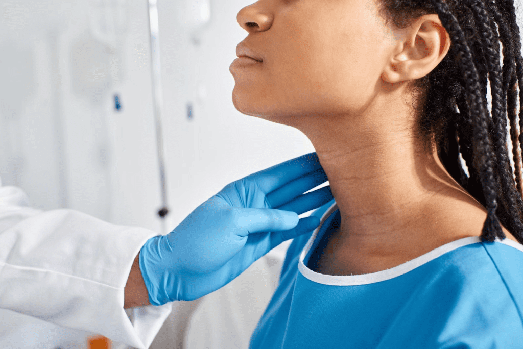
Diagnosing involves looking at images, patient history, and sometimes team work. Radiologists mainly use ultrasound for thyroid nodule checks.
They analyze the nodule’s details to figure out what it is. Their skill in reading images makes diagnosis more accurate.
| Diagnostic Factor | Description | Importance in Diagnosis |
| Nodule Size | Larger nodules are more likely to be malignant. | High |
| Nodule Composition | Solid nodules are more suspicious than cystic ones. | High |
| Calcification Patterns | Microcalcifications are often associated with malignancy. | Very High |
Collaboration with Endocrinologists and Surgeons
Radiologists team up with endocrinologists and surgeons for better care. This teamwork is key for:
- Talking about image findings and what they mean.
- Deciding on more tests or procedures.
- Planning the best treatment based on the diagnosis.
Together, they give a more accurate diagnosis and a treatment plan that fits the patient.
Identifying a Suspicious Thyroid Nodule: Warning Signs
Spotting suspicious thyroid nodules involves looking at several key features. Doctors use these signs to guess if a nodule might be cancerous.
Size and Growth Pattern
The size of a thyroid nodule is key in checking for cancer. Bigger nodules are more likely to be cancerous, and fast growth is a red flag. Watching how a nodule changes size is very important for catching cancer early.
- Nodules over 1 cm need extra attention.
- Fast growth can mean cancer.
Appearance and Composition
The look and makeup of a thyroid nodule can tell us a lot. Solid nodules are more worrisome than cystic ones because they might be cancerous.
- Solid nodules that look darker than the rest are more suspicious.
- Nodules with uneven edges or a bumpy look might be cancerous.
Calcification Patterns
Calcifications in a thyroid nodule are a big warning sign. Microcalcifications, tiny bright spots on ultrasound, are very concerning.
- Microcalcifications often mean papillary thyroid carcinoma.
- Coarse calcifications, though less specific, can also hint at cancer.
Vascularity and Blood Flow Features
The blood flow in a thyroid nodule can also give clues. More blood flow, mainly in solid parts, might mean cancer.
- Nodules with blood inside are more suspicious.
- Doppler ultrasound helps check the risk of cancer.
Ultrasound: The Primary Tool for Thyroid Imaging
Ultrasound has changed how we look at the thyroid gland. It’s safe and works well. It helps us see thyroid nodules clearly, which is key for treatment.
How Thyroid Ultrasound Works
Ultrasound uses sound waves to show the thyroid gland and nearby areas. It’s a non-invasive way to check thyroid nodules.
A transducer sends sound waves that bounce off the gland. These echoes turn into images on a screen.
What Happens During an Ultrasound Examination
The patient lies on their back with their neck up. A gel is put on the skin to help sound waves move.
The radiologist moves the transducer to get images from different angles. Lymph node mapping is done during every ultrasound to check for more testing needs.
Advantages of Ultrasound for Thyroid Assessment
Ultrasound is great for thyroid checks because it’s non-invasive and doesn’t use radiation. It shows what’s inside nodules, like size and blood flow, which helps figure out if they’re cancerous.
- It gives clear images of thyroid nodules.
- Ultrasound helps guide biopsies for suspicious nodules.
- It’s affordable and easy to find.
Limitations of Ultrasound Technology
Ultrasound is very useful but has some downsides. The quality of images depends on the person doing it. There can also be different opinions on what the images mean.
It might not show how far the disease has spread or if it’s touching other areas. Sometimes, other tests like CT or MRI are needed.
The TI-RADS Classification System
Knowing about TI-RADS is key for both doctors and patients when it comes to thyroid cancer. The Thyroid Imaging Reporting and Data System, or TI-RADS, helps figure out the risk of cancer in thyroid nodules. It does this by looking at ultrasound features.
Understanding the Thyroid Imaging Reporting and Data System
The TI-RADS system was made to make thyroid nodule ultrasound reports easier to understand. It sorts nodules into categories based on things like what they’re made of, how they look, and if they have calcifications. This helps doctors decide what to do next.
The Five TI-RADS Categories
TI-RADS breaks down thyroid nodules into five levels of risk: TR1 (Benign), TR2 (Not Suspicious), TR3 (Mildly Suspicious), TR4 (Moderately Suspicious), and TR5 (Highly Suspicious). Each level shows a different risk of cancer, from very low to very high.
- TR1: These are benign nodules, usually cystic or spongiform, with a very low cancer risk.
- TR2: These are not suspicious, with a low risk.
- TR3: These are mildly suspicious, with an intermediate risk.
- TR4: These are moderately suspicious, with a higher risk.
- TR5: These are highly suspicious, with a high risk of cancer.
How Radiologists Apply TI-RADS in Practice
Radiologists use TI-RADS during ultrasound exams to check thyroid nodules. They look at the nodule’s features and give it a TI-RADS score. This score helps doctors know what to do next, like follow-up, biopsy, or other treatments.
Other Imaging Techniques for Thyroid Cancer Detection
Ultrasound is not the only tool used to find thyroid cancer. Other imaging methods give more details. They help doctors see how far the cancer has spread and plan treatment.
CT Scans and MRI
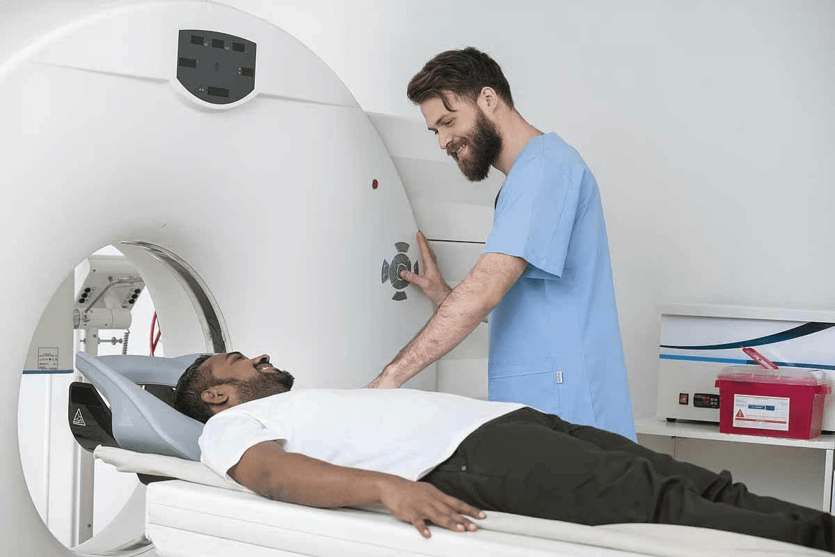
CT scans and MRI are key in checking if thyroid cancer has spread. CT scans show detailed pictures of the tumor and its surroundings. MRI is great for seeing soft tissues, which helps in checking if cancer has invaded important areas.
These scans are vital before surgery. They help doctors decide the best way to operate. They also check for cancer return after treatment.
Nuclear Medicine Scans
Nuclear medicine scans, like iodine-131 scans, find thyroid cancer, even if it has spread. They use radioactive material to spot cancerous areas. Thyroid cancer cells take up iodine, making it a good tracer.
These scans are essential for checking the whole body. They find cancer in distant parts and see if treatment is working.
PET Scans for Advanced Cases
PET scans are for thyroid cancer that’s hard to treat. PET scans use a radioactive sugar to find active cancer cells. They’re very good at finding cancer that has come back or spread.
PET scans help plan treatment. They decide if more surgery, chemotherapy, or other treatments are needed.
How Accurate Are Radiologists at Detecting Thyroid Cancer?
It’s important to know how well radiologists can spot thyroid cancer. They play a key role in finding cancerous nodules. Their skill can greatly affect how well a patient does.
Sensitivity and Specificity Rates
How good radiologists are at spotting thyroid cancer is measured in two ways. Sensitivity is about finding those with cancer. Specificity is about not missing those without it. Advanced imaging, like ultrasound, helps them do this well.
For example, a study might say radiologists are 90% sensitive and 95% specific with ultrasound. This means they’re good at finding both cancer and non-cancer nodules.
False Positives and False Negatives
Even with high accuracy, radiologists can make mistakes. A false positive is when a non-cancerous nodule looks like cancer. A false negative is when a cancerous nodule looks like it’s not. These mistakes can worry patients and affect their care.
Things like the imaging quality, the radiologist’s experience, and the nodule’s details can cause these errors. Better training and technology can help reduce them.
Factors Affecting Diagnostic Accuracy
Many things can affect how well radiologists can diagnose thyroid cancer. The quality of the imaging, the radiologist’s skill in thyroid imaging, and the nodule’s size and other details are important.
- The quality and resolution of the imaging equipment
- The radiologist’s experience and specialized training
- The characteristics of the thyroid nodule itself
Knowing these factors helps healthcare providers work to improve accuracy and better care for patients.
When Imaging Alone Isn’t Enough
Radiological assessments are key in spotting thyroid nodules. But sometimes, just looking at images isn’t enough. These methods give us info on size, location, and type of nodules. Yet, they can’t always tell if a nodule is cancerous or not.
Limitations of Radiological Assessment
Even with advanced imaging like ultrasound, CT scans, and MRI, there are limits. For example, some nodule features, like being cystic or solid, hint at cancer. But they’re not sure signs.
Key limitations include:
- It’s hard to tell if a nodule is cancerous just by looking at images
- These methods can’t fully check how well a nodule works
- Doctors might see things differently when looking at images
When Fine Needle Aspiration Is Recommended
When images look suspicious or unclear, a fine-needle aspiration biopsy (FNAB) is often suggested. This method uses a thin needle to take cell samples from the nodule for lab tests.
The decision to perform FNAB is typically based on:
- The size and look of the nodule on images
- Ultrasound features that might suggest cancer, like being dark or having tiny spots
- Things about the patient, like past radiation or family cancer history
| Characteristic | Benign Features | Suspicious Features |
| Echogenicity | Hyperechoic or isoechoic | Hypoechoic |
| Margins | Smooth | Irregular or lobulated |
| Calcifications | Absent or large, coarse calcifications | Microcalcifications |
The Importance of Tissue Diagnosis
Getting a tissue diagnosis through FNAB is key for a clear diagnosis. It helps decide the best treatment, like surgery, watching closely, or other options.
FNAB results help diagnose thyroid cancer and figure out the cancer type. This info is vital for knowing the outlook and planning treatment.
The Fine Needle Aspiration Biopsy Process
Diagnosing thyroid cancer often starts with a fine-needle aspiration biopsy. This method helps find out if a thyroid nodule is cancerous or not.
How FNA Is Performed
A thin needle is used to take cell samples from thyroid nodules. This procedure usually happens in a doctor’s office or clinic. The area is cleaned, and sometimes a numbing cream is applied to make it less painful.
Ultrasound guidance is often used to find the nodule accurately. An ultrasound machine helps locate the nodule and guide the needle.
The Role of Ultrasound Guidance
Ultrasound guidance is key in fine-needle aspiration biopsy. It allows for real-time imaging, helping the doctor pinpoint the nodule and avoid other areas.
Using ultrasound makes the biopsy more accurate. It’s very helpful for small or hard-to-reach nodules.
What to Expect During the Procedure
The patient lies on their back with their neck slightly up during the biopsy. The doctor uses ultrasound to guide the needle into the nodule.
They might take several samples to get enough for analysis. The whole process usually takes 15 to 30 minutes.
Potential Complications and Recovery
While generally safe, there are risks like bleeding or infection at the site.
| Potential Complications | Frequency | Management |
| Bleeding | Rare | Apply pressure; may require medical attention if severe |
| Infection | Very Rare | Antibiotics; monitor for signs of infection |
| Pain or Discomfort | Common | Over-the-counter pain relievers; usually resolves quickly |
Most people can go back to normal activities right after. Some might feel a bit of pain or swelling, but it usually goes away quickly.
Understanding Your Thyroid Radiology Report
Understanding your thyroid radiology report is a big step. It helps you know what to do next. This report gives you details about your thyroid health, like if there are nodules or cancer. Knowing what it says can ease your worries and help you make smart choices about your health.
Common Terminology in Thyroid Imaging Reports
Thyroid imaging reports use special terms that might be new to you. Words like “hypoechoic,” “hyperechoic,” “cystic,” and “solid” describe thyroid nodules. Hypoechoic nodules look darker on ultrasound and might be more likely to be cancerous. On the other hand, hyperechoic nodules are brighter and seem less worrisome.
Terms like “TI-RADS” help rate how likely it is for a nodule to be cancerous. Knowing these terms can make your report easier to understand.
What “Indeterminate” or “Suspicious” Findings Mean
Getting a report with “indeterminate” or “suspicious” findings can be scary. Indeterminate findings mean the nodule’s look isn’t clearly good or bad, so more tests are needed. Suspicious findings show features that might suggest cancer but aren’t sure.
It’s important to remember that these findings don’t always mean you have cancer. You might need a biopsy to figure out what the nodule is.
Questions to Ask Your Doctor About Your Results
When talking to your doctor about your report, it’s good to have questions ready. Ask things like “What do my test results mean?”, “What’s next?”, “Do I need more tests?”, and “What are the risks and benefits of what you’re suggesting?”
Being active and informed helps you handle your diagnosis and treatment better.
From Imaging to Diagnosis: The Complete Thyroid Cancer Pathway
The journey to diagnose thyroid cancer starts with imaging, like ultrasound. Then, doctors do clinical checks and might need more tests.
Typical Diagnostic Timeline
The time it takes to diagnose thyroid cancer varies. It depends on how complex the case is and if more tests are needed. It can be a few days to several weeks from first imaging to diagnosis.
Patients might have to go through many tests, like a fine-needle aspiration biopsy. This helps figure out what the thyroid nodule is. A multidisciplinary team of doctors works together to get all the needed info.
Treatment Decisions
Treatment decisions for thyroid cancer depend on several factors. These include the diagnosis, cancer stage, and patient health. Specialists like endocrinologists, surgeons, and oncologists come together to decide the best treatment.
The treatment plan might include surgery, radioactive iodine therapy, or other options. The aim is to treat the cancer well while keeping side effects low.
Multidisciplinary Tumor Board Approach
The multidisciplinary tumor board is key in thyroid cancer care. This team includes different specialists who discuss each case. They talk about the best treatment options and make suggestions.
It ensures patients get care that fits their needs. The team’s work helps make diagnoses more accurate and treatments more effective.
Advances in Thyroid Cancer Imaging Technology
New technologies in thyroid cancer imaging are giving doctors better tools for finding cancer. These tools make it easier to spot cancer early and accurately.
Artificial Intelligence and Machine Learning
Artificial intelligence (AI) and machine learning (ML) are changing how we look at thyroid cancer images. They help doctors see things they might miss. Studies show AI can make cancer detection more accurate.
AI and ML help in many ways:
- They make images clearer
- They find patterns and oddities
- They predict how a patient might do
Experts say AI could cut down on unnecessary biopsies and improve care.
This tech is a big step towards catching thyroid cancer early.
Molecular Imaging Techniques
Molecular imaging gives us new ways to understand thyroid cancer. Tools like PET scans and special ultrasound agents help spot cancer cells and track treatment.
The good things about molecular imaging are:
- It makes diagnosis more accurate
- It shows more about tumor biology
- It helps see how well treatment is working
Elastography and Other Emerging Methods
Elastography, which checks tissue stiffness, is becoming important in thyroid cancer imaging. It can tell if a nodule is cancerous or not, helping doctors make better choices.
Other new methods, like contrast-enhanced ultrasound and advanced MRI, are also being looked at. They might help make thyroid cancer imaging even better.
In summary, new tech in thyroid cancer imaging is making a big difference. It’s helping doctors diagnose and treat cancer more effectively. As these technologies get better, they promise to help patients even more.
Living with a Suspicious Thyroid Nodule
Living with a suspicious thyroid nodule can be tough. It requires patience, understanding, and the right medical advice. Getting a diagnosis can make patients worried about thyroid cancer.
Active Surveillance Protocols
Many patients start with active surveillance for a suspicious thyroid nodule. This means regular ultrasound checks to watch the nodule’s size and shape over time.
- Regular ultrasound examinations
- Assessment of nodule growth or changes
- Adjustments to the surveillance plan as needed
Active surveillance is often chosen for small nodules with low cancer risk. It aims to avoid surgery while keeping a close eye on the nodule for cancer signs.
Psychological Impact of “Watching and Waiting”
The waiting period can be hard on patients. The uncertainty about the nodule’s nature and cancer risk can cause a lot of stress and worry.
Support mechanisms are key during this time. This includes counseling, support groups, and clear talks with doctors about the surveillance plan and any updates.
When to Consider More Aggressive Intervention
While active surveillance is good for some, more aggressive steps might be needed in certain cases. This includes nodules that grow a lot, show suspicious ultrasound signs, or if there’s a history of radiation or family cancer history.
- Nodule growth or suspicious ultrasound features
- Patient’s medical or family history
- Patient preference after thorough discussion
Support Resources for Patients
Dealing with a suspicious thyroid nodule can be tough, but there are many support resources out there. These include online forums, patient groups, and educational materials from trusted health organizations.
Patients are encouraged to look for these resources. They help understand the condition and the management options. Being informed and supported helps patients deal with the challenges of a suspicious thyroid nodule better.
Conclusion
Getting a correct diagnosis for thyroid cancer is key for good treatment and results. Radiologists are very important in finding suspicious thyroid nodules. They use imaging, like ultrasound, to do this.
The Thyroid Imaging Reporting and Data System (TI-RADS) helps radiologists figure out the risk of cancer. Imaging is a big part of finding thyroid cancer. But, it’s often used with a biopsy to confirm cancer.
A radiologist who specializes in thyroid imaging can give important insights. They help decide if more tests or treatment are needed. Knowing how imaging helps in thyroid cancer diagnosis helps patients make better choices.
Using new imaging tech and focusing on the patient can lead to better results. This is true for both thyroid nodules and cancer.
FAQ
What is the role of radiologists in thyroid cancer diagnosis?
Radiologists are key in finding thyroid cancer. They look at images like ultrasounds. This helps spot and check thyroid nodules for risk.
What are the warning signs of a suspicious thyroid nodule?
Signs include size, growth, look, and how it’s made. Also, calcification patterns and blood flow are important.
How does ultrasound work for thyroid imaging?
Ultrasound sends sound waves to see the thyroid gland. This lets radiologists check nodules and their details.
What is the TI-RADS classification system?
TI-RADS is a system for thyroid nodules. It shows how likely a nodule is to be cancerous, from low to high risk.
What is fine-needle aspiration biopsy, and when is it recommended?
Fine-needle aspiration biopsy takes cells from a nodule with a thin needle. It’s used when ultrasound results are unclear or show a risk.
How accurate are radiologists at detecting thyroid cancer?
Radiologists’ accuracy varies. It depends on image quality, nodule details, and their skill.
What are the limitations of radiological assessment in thyroid cancer diagnosis?
Radiology can miss cancer or say it’s there when it’s not. It can’t confirm cancer without a tissue sample.
What is the typical diagnostic timeline for thyroid cancer?
The timeline for thyroid cancer diagnosis varies. It includes imaging, biopsy, and then planning treatment.
How are treatment decisions made for thyroid cancer?
A team of doctors decides treatment. They consider the diagnosis, cancer stage, and patient health.
What are the latest advances in thyroid cancer imaging technology?
New tech includes artificial intelligence and molecular imaging. These can make diagnosis better and help patients more.
What is active surveillance for thyroid cancer, and when is it recommended?
Active surveillance means watching low-risk thyroid cancer closely. It avoids unnecessary surgery. It’s for nodules or cancers that are not high-risk.
How can patients understand their thyroid radiology report?
Patients should ask their doctor about their report. They should understand any unclear findings and what to do next.


