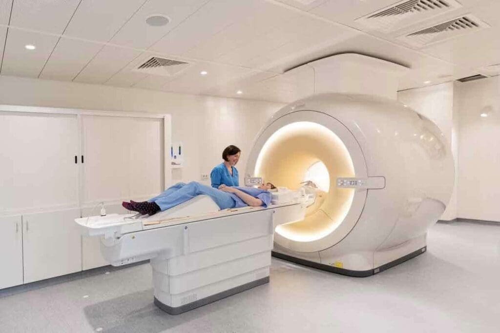
Doctors often use X-rays and MRIs to check for bone injuries. At Liv Hospital, we use top-notch MRI tech for accurate diagnoses and care. MRI scans show more detail inside the body, like soft tissues, which are key in complex injuries. X-rays are good for first checks, but MRI gives a deeper look at bone injuries. It spots small fractures, bone bruises, and soft tissue harm.Can mri see broken bones: What MRI shows that X-rays can’t for detecting fractures.
Key Takeaways
- MRI scans can detect subtle fractures and soft tissue injuries that X-rays may miss.
- Advanced MRI technology provides a more detailed view of internal body structures.
- Liv Hospital is committed to providing world-class care and precision diagnostics.
- MRI is useful for complex injuries and soft tissue damage.
- Our team uses MRI technology for detailed patient care.
Understanding X-Ray and MRI Technology Basics
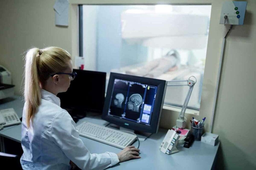
To understand X-ray and MRI, we need to know how they work. Medical imaging is key in diagnosing and treating many health issues, like bone fractures. We use X-rays and MRIs to see inside our bodies.
How X-Ray Imaging Works
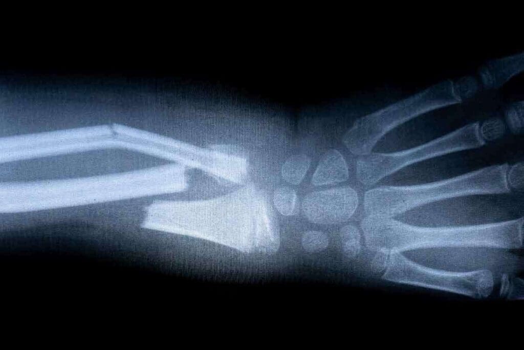
X-ray imaging uses special radiation to show bones and other body parts. When an X-ray beam hits the body, different parts absorb it differently. Bones, being denser, absorb more and look white on the image. Softer tissues show up in gray or black.
This method is great for spotting bone fractures. A break in the bone looks like a dark line or gap.
For instance, if you’re wondering can MRI show broken bones, it’s important to know. While X-rays are often the first choice for fractures, they’re not perfect. They can miss soft tissue injuries or complex fractures.
How MRI Technology Functions
MRI uses a strong magnetic field and radio waves to create images. Unlike X-rays, MRI doesn’t use harmful radiation. It’s safer for some patients. MRI is great at showing soft tissues like tendons and ligaments, along with bones.
When thinking about what does an MRI show that an X-ray does not, MRI’s strengths are clear. It can see soft tissues and bone marrow details that X-rays can’t. This makes MRI essential for diagnosing complex injuries or conditions.
Key Differences in Imaging Principles
The main difference between X-ray and MRI is how they work. X-rays depend on material density, making them good for bones. MRI uses hydrogen atoms’ magnetic properties to show both soft and hard tissues. This means X-rays are quick for initial bone fracture checks, but MRI offers a more detailed view, including soft tissue injuries.
- X-rays are fast and effective for initial bone fracture assessments.
- MRI provides detailed images of both bones and soft tissues.
- The choice between X-ray and MRI depends on the specific diagnostic needs.
For more info on picking the right imaging, check out Excel Spine’s guide on X-ray vs CT vs MRI. Knowing these technologies helps us make better healthcare choices.
Can MRI See Broken Bones? The Definitive Answer
Understanding MRI’s advanced imaging is key to knowing if it can spot broken bones. MRI is a top tool for finding fractures when X-Rays don’t show anything.
MRI’s Capability in Fracture Detection
MRI is great at finding broken bones because it shows both bone and soft tissue clearly. It can spot fractures that X-Rays miss, like stress fractures and tiny cracks. This is very important for quick and correct diagnosis.
In sports injuries, MRI gives detailed views of bones and soft tissue. This helps doctors understand how bad the injury is.
Comparing Sensitivity with X-Rays
MRI is more sensitive than X-Rays for finding fractures. X-Rays are often used first because they’re easy to get and cheap. But, they might not catch all fractures. MRI can see bone marrow edema, which X-Rays can’t, making it better for finding hidden fractures.
When MRIs Are Preferred for Bone Imaging
MRIs are chosen over X-Rays in many situations. For example, if X-Rays don’t show a fracture but doctors think there might be one, MRI is used. Also, for complex fractures or checking how a fracture is healing, MRI gives important info for treatment plans.
Knowing MRI’s strengths in finding fractures helps doctors choose the best imaging for patients. This ensures accurate diagnosis and effective treatment.
Types of Fractures Better Visualized with MRI
MRI is great for spotting bone marrow changes. This makes it key for finding certain fractures that X-rays can’t catch.
Stress Fractures and Hairline Cracks
Stress fractures and hairline cracks are easier to see with MRI. These injuries happen a lot in athletes and people who do the same thing over and over. MRI finds bone marrow edema early, helping to treat them fast.
- Early Detection: MRI spots stress fractures early, so they can be treated right away.
- Detailed Imaging: MRI’s clear pictures show how big the fracture is.
Occult Fractures Not Visible on X-Ray
Occult fractures, which X-rays can’t see, are found with MRI. These fractures happen from trauma or osteoporosis.
Using MRI for these fractures has big benefits:
- It finds fractures X-rays miss.
- It checks for damage to soft tissues too.
Complex Fracture Patterns
Complex fractures, with many pieces or broken into tiny bits, are clearer on MRI. The detailed images help plan surgery.
Using MRI for these fractures helps doctors give better diagnoses. They can then make treatment plans that work better, helping patients get better faster.
Bone Marrow Edema and Contusions: What Only MRI Can Reveal
Bone marrow edema and contusions show bone trauma well. MRI can spot these changes with great sensitivity. When a bone gets hurt, the marrow can swell or get inflamed. These signs are hard to see on X-rays, making MRI key for diagnosis.
Understanding Bone Bruises
A bone bruise happens when the bone marrow gets hurt, usually from trauma. This injury can come from a direct hit or from twisting or pulling. MRI is very good at seeing changes in bone marrow, spotting bruises that X-rays miss.
Bone bruises on MRI look like areas of low signal on T1 images and high on T2. This shows edema or bleeding in the marrow.
Early Detection of Bone Trauma
Spotting bone trauma early is key for good care and avoiding more harm. MRI can find bone marrow edema and contusions early. This helps doctors decide on treatment, like if to immobilize or avoid certain activities.
- Prompt Diagnosis: MRI helps quickly find bone injuries, lowering the chance of problems.
- Guiding Treatment: MRI info helps doctors make the right treatment plans for patients.
- Monitoring Progress: MRI scans can track healing, helping adjust treatment as needed.
Clinical Significance of Marrow Changes
Finding bone marrow edema and contusions by MRI is very important. It helps diagnose injuries and understand how bad they are. This is key for knowing how well a patient will recover and planning rehab.
MRI shows both bone and soft tissue clearly. This lets doctors make a full treatment plan, covering all injury aspects.
Soft Tissue Injuries Associated with Fractures
Soft tissue injuries often happen with fractures. It’s important to check them well for good treatment. The soft tissues around a fracture can get hurt, like ligaments, tendons, and cartilage. Knowing how bad these injuries are helps make a good treatment plan.
Ligament and Tendon Damage Assessment
MRI is key in checking ligament and tendon damage with fractures. It shows detailed images of these soft tissues. This helps doctors see tears, strains, and other injuries not seen on X-rays. This info is key for choosing the right treatment.
For example, MRI can spot ligament injuries in complex fractures that might need surgery. It also checks tendon damage, helping plan the right rehab.
Cartilage Injuries Accompanying Fractures
Cartilage injuries often happen with fractures, mainly in joints that bear weight. MRI can see cartilage injuries like osteochondral lesions or thinning. This is important for knowing the long-term risks, like osteoarthritis.
By checking cartilage health, doctors can make treatment plans that fix both the fracture and cartilage injuries. This helps patients get better faster.
Comprehensive Injury Evaluation
It’s important to check both bone and soft tissue injuries well for fracture care. MRI gives a full view of the injury. This helps doctors make a treatment plan that covers all injury parts.
We use MRI to see all injury details, making sure nothing is missed. This way, we can give care that fits each patient’s needs.
| Injury Type | MRI Findings | Clinical Significance |
| Ligament Damage | Tears or strains visible on MRI | Impacts joint stability and requires appropriate treatment |
| Tendon Damage | Tendon tears or tendinopathy | Affects muscle function and requires rehabilitation |
| Cartilage Injuries | Osteochondral lesions or cartilage thinning | Predicts possible osteoarthritis and guides treatment |
Clinical Scenarios Where MRI Outperforms X-Ray
MRI is better than X-ray in many cases, thanks to its advanced imaging. It’s great for finding small or complex fractures. We’ll look at how MRI beats X-ray in sports injuries, kids’ bone injuries, and for older adults.
Sports Injuries and Subtle Fractures
In sports medicine, MRI is key for spotting small fractures that X-ray can’t see. Athletes often get stress fractures or tiny cracks. MRI finds these early, helping to avoid more harm.
A study showed MRI found 95% of stress fractures, while X-ray found only 40%. This proves MRI is better for sports injuries.
Pediatric Bone Injuries
In kids, MRI is best for checking bone injuries. Kids’ bones are growing, and their injuries need special care. MRI is safe and shows bones and soft tissues well.
For hidden fractures, MRI gives a clear diagnosis. This helps doctors plan the right treatment.
Geriatric Fracture Assessment
In older adults, finding fractures is hard because of osteoporosis and complex bones. MRI’s detailed images help spot fractures X-ray can’t see.
A study showed MRI was better than X-ray at finding fractures in the elderly. This is important for them.
| Clinical Scenario | MRI Advantages | X-Ray Limitations |
| Sports Injuries | Detects subtle fractures, images soft tissues | May miss hairline cracks, limited soft tissue visualization |
| Pediatric Bone Injuries | No radiation, detailed bone and soft tissue imaging | Radiation exposure, may not detect complex pediatric fractures |
| Geriatric Fracture Assessment | Identifies fractures in osteoporotic bones, detailed imaging | Difficulty detecting fractures in low-density bones |
Understanding when MRI is better than X-ray helps us see its value. It’s key for finding small fractures in athletes, checking kids’ bones, and looking at complex fractures in older adults. MRI gives doctors the info they need for better care.
Limitations of X-Rays in Fracture Diagnosis
X-rays are useful but have limits that make diagnosing fractures tricky, mainly in certain bones. These issues can lead to unclear or wrong diagnoses. This can affect how we treat patients.
Anatomical Challenges and Overlapping Structures
Complex areas like the spine, pelvis, and wrist are hard to image with X-rays. This is because many bones overlap, making it hard to see fractures clearly. A radiologist notes that X-rays might miss fractures in these areas because of bone overlap.
We often see cases where X-rays can’t give a clear diagnosis in these complex areas.
Sensitivity Issues in Specific Bone Types
X-rays work differently on different bones. For example, finding stress fractures or small cracks in bones like the scaphoid or femoral neck is tough with X-rays alone. In these cases, we use MRI for a more precise diagnosis.
A study found that MRI is better than X-rays for spotting some fractures, even in the early stages. This shows why we might choose other imaging methods when X-rays aren’t clear.
When X-Ray Results Are Inconclusive
So, when do we know X-rays aren’t enough? If a patient keeps feeling pain or discomfort after a negative or unclear X-ray, we suggest more tests. MRI is great for showing detailed images of bones and soft tissues. It can spot fractures that X-rays miss.
In summary, while X-rays are helpful in diagnosing fractures, they have their limits. By understanding these and knowing when to use MRI, we can give better diagnoses and treatments to our patients.
Current Research on MRI’s Role in Fracture Classification
Medical research has focused on MRI’s role in fracture classification. MRI technology is being explored for its ability to detect and classify fractures. Recent studies have shown its effectiveness in these areas.
Recent Studies on Diagnostic Accuracy
Recent studies have found MRI to significantly improve fracture classification accuracy. A study on the National Center for Biotechnology Information website highlights MRI’s advancements. It shows MRI can be as good as CT scans in some cases, giving detailed images of bones and soft tissues.
MRI can detect fractures X-rays can’t. Can an MRI show a fracture that an X-ray can’t? Yes, it can. MRI’s sensitivity to bone marrow and soft tissues helps spot fractures and injuries X-rays might miss.
Advances in MRI Protocols for Bone Imaging
New MRI protocols have greatly improved bone imaging. Techniques and sequences have been developed to better see bone structures and marrow. These advancements make MRI a valuable tool for complex fracture assessment and treatment planning.
Does MRI show bone fractures more effectively than X-ray? Often, yes. MRI gives a detailed view of injuries, including bone and soft tissue damage. This is key when the injury’s extent is unclear.
Comparison with CT Scans for Fracture Detection
Comparing MRI to CT scans, both have their advantages. CT scans provide detailed bone images, but MRI also shows soft tissue injuries. What does MRI show that X-ray doesn’t? MRI details soft tissue conditions, like ligaments, tendons, and cartilage, which is vital for full injury assessment.
In summary, MRI is proving valuable in fracture classification, thanks to its accuracy and advanced imaging. As MRI techniques improve, so will fracture diagnosis and treatment.
International Standards in Medical Imaging: Liv Hospital’s Approach
At Liv Hospital, we aim to offer top-notch medical imaging services. We follow global best practices. Accurate diagnoses are key to good treatment plans, and we focus on this.
Academic Protocols and Multidisciplinary Services
Our team at Liv Hospital uses advanced academic protocols in imaging. We work together to give full care, from start to finish. This helps us tackle tough cases, like finding fractures not seen on X-rays.
Studies show MRI is very good at finding fractures. This is backed by research on PMC.
Patient-Centered Imaging Experience
We make sure our imaging services are not just accurate but also comfortable. Our facilities are welcoming for patients from around the world.
Ethical Considerations in Diagnostic Imaging
Liv Hospital values ethical imaging practices. We care deeply about patient privacy and safety. We follow international rules to keep our standards high.
We combine knowledge, teamwork, and care for our patients. Our MRI tech and expertise help us give the best insights. Whether it’s finding broken bones or complex injuries, we lead the way.
Conclusion: Making Informed Decisions About Bone Imaging
Understanding how different imaging tools work is key when diagnosing bone injuries. We’ve seen how MRI is better than X-ray at spotting small fractures and soft tissue damage. MRI can show things like bone marrow edema, stress fractures, and complex fractures that X-rays can’t.
Can MRI find broken bones that X-rays miss? Yes, it’s great at finding hidden fractures and checking soft tissue injuries. This is very important for making the right treatment plans. At Liv Hospital, we use MRI to give accurate diagnoses and the best care for our patients.
Knowing what MRI can do helps doctors choose the right imaging tests. Will MRI find a broken bone if X-ray can’t? Often, yes, for fractures that X-rays can’t see. Our goal is to offer top-notch healthcare with advanced imaging and full support for our patients.
FAQ
What does an MRI show that an X-ray can’t when it comes to broken bones?
MRI shows detailed images of bones and soft tissues. It’s great for finding complex injuries like small fractures and soft tissue damage. These things might not show up on X-rays.
Can MRI see broken bones?
Yes, MRI is very good at finding broken bones. It’s great when X-rays aren’t clear. It spots stress fractures, hairline cracks, and other fractures X-rays miss.
Will an MRI show a broken bone?
Yes, MRI can spot broken bones, even if X-rays don’t. It’s very good at finding fractures because it looks at bone marrow changes.
Do MRIs show broken bones?
Yes, MRIs can show broken bones. They use advanced imaging to find fractures, even in complex cases or when X-rays aren’t clear.
Does MRI show bone fractures?
Yes, MRI can show bone fractures. It’s great for finding stress fractures, occult fractures, and other fractures X-rays can’t see.
What types of fractures are better visualized with MRI?
MRI is better for stress fractures, hairline cracks, and occult fractures. It’s also good for complex fracture patterns and soft tissue injuries with fractures.
Can MRI detect bone marrow edema and contusions?
Yes, MRI can find bone marrow edema and contusions. These signs of bone trauma are often missed by X-rays. MRI is a key diagnostic tool.
How does MRI compare to X-ray in diagnosing fractures?
MRI is more sensitive than X-ray for fractures, often when X-rays are unclear. MRI shows detailed images of bones and soft tissues, making it a better diagnostic tool.
When is MRI preferred over X-ray for bone imaging?
MRI is preferred for complex injuries or unclear X-ray results. It’s very useful in sports medicine, pediatric care, and for older adults with fractures.
Can MRI evaluate soft tissue injuries associated with fractures?
Yes, MRI can fully check soft tissue injuries like ligament, tendon, and cartilage damage. This is key for making effective treatment plans.
Reference
- Kwee, T. C., & Kwee, R. M. (2020). MRI in acute bone fractures: how useful is it? European Radiology, 30(11), 6215–6224. https://pubmed.ncbi.nlm.nih.gov/32122881/
- Yang, Q., Zhang, S., Zhou, Y., & Niu, X. (2022). MRI for detection of occult fractures in patients with suspected extremity trauma and negative initial radiographs: a systematic review and meta-analysis. European Radiology, 32(2), 1296–1307. https://pubmed.ncbi.nlm.nih.gov/34548610/
- Cahir, J. G., & Daffner, R. H. (2019). Occult fractures. Radiology Clinics of North America, 57(4), 689–703. https://pubmed.ncbi.nlm.nih.gov/31224670/


