Last Updated on November 26, 2025 by Bilal Hasdemir

Can You See Cancer in the Abdomen with Ultrasound? Here’s What You Need to Know
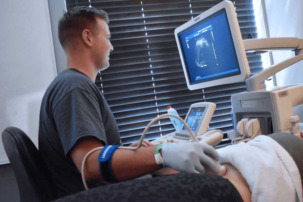
Did you know that ultrasound technology is key in finding many cancers, including those in the abdomen? If you’ve ever wondered, “can you see cancer in the abdomen with ultrasound?, the answer is yes ” to some extent. Ultrasounds are commonly used to check the belly for cancer signs and other health issues.
Finding cancer early is very important for treatment success. Ultrasounds help detect early signs by using sound waves to create clear images of internal organs. This helps doctors identify any irregularities that might suggest cancer.
Ultrasounds are especially useful for examining the liver, gallbladder, and kidneys. They offer a fast, safe, and non-invasive way to assess organ health ” helping to catch potential problems early.
Key Takeaways
- Ultrasounds are a vital tool in detecting abdominal cancers.
- Early detection significantly improves cancer treatment outcomes.
- Ultrasound technology uses high-frequency sound waves to create images of internal organs.
- Organs such as the liver, gallbladder, and kidneys can be effectively examined using ultrasounds.
- Ultrasounds offer a non-invasive method for assessing organ health.
Understanding Ultrasound Technology in Cancer Detection
Ultrasound technology uses sound waves to help find cancer. It’s a non-invasive way to see inside the body. This helps us spot tumors and check how treatments are working.
How Ultrasound Imaging Works
Ultrasound sends sound waves into the body. These waves bounce off and come back to the device. This creates detailed images of what’s inside.
The technology works because sound waves move at different speeds in different tissues. This lets ultrasound machines show us what’s inside. It helps us find and track cancer.
Types of Ultrasound Techniques Used for Cancer Screening
There are many ultrasound techniques for cancer screening. Each one is used in different ways:
- Conventional Ultrasound: Gives us 2D images of organs and tissues. It helps find tumors and see how big they are.
- Doppler Ultrasound: Shows how blood flows through vessels. It helps spot tumors by looking at blood flow.
- Contrast-Enhanced Ultrasound: Uses special agents to make blood vessels and tissue clearer. This helps find tumors better.
- Elastography: Checks how stiff tissues are. It helps tell if a lump is cancerous or not.
Advantages of Ultrasound Over Other Imaging Methods
Ultrasound has many benefits for finding and tracking cancer:
- Non-invasive: It doesn’t need to go inside the body or use radiation.
- Real-time Imaging: Gives us images right away. This lets us see how things are changing.
- Cost-effective: It’s often cheaper than MRI or CT scans.
- Wide Availability: Ultrasound machines are in many hospitals. This makes it easy for patients to get it.
Thanks to these benefits, ultrasound is a key tool in finding cancer early. It also helps us see how well treatments are working.
The Role of Ultrasound in Cancer Diagnosis

Ultrasound technology is key in finding cancers. It’s a non-invasive way to spot tumors. We use it to find and watch cancer growths, helping plan treatments.
When Doctors Order Ultrasounds for Cancer Screening
Doctors often start with ultrasounds for some cancers. This is true for cancers in the abdomen or thyroid. They choose ultrasound based on the patient’s history and symptoms. For example, those with cancer history or signs of growths might get an ultrasound.
- Identifying tumors and assessing their size and location
- Guiding biopsy procedures for definitive diagnosis
- Monitoring the effectiveness of cancer treatment
Ultrasound as a First-Line or Supplementary Diagnostic Tool
Ultrasound can be the first or a second step in diagnosis. It depends on the situation. Its flexibility makes it a key tool in cancer diagnosis.
For thyroid nodules, ultrasound is often the first test. It checks the nodule’s details and if a biopsy is needed.
The Diagnostic Process: From Ultrasound to Diagnosis
The process starts with an ultrasound. It gives first images of organs or tissues. If it finds something suspicious, more tests like biopsies might follow. We help patients understand each step and why it’s done.
- Ultrasound examination to identify possible tumors or abnormalities
- Further diagnostic testing, such as biopsy or additional imaging, if suspicious findings are detected
- Diagnosis and staging of cancer based on the results of all diagnostic tests
- Development of a treatment plan tailored to the patient’s specific needs
Can You See Cancer in the Abdomen with Ultrasound?
Ultrasound can spot cancer in the abdomen, but it’s not always easy. It works well for some cancers but not all. The type and location of the cancer matter a lot.
Abdominal Cancers Detectable via Ultrasound
Ultrasound is great for finding cancers in the liver, gallbladder, and kidneys. Liver cancer shows up as changes in the liver’s texture and tumors. Gallbladder cancer is seen as oddities in the gallbladder wall.
| Organ | Cancer Type | Ultrasound Characteristics |
|---|---|---|
| Liver | Hepatocellular Carcinoma | Hypoechoic or hyperechoic masses |
| Gallbladder | Adenocarcinoma | Wall thickening, polyps, or masses |
| Kidney | Renal Cell Carcinoma | Hypoechoic or isoechoic masses |
Limitations of Abdominal Ultrasound for Cancer Detection
Ultrasound has its limits. It misses some cancers, like small ones or those in hard-to-see spots. Pancreatic cancer is tough to find early because of where it is and the gas around it.
Case Studies: When Abdominal Ultrasounds Revealed Cancer

Ultrasound has helped find cancers in many cases. For example, a patient with liver disease got an ultrasound that found a liver mass. This was later confirmed as cancer. Finding it early helped with treatment.
Ultrasound has been key in spotting cancers early, which has helped patients a lot. As technology gets better, ultrasound will likely play an even bigger role in finding cancer.
Liver Cancer Detection Through Ultrasound
Ultrasound imaging is key in finding liver cancer early. It’s a non-invasive way to spot liver tumors early. This helps improve treatment results.
Tumor Characteristics on Ultrasound
Liver tumors show unique signs on ultrasound. These signs include:
- Hyperechoic or hypoechoic patterns, showing the tumor’s density.
- Size and shape, which hint at the tumor’s danger level.
- Margins, which are either clear or unclear, giving hints about the tumor.
Screening Protocols for High-Risk Patients
People at high risk, like those with hepatitis B or C, need regular checks. We suggest:
- Ultrasound tests every six months for those at high risk.
- Checking alpha-fetoprotein (AFP) levels to add to ultrasound results.
- Changing how often to screen based on risk and past results.
Accuracy Rates for Liver Cancer Detection
Ultrasound is very good at finding liver cancer, even better with other tests. Research shows:
- Ultrasound is very sensitive in spotting liver cancer, mainly in those at high risk.
- Using ultrasound and AFP tests together makes finding cancer even better.
- Finding cancer early through regular checks leads to better treatment and survival.
We stress the need for regular screening and ultrasound use in fighting liver cancer.
Pancreatic Cancer and Ultrasound Imaging
Finding pancreatic cancer early is tough, but ultrasound helps a lot. The pancreas is deep inside the body, making it hard to spot cancer early. But, new ultrasound tech has made it easier to find cancer.
Challenges in Detecting Pancreatic Cancer
Ultrasound has its limits when it comes to finding pancreatic cancer. The pancreas is hidden behind the stomach and intestines. Also, gas in the intestines can mess with the ultrasound signals, making images unclear. Plus, cancer symptoms often don’t show up until it’s too late.
Key challenges include:
- Deep location of the pancreas
- Interference from intestinal gas
- Lack of early symptoms
Endoscopic Ultrasound for Pancreatic Tumors
Endoscopic ultrasound (EUS) is a big help in finding pancreatic cancer. It uses a special probe through an endoscope to see the pancreas clearly. This method avoids the problem of gas in the intestines, making it better for spotting tumors.
EUS is great for:
- Finding small tumors that other tests miss
- Helping to get tissue samples for tests
- Accurately checking how far the cancer has spread
When Pancreatic Cancer Is Visible on Ultrasound
Ultrasound can spot pancreatic cancer when the tumor is big enough. How easy it is to see the tumor also depends on where it is and the skill of the person doing the ultrasound. Tumors over 2-3 cm are usually easier to find.
Factors influencing visibility include:
- Tumor size
- Tumor location
- Operator expertise
Kidney and Bladder Cancer Visualization
Kidney and bladder cancers can be seen with ultrasound imaging. This method is non-invasive. Ultrasound technology has improved a lot. It can now find many types of cancers, including kidney and bladder ones. We will look at how ultrasounds help diagnose these cancers.
Renal Mass Identification Through Ultrasound
Ultrasound is great for finding renal masses, which might mean kidney cancer. It uses sound waves to make kidney images. These images help doctors spot problems like tumors or cysts. Renal mass identification through ultrasound is often the first step in diagnosing kidney cancer.
We check the size, location, and details of renal masses with ultrasound. This info is key for deciding what to do next. It might be more tests or treatment right away.
Bladder Cancer Screening with Ultrasound
Ultrasound is also used for bladder cancer screening. It can spot tumors or other bladder issues. It’s a helpful tool, even for those who can’t have more invasive tests.
During the ultrasound, we look for cancer signs in the bladder. Early detection is key for good treatment. Ultrasound is quick and easy for checking the bladder.
Follow-Up Testing After Suspicious Findings
If an ultrasound finds something suspicious, more tests are needed. This might include CT scans, MRIs, or biopsies. Follow-up testing is important to confirm cancer and plan treatment.
We help patients understand their ultrasound results and what comes next. Knowing about their diagnosis and treatment options helps them make informed choices.
Breast Cancer Detection with Ultrasound
Ultrasound imaging is key in finding breast cancer, mainly in women with dense breasts. We use it along with mammography for a better diagnosis.
Comparing Breast Ultrasound and Mammography
Mammography is the usual tool for breast cancer screening. But ultrasound is better for dense breasts where mammography fails. Ultrasound can tell solid masses from cysts, cutting down on extra tests.
A study showed combining ultrasound and mammography boosts cancer detection in dense breasts. We suggest a tailored approach, taking into account the patient’s risk and breast density for choosing between ultrasound and mammography.
| Characteristics | Mammography | Ultrasound |
|---|---|---|
| Detection in Dense Tissue | Limited | Effective |
| Cyst vs. Solid Mass Differentiation | Limited | Highly Effective |
| Radiation Exposure | Yes | No |
Characteristics of Breast Cancer on Ultrasound Images
Breast cancer on ultrasound looks like a hypoechoic mass with irregular edges. Microcalcifications, posterior shadowing, and a taller-than-wide shape hint at cancer. We look at these signs to guess if it’s cancer.
Ultrasound-Guided Breast Biopsies
Ultrasound-guided biopsy is a safe way to get tissue samples. This method is great for lesions seen on ultrasound, giving a clear diagnosis without surgery.
We guide the biopsy needle with ultrasound for accurate placement. This lowers risks and boosts accuracy. It helps us give a quick and accurate diagnosis, leading to the right treatment.
Thyroid Cancer and Ultrasound Screening
Ultrasound technology is key in finding thyroid cancer. It gives us detailed images to check thyroid nodules. We use it to look at the thyroid gland, mainly when nodules are found.
Thyroid Nodule Assessment
Thyroid nodules are common, but not all are cancerous. Ultrasound helps us see how big, what they’re made of, and their shape. The American Thyroid Association says certain signs, like being darker or having spots, mean they might be cancerous.
What Thyroid Cancer Looks Like on Ultrasound
Thyroid cancer shows up as a nodule with certain signs on ultrasound. These include being solid, darker, and having irregular shapes. We also look for signs it’s spreading, which means it’s aggressive.
Thyroid Ultrasound Color Patterns and Their Meaning
Ultrasound often uses Doppler imaging to show blood flow in nodules. The colors on Doppler ultrasound tell us a lot. For example, if a nodule has a lot of blood flow, it might be cancerous.
Knowing what thyroid cancer looks like on ultrasound is key for early treatment. By combining ultrasound results with other tests, we can find and treat thyroid cancer well.
Gynecological Cancers and Ultrasound Detection
Ultrasound is key in finding gynecological cancers. It helps spot cancers in the female reproductive system. This includes ovarian, uterine, endometrial, and cervical cancers.
Ovarian Cancer Ultrasound Imaging
Ovarian cancer is hard to catch early because its symptoms are vague. But, ultrasound can help find it early. We use transvaginal ultrasounds to see the ovaries closely.
Ultrasound looks for tumors with odd shapes, solid parts, and lots of blood vessels. Our experts study these signs to guess if cancer is there.
Uterine and Endometrial Cancer Detection
We use ultrasound and other tests to find uterine and endometrial cancers. It checks the endometrium’s thickness and looks for oddities in the uterus.
Signs of uterine and endometrial cancer on ultrasound include a thick endometrium, mixed textures, and tumors in the uterus.
Cervical Cancer and Transvaginal Ultrasound
Ultrasound isn’t the main way to find cervical cancer. But, it gives us clues about the cancer’s size and spread. It helps us see how big the tumor is and if it’s growing into nearby tissues.
| Type of Cancer | Ultrasound Characteristics | Diagnostic Method |
|---|---|---|
| Ovarian Cancer | Irregular shapes, solid components, increased vascularity | Transvaginal Ultrasound |
| Uterine/Endometrial Cancer | Irregularly thickened endometrium, heterogeneous echotexture | Transvaginal Ultrasound |
| Cervical Cancer | Tumor size, invasion into surrounding tissues | Transvaginal Ultrasound (supplementary) |
Ultrasound helps us find and diagnose gynecological cancers better. This leads to better care for our patients.
Interpreting Ultrasound Images: Colors and Patterns
Ultrasound technology gives us clues about masses through colors and patterns. We look at ultrasound images to find signs of cancer. It’s key for making the right diagnosis and treatment plan.
What Do the Colors on an Ultrasound Mean?
Ultrasound images use Doppler to show blood flow. Red and blue colors show blood flow direction. Red means blood is moving towards the transducer, and blue means it’s moving away.
This info is important because tumors often have more blood flow. This is because they grow new blood vessels to feed themselves.
Characteristics of Malignant vs. Benign Masses
Malignant and benign masses look different on ultrasound. Malignant ones have irregular shapes and are taller than wide. They also have a mix of echoes.
Benign masses are rounder, wider, and have a uniform echo. Knowing these differences helps us figure out if a mass is cancerous or not.
Common Visual Indicators of Cancer on Ultrasound
Ultrasound can show signs of cancer. Darker than normal lesions and microcalcifications are two signs. A “halo” around a lesion also points to cancer.
Spotting these signs helps us catch cancer early. Then, we can suggest more tests or treatment.
Limitations of Ultrasound in Cancer Detection
Ultrasound technology has changed how we find cancer. Yet, it has its limits. Knowing these helps us do better for our patients.
Types of Cancers Difficult to Detect with Ultrasound
Some cancers are hard to spot with ultrasound. This is because of where they are or what they are like. For example, cancers in the pancreas or deep inside the body are tough to see.
- Pancreatic cancer, due to its deep location and the presence of gas in the intestines
- Lung cancer, as ultrasound has limited ability to penetrate air-filled structures
- Certain types of ovarian cancer, especially if they are small or located in a hard-to-reach area
Knowing these challenges helps us use ultrasound better with other tests.
Patient Factors Affecting Ultrasound Accuracy
Things about the patient can affect how well ultrasound works. For instance, being overweight can make images less clear, making it harder to find problems.
| Patient Factor | Impact on Ultrasound |
|---|---|
| Obesity | Reduced image quality due to increased tissue depth |
| Presence of gas in the intestines | Interference with sound waves, obscuring underlying structures |
| Surgical scars or dressings | Potential interference with probe placement and image quality |
Knowing these things helps us plan our tests better.
False Positives and False Negatives
Ultrasound can sometimes give wrong results. A wrong positive can cause worry and more tests. A wrong negative can make us think everything is okay when it’s not.
“The accuracy of ultrasound diagnosis depends on various factors, including the skill of the operator and the quality of the equipment.”
To avoid these problems, we often use ultrasound with other tests and doctor’s checks.
By knowing the limits of ultrasound, we can keep getting better at finding and treating cancer. This helps us give our patients the best care possible.
Complementary Diagnostic Methods
Using different diagnostic methods is key in finding cancer early. Ultrasound is a first step, but we often use more to confirm or get more details.
When Additional Imaging Is Necessary
More imaging is needed when ultrasound finds something suspicious. Or when we’re not sure how big the disease is. We also use these methods to check if treatment is working or if cancer comes back.
CT scans, MRIs, and PET scans are often used. CT scans show detailed cross-sections. MRIs are great for soft tissues. PET scans spot areas with high activity, which might mean cancer.
CT Scans, MRIs, and PET Scans for Cancer Detection
CT scans are good for finding cancers in the lungs, liver, and pancreas. MRIs help with brain, spine, and prostate cancers. They also check the abdomen and pelvis. PET scans find cancer spread and total cancer load.
We use these scans alone or together, based on the case. For example, a PET-CT scan combines PET’s function with CT’s anatomy for a better view.
The Importance of Biopsy for Definitive Diagnosis
Imaging is vital for finding and staging cancer, but biopsy is the best way to confirm it. A biopsy takes a tumor sample for microscopic cancer cell check.
Biopsy results tell us if there’s cancer, what type, and how to treat it. Sometimes, we do genetic tests on the sample to learn more about the cancer and how it might react to treatments.
Conclusion: The Future of Ultrasound in Cancer Detection
Medical technology is advancing fast, making ultrasound a key tool in cancer detection. The future looks bright for ultrasound in finding cancers early and accurately.
Ultrasound technology is getting better at spotting cancers. Soon, it will be even more important in catching cancers early. This could lead to better treatment results.
New ultrasound imaging tech is making it possible to see smaller tumors. This means better care for patients and more effective treatments.
Ultrasound’s role in cancer detection is set to grow. Ongoing research aims to make it even better. We can look forward to big improvements in diagnosing and treating cancer.
FAQ
What cancers can be detected using ultrasound?
Ultrasound can spot many cancers. This includes liver, pancreatic, kidney, bladder, breast, thyroid, ovarian, uterine, endometrial, and cervical cancers.
Can you see cancer in the abdomen with ultrasound?
Yes, ultrasound can find cancers in the abdomen. This includes liver, pancreatic, and kidney cancers.
How does ultrasound imaging work?
Ultrasound uses sound waves to make images of the body’s inside. It helps find tumors, including cancerous ones.
What are the advantages of using ultrasound for cancer detection?
Ultrasound is non-invasive and painless. It’s also affordable and shows real-time images. This makes it great for finding and diagnosing cancer.
Can ultrasound detect liver cancer?
Yes, ultrasound can find liver cancer. It’s often used to screen high-risk patients.
What are the characteristic features of liver tumors on ultrasound?
Liver tumors can look hypoechoic or hyperechoic on ultrasound. Their look can tell if they’re benign or malignant.
Can ultrasound detect pancreatic cancer?
Ultrasound can find pancreatic cancer. But it’s hard because of the pancreas’s location and gas in intestines.
What is endoscopic ultrasound, and how is it used for pancreatic cancer detection?
Endoscopic ultrasound uses a probe through an endoscope. It gets detailed images of the pancreas and nearby tissues. This helps accurately spot pancreatic cancer.
Can ultrasound detect kidney and bladder cancers?
Yes, ultrasound can find kidney and bladder cancers. It spots renal masses and bladder lesions.
How is breast cancer detected using ultrasound?
Breast ultrasound checks breast lesions. It helps tell if they’re benign or malignant.
What are the characteristic features of breast cancer on ultrasound images?
Breast cancer looks like a hypoechoic or irregular mass on ultrasound. It might have spiculations, angular margins, and posterior shadowing.
Can ultrasound detect thyroid cancer?
Yes, ultrasound can find thyroid cancer. It looks at thyroid nodules and their characteristics.
What do the colors on a thyroid ultrasound mean?
Thyroid ultrasound colors show blood flow. Red means flow towards the probe, and blue means away.
Can ultrasound detect gynecological cancers?
Yes, ultrasound can find gynecological cancers. This includes ovarian, uterine, endometrial, and cervical cancers. It’s best with transvaginal ultrasound.
How do you interpret ultrasound images?
Understanding ultrasound images means knowing normal and abnormal tissues. You also need to know about colors and patterns.
What are the limitations of using ultrasound for cancer detection?
Ultrasound has limits. It can miss small or hard-to-reach cancers. Patient factors can also affect image quality.
When is additional imaging necessary after an ultrasound?
More imaging, like CT, MRI, or PET scans, might be needed. This is to check ultrasound findings or get more cancer details.
Why is biopsy necessary for a definitive cancer diagnosis?
Biopsy is key for a cancer diagnosis. It gets a tissue sample for detailed examination.
References
- Chiu, S., Staley, H., Jeevananthan, P., et al. (2025). Ovarian Cancer Screening: Recommendations and Future Prospects. Rofo. https://doi.org/10.1055/a-2589-5696
- National Comprehensive Cancer Network. (2025). NCCN Clinical Practice Guidelines in Oncology: Ovarian Cancer. https://www.nccn.org/guidelines/guidelines-detail?category=1&id=1459
- British Gynaecological Cancer Society. (2017). BGCS Ovarian Cancer Guidelines. https://www.bgcs.org.uk/wp-content/uploads/2019/05/BGCS-Guidelines-Ovarian-Guidelines-2017.pdf


