Last Updated on November 27, 2025 by Bilal Hasdemir
PET scans are key in finding cancer in the body. They help spot hypermetabolic lymph nodes, which might mean cancer. Studies show PET/CT radiomics can tell if axillary lymph nodes are cancer-free after chemotherapy in breast cancer patients. Doctors often look for signs of cancerous lymph nodes on a PET scan to guide treatment decisions.
Knowing how lymph nodes work is important for cancer treatment. PET scans can see when lymph nodes are very active. This helps doctors understand how far cancer has spread. It helps them decide the best treatment.
Key Takeaways
- PET scans can identify hypermetabolic lymph nodes, potentially indicating cancer.
- Understanding lymph node metabolism is vital for cancer staging.
- PET/CT radiomics can predict lymph node status after chemotherapy.
- Hypermetabolic lymph nodes play a significant role in cancer diagnosis.
- PET scans aid in guiding cancer treatment decisions.
Understanding PET Scans and Their Role in Cancer Detection

PET scans play a crucial role in detecting cancer by revealing the activity levels of lymph nodes and tissues. This helps spot cancer cells and see how far cancer has spread.
What is a PET Scan?
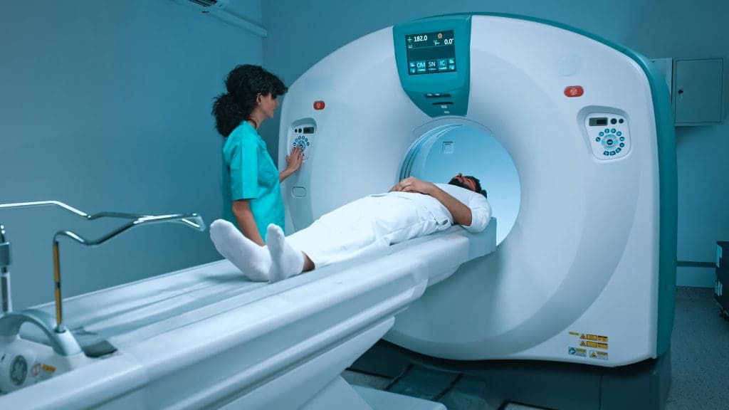
A PET scan is a detailed imaging method that shows how cells work. It’s different from other scans because it looks at how cells function, not just their shape. This makes it great for finding cancer because it shows where cells are working too hard.
How PET Scans Work
PET scans use a tiny bit of radioactive tracer, like Fluorodeoxyglucose (FDG), in the body. Cancer cells take up more of this tracer because they work harder. This makes them show up clearly on the scan.
The steps of a PET scan are:
- Putting the radioactive tracer into the body
- Cancer cells taking up more of the tracer
- Scanning to see where the tracer is
- Looking at the scan to find active areas
Advantages of PET Scans in Cancer Detection
PET scans offer numerous advantages in cancer detection, such as:
- Early Detection: They can find cancer early, even before it’s seen on other scans.
- Accurate Staging: They help figure out how far cancer has spread by looking at how active tissues are.
- Monitoring Treatment Response: They check if cancer treatments are working, helping to change plans if needed.
Experts say, “PET scans have changed oncology by showing how cancer cells work. This helps doctors diagnose and plan treatment better.
Limitations of PET Scan Technology
Even though PET scans are very useful, they have some downsides. These include:
- False Positives: Some conditions can make it look like there’s cancer when there isn’t.
- False Negatives: Some tumors might not show up because they don’t use much tracer.
- Radiation Exposure: PET scans do involve some radiation.
Knowing these limitations helps doctors understand PET scan results better. This is important for making the best decisions for patients.
The Lymphatic System: Structure and Function
The lymphatic system is a complex network of tissues and organs. It helps protect the body from harmful substances and abnormal cells. Lymph nodes act as filters, playing a key role in immune function.
Anatomy of the Lymphatic System
The lymphatic system includes lymphoid organs, lymph nodes, and vessels. Lymph nodes are bean-shaped and found throughout the body. They are important for immune cell activation.
Lymphatic vessels carry lymph fluid, which has white blood cells, around the body.
Role of Lymph Nodes in Immune Function
Lymph nodes are vital for immune surveillance. They trap pathogens and activate immune cells. Inside, you’ll find lymphocytes, like B cells and T cells, which are key for immune responses.
When lymph nodes find pathogens, they start an immune response to fight off the threat.
Normal vs. Abnormal Lymph Nodes
Normal lymph nodes are small and not tender. But, abnormal lymph nodes can grow, become tender, or inflamed. This can happen due to infection, inflammation, or cancer.
Lymphadenopathy, or lymph node enlargement, is a sign that needs medical attention.
Common Locations of Lymphadenopathy
Lymphadenopathy can show up in the neck, armpits, and groin. The size and feel of enlarged lymph nodes can hint at the cause. It could be lymph node inflammation from an infection or lymph node enlargement linked to cancer.
What Are Hypermetabolic Lymph Nodes?
“Hypermetabolic lymph nodes” are lymph nodes that show high activity on PET scans. This high activity is often linked to diseases like cancer.
Definition and Characteristics
These nodes take up more of the radioactive tracer used in PET scans, like FDG (fluorodeoxyglucose). This means they are very active metabolically. Knowing about these nodes can help understand the disease.
Metabolic Activity in Normal vs. Hypermetabolic Nodes
Normal lymph nodes don’t take up much FDG, showing low activity. But hypermetabolic nodes take up a lot, showing high activity. This difference is key for diagnosing and treating diseases, including cancer.
SUV Values and Their Significance
SUV (Standardized Uptake Value) measures how much tracer a node takes up. Higher SUV values mean more activity. For hypermetabolic nodes, SUV values help doctors understand the disease’s severity and how well treatments are working.
Distribution Patterns of Hypermetabolic Lymph Nodes
The way hypermetabolic nodes spread can tell us a lot about the disease. A group of nodes in one area might mean a local problem. But nodes all over could point to a widespread disease. Knowing this helps doctors make the right diagnosis and treatment plan.
How Cancerous Lymph Nodes Appear on PET Scans
PET scans are key in spotting cancerous lymph nodes. They show the nodes’ activity. This helps in planning treatment and understanding the cancer’s spread.
Typical Imaging Features of Malignant Lymph Nodes
Cancerous lymph nodes show up bright on PET scans. This is because they use a lot of energy. The brightness level, or SUV, tells us how aggressive the cancer is.
The way these nodes light up can also tell us a lot. A focused light might mean cancer. But a spread-out light could mean infection or inflammation.
FDG Uptake Patterns in Cancerous Nodes
The way FDG uptake looks in cancerous lymph nodes can differ. Some might light up evenly, while others might not. But cancerous nodes usually light up more than healthy ones.
It’s important to look at the whole picture when seeing these lights. For example, someone with cancer is more likely to have cancerous nodes if they light up a lot.
Size and Shape Characteristics
The size and shape of lymph nodes are also clues on PET scans. Cancerous nodes are often bigger and not perfectly round.
But, size alone isn’t enough. Some cancerous nodes are small, and some big nodes might not be cancer.
Limitations in Identifying Cancerous Lymph Nodes
Even with PET scans, there are limits. Small nodes or those that don’t light up much might not show up. Also, sometimes, inflammation or infection can look like cancer.
So, it’s best to look at PET scans with other info and tests. This way, we can make sure we’re diagnosing and staging correctly.
Common Causes of Hypermetabolic Lymph Nodes
Hypermetabolic lymph nodes on a PET scan can have many causes. These range from cancer to non-cancerous conditions. Knowing the causes helps doctors diagnose and plan treatment.
Malignant Causes
Cancerous lymph nodes show up because of fast-growing cancer cells. This is true for cancers like lymphoma, breast, lung, and melanoma. These cancers make lymph nodes light up on PET scans.
The FDG uptake in these nodes is high. This means they show up brightly on scans. Where these nodes are can tell doctors how far and how advanced the cancer is.
Non-Malignant Causes
Not all hypermetabolic lymph nodes are cancer. Infectious and inflammatory conditions can also cause them. For example, tuberculosis, sarcoidosis, and inflammation can make lymph nodes light up.
Autoimmune diseases can also cause hypermetabolic lymph nodes. It’s important to look at the pattern of FDG uptake and the patient’s history to tell these apart from cancer.
In summary, while cancer is a big worry with hypermetabolic lymph nodes, other causes are important too. Doctors need to look at the patient’s history, scan results, and sometimes take a biopsy to figure out what’s going on.
Differentiating Between Cancerous and Reactive Lymph Nodes
Telling cancerous from reactive lymph nodes is a big challenge in medicine. Getting it right is key for the right treatment and care.
Key Distinguishing Features
There are key signs to tell cancerous from reactive lymph nodes. Cancerous nodes usually have higher metabolic activity on PET scans. This is shown by FDG uptake. Reactive nodes have mild to moderate FDG uptake.
The shape and size of lymph nodes also matter. Cancerous nodes are larger and irregular. Reactive nodes are smaller and oval.
Patterns of FDG Uptake
The way FDG uptake happens is important too. Cancerous nodes have intense, homogeneous uptake. Reactive nodes show more variable uptake.
Size and Distribution Considerations
The size and where lymph nodes are located matter too. Cancerous nodes are larger than 1 cm and may be clustered or confluent. Reactive nodes are smaller and more scattered.
Clinical Context and History
The patient’s history and current situation are also important. Things like patient age, medical history, and symptoms help a lot.
For example, someone with a cancer history is more likely to have cancerous lymph nodes. But someone with a recent infection might have reactive nodes.
Lymphadenopathy: Types and Presentations
Lymphadenopathy can show up in many ways, depending on the cause and where the lymph nodes are affected. It’s a term for when lymph nodes get bigger. Knowing the different types helps doctors diagnose and treat better.
Localized vs. Generalized Lymphadenopathy
Lymphadenopathy can be either localized or generalized. Localized lymphadenopathy means nodes in one area are swollen, often due to a local infection. On the other hand, generalized lymphadenopathy means nodes all over are swollen, pointing to a bigger issue.
Acute vs. Chronic Lymphadenopathy
How long lymphadenopathy lasts is key. Acute lymphadenopathy is short-term, like with infections. Chronic lymphadenopathy lasts longer and might mean something serious like cancer.
Regional Patterns and Their Clinical Significance
Where lymphadenopathy happens can tell doctors a lot. Neck lymph nodes might mean head or neck cancer. Axillary nodes could point to breast cancer. Knowing these patterns helps doctors figure out what to do next.
Symptoms Associated with Abnormal Lymph Nodes
People with lymphadenopathy might feel pain or tenderness, fever, night sweats, or lose weight. These symptoms, with big or lasting lymphadenopathy, need a doctor’s check-up to find the cause.
PET Scan Protocols for Evaluating Lymph Nodes
To check lymph nodes well, PET scan protocols need careful planning. This includes several steps for accurate results.
Patient Preparation
Getting ready for a PET scan is key. Patients often fast before to reduce glucose in non-target areas. They also avoid hard exercise and drink lots of water.
Imaging Techniques
PET scans use a radioactive tracer, like Fluorodeoxyglucose (FDG), to find cancer in lymph nodes. Advanced scanners show these areas clearly. This helps doctors see if lymph nodes are involved.
Interpretation Criteria
Reading PET scan images needs skill and knowledge. The Standardized Uptake Value (SUV) shows how much tracer is in lymph nodes. A high SUV might mean cancer.
Hybrid Imaging: PET/CT and PET/MRI
Hybrid imaging, like PET/CT and PET/MRI, mixes PET’s function with CT or MRI’s anatomy. This combo gives a clearer view of lymph nodes’ activity and location.
Using PET scan protocols, including hybrid imaging, has boosted lymph node checks in cancer patients. This way, doctors can make better diagnoses and treatment plans.
Sensitivity and Specificity of PET Scans for Lymph Node Assessment
Understanding PET scan sensitivity and specificity is key to their use in lymph node evaluation. PET scans are vital in oncology for spotting cancer in lymph nodes.
Detection Rates for Different Cancer Types
PET scans’ ability to find cancer in lymph nodes varies by cancer type. For example, they are very good at spotting it in lymphoma and lung cancer. Sensitivity rates for these cancers are often 80% to over 90%.
But, for cancers like prostate cancer or breast cancer, sensitivity can be lower. This is because these cancers might not show up as much on PET scans.
PET scans’ specificity also changes. This is because things like infections can cause false positives. So, it’s important to look at the whole picture when reading PET scan results.
False Positive and False Negative Results
False positives on PET scans can happen for many reasons. This includes infectious diseases, granulomatous diseases, and inflammatory processes. On the other hand, false negatives can occur if the cancer is small or not very active.
It’s vital for doctors to know these limitations. This helps them understand PET scan results better and make the right treatment plans.
Comparison with Other Imaging Modalities
When looking at PET scans for lymph node assessment, comparing them to other imaging is important. This includes CT scans, MRI, and ultrasound. PET scans give metabolic info, which can help spot cancer.
But, other imaging might give clearer pictures or be better for certain cancers or patients. So, the right imaging choice depends on the situation and what’s being looked for.
Differential Diagnosis of Hypermetabolic Lymph Nodes
Identifying the cause of hypermetabolic lymph nodes is key. This involves looking at many clinical factors and test results.
A Systematic Approach to Diagnosis
When diagnosing hypermetabolic lymph nodes, many causes must be considered. This includes both cancer and non-cancer conditions that can cause lymph nodes to be more active.
- Malignant Causes: Lymphoma, metastatic cancer
- Non-Malignant Causes: Infections, inflammatory conditions, granulomatous diseases
It’s important to know the patient’s history, symptoms, and test results. This helps narrow down the possible causes.
Common Diagnostic Pitfalls
There are challenges in diagnosing hypermetabolic lymph nodes. These include:
- False-positive results from inflammation or infections
- False-negative results in low-grade cancers or small tumors
- Different levels of FDG uptake in various cancers
Knowing these challenges helps doctors better understand PET scan results.
Role of Clinical Correlation
Clinical correlation is vital in diagnosing hypermetabolic lymph nodes. It combines imaging results with the patient’s symptoms, lab tests, and other diagnostic info.
For example, a patient with cancer history might likely have metastatic disease causing hypermetabolic lymph nodes.
When to Consider Biopsy
Biopsy is needed when the diagnosis is unclear or when it affects treatment plans. The decision to biopsy depends on the patient’s health, the chance of cancer, and the biopsy risks.
In summary, diagnosing hypermetabolic lymph nodes needs a detailed and systematic approach. It involves clinical correlation and awareness of common diagnostic challenges.
Clinical Significance of Hypermetabolic Lymph Nodes
Understanding hypermetabolic lymph nodes is key for accurate cancer staging and treatment planning. These nodes, seen through PET scans, are very important for patient care.
Implications for Cancer Staging
Hypermetabolic lymph nodes are vital in cancer staging. Their presence can change a patient’s cancer stage, affecting treatment. Accurate staging is essential for knowing how far the disease has spread and for planning treatment.
Accurate staging helps find patients who need more aggressive or targeted treatments. It also helps avoid unnecessary treatments by showing if the disease is more localized.
Impact on Treatment Planning
The discovery of hypermetabolic lymph nodes affects treatment planning. If these nodes are near a primary tumor, it might mean a bigger surgery or more areas treated.
Radiation therapy plans may also change based on these nodes. This ensures all areas at risk get enough treatment.
Prognostic Value
Hypermetabolic lymph nodes are important for predicting patient outcomes. The number and location of these nodes give clues about the patient’s prognosis. More nodes usually mean a worse prognosis.
- The presence of hypermetabolic lymph nodes indicates a more advanced disease.
- The distribution pattern of these nodes can offer insights into the disease’s spread.
- Monitoring changes in these nodes over time can help in assessing treatment response.
Role in Recurrence Detection
Hypermetabolic lymph nodes are also key in detecting cancer recurrence. During follow-up, PET scans can spot new or growing lymph nodes that might mean cancer is back.
Early detection of recurrence allows for timely treatment, which can improve outcomes. It’s important for cancer patients to have regular check-ups to catch recurrence early.
Diagnostic Approach to Hypermetabolic Lymph Nodes
Diagnosing hypermetabolic lymph nodes involves several steps. These include imaging, biopsy, and molecular testing. When these nodes show up on a PET scan, more tests are needed to find out why.
Initial Evaluation
The first step is a detailed medical history and physical check-up. This helps find out if the lymph nodes are swollen due to infection or cancer. The PET scan’s findings, like the SUV value, help decide what to do next.
Follow-up Imaging
More imaging might be needed to see how the lymph nodes are doing. Serial PET scans are helpful here. They show if treatment is working or if the nodes are changing.
Biopsy Considerations
If tests are not clear, a biopsy might be needed. A biopsy takes tissue for lab tests. This is key for spotting cancer or other issues in the lymph nodes.
Molecular and Genetic Testing
Molecular and genetic testing offer more clues. They can find specific cancer markers. This helps doctors plan the best treatment for each patient.
Putting together info from all these tests helps doctors understand hypermetabolic lymph nodes well. They can then plan the right treatment.
Treatment Strategies for Patients with Hypermetabolic Lymph Nodes
Getting the right treatment for hypermetabolic lymph nodes starts with a correct diagnosis. Doctors use a mix of treatments to manage these nodes.
Treatment Based on Underlying Cause
The treatment plan for hypermetabolic lymph nodes depends on the cause. Malignant causes need strong treatments like surgery, radiation, and systemic therapy. On the other hand, non-malignant causes might get milder treatments that focus on the root problem.
Surgical Approaches
Surgery is key in treating hypermetabolic lymph nodes, mainly for cancer. Surgical methods include:
- Biopsy for diagnosis
- Lymph node removal for treatment
- Reducing big tumor sizes
Radiation Therapy
Radiation therapy is also important for treating hypermetabolic lymph nodes, mostly for cancer. It’s used:
- As a main treatment to shrink tumors
- With surgery to get rid of leftover cancer
- To ease symptoms
Systemic Therapy Options
Systemic therapy, like chemotherapy, targeted therapy, and immunotherapy, is used for cancer-related hypermetabolic lymph nodes. The choice depends on the cancer type, stage, and the patient’s health.
“Choosing the right treatment for hypermetabolic lymph nodes needs a team effort. Doctors, surgeons, and radiologists work together to find the best plan.”
Expert Opinion
Healthcare teams use surgery, radiation, and systemic therapies to treat hypermetabolic lymph nodes. This approach helps patients get the best care.
Conclusion
Understanding hypermetabolic lymph nodes is key for accurate cancer diagnosis and treatment planning. PET scans help spot these nodes, which may show cancer activity. They look at the nodes’ metabolic activity, size, and how they spread.
It’s important to correctly read PET scan results to tell cancerous nodes from reactive ones. This helps figure out the cancer stage, plan treatments, and predict outcomes. By using PET scan data with other medical info, doctors get a better picture of the disease.
PET scans are very important for finding and treating cancer. They help doctors see where cancer might spread, check how treatments work, and find cancer coming back. As cancer treatment gets better, PET scans will keep being a big part of caring for patients.
FAQ
What are the implications of hypermetabolic lymph nodes for cancer staging and treatment planning?
Hypermetabolic lymph nodes are important for cancer staging and planning treatment. Their presence and extent affect cancer stage, prognosis, and treatment options.
How do PET/CT and PET/MRI hybrid imaging modalities contribute to lymph node evaluation?
PET/CT and PET/MRI combine metabolic info from PET scans with detailed anatomy from CT or MRI. This combo improves lymph node evaluation, helping to accurately identify hypermetabolic nodes.
What is the role of biopsy in the diagnosis of hypermetabolic lymph nodes?
Biopsy is key for diagnosing hypermetabolic lymph nodes, when the cause is unclear or suspected to be cancer. It confirms the diagnosis, guiding treatment.
How are hypermetabolic lymph nodes managed and treated?
Treatment for hypermetabolic lymph nodes varies by cause. It might include surgery, radiation, or systemic therapy. The choice depends on the diagnosis and situation.
What are the common causes of hypermetabolic lymph nodes?
Hypermetabolic lymph nodes can come from cancer or non-cancer reasons. Cancer types like lymphoma and metastasis are causes. Non-cancer reasons include infections and inflammation.
Can PET scans differentiate between cancerous and reactive lymph nodes?
PET scans can find hypermetabolic lymph nodes but can’t always tell if they’re cancerous or not. The size, pattern, and context of the nodes are key to making this call.
What is the significance of SUV values in PET scans for lymph node evaluation?
SUV values show how much tracer a tissue takes up. For lymph nodes, higher values mean more activity. This can mean cancer or inflammation.
How do PET scans work in detecting hypermetabolic lymph nodes?
PET scans use a radioactive tracer, like FDG, injected into the body. This tracer goes to active areas, like cancer. The scan then shows these areas, helping spot hypermetabolic lymph nodes.
What are hypermetabolic lymph nodes, and how are they detected?
Hypermetabolic lymph nodes show high activity, often due to cancer or disease. They are found using PET scans, which check tissue activity.


