Last Updated on October 22, 2025 by mcelik

Positron Emission Tomography (PET) scans are a key tool in finding cancer. But, they have limitations. They can miss Cancers not detected PET scan, leaving patients with undiagnosed conditions.
The main problem with PET scans is their blind spots. Some cancers can’t be found. This is a big issue for both patients and doctors, affecting treatment plans and outcomes.

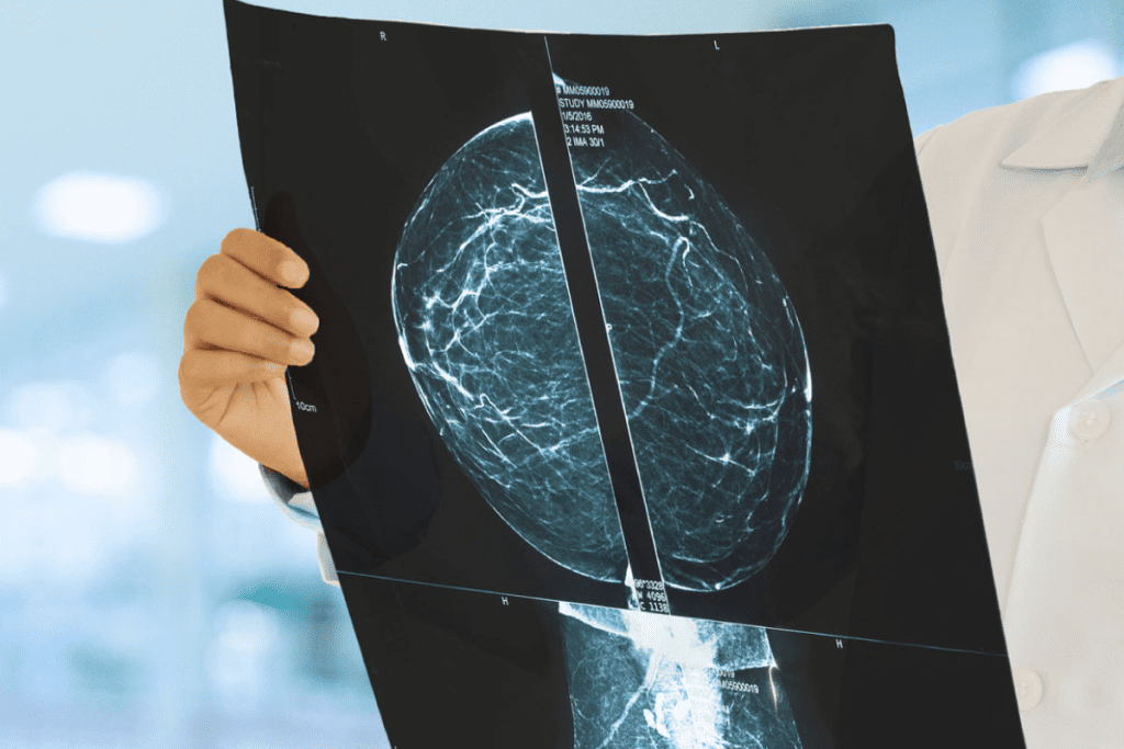
PET scan technology is key in finding cancer and checking how treatments work. It uses radiotracers to spot cancer cells by their activity.
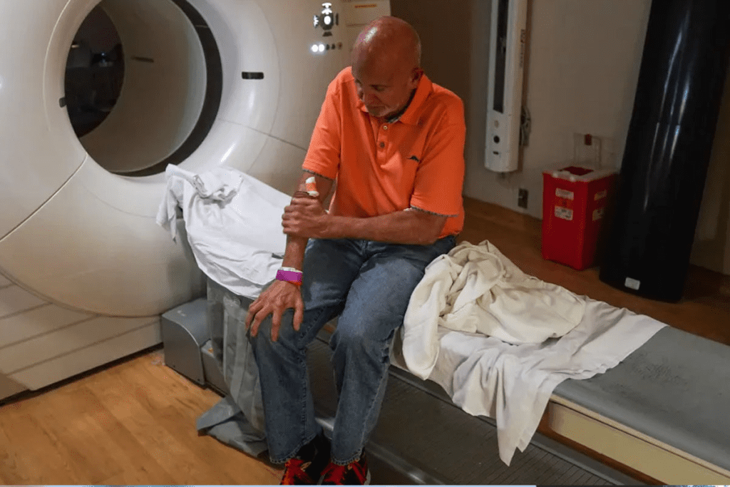
PET imaging catches on the fact that cancer cells burn energy faster than regular cells. A tiny amount of radioactive tracer is given, which goes to active areas like tumors.
The tracer sends out positrons that meet electrons, making gamma rays. These rays are caught by the PET scanner. This info makes detailed images of the body’s activity.
Fluorodeoxyglucose (FDG) is the main tracer in PET scans. It’s a sugar molecule with a radioactive tag. Cancer cells take up more FDG than normal cells, making tumors visible.
But, PET scans can miss some cancers because not all are active. This limits how well they can detect cancer.
Today’s PET scanners often team up with CT or MRI. This mix gives both the PET’s metabolic info and the CT or MRI’s body details.
This combo makes cancer diagnosis and planning better. It gives a clearer picture of where the tumor is and how active it is.
| Imaging Modality | Primary Use | Benefits |
| PET | Cancer detection, staging, and monitoring treatment response | Provides metabolic information, high sensitivity for detecting cancerous tissues |
| CT | Anatomical imaging, detecting structural abnormalities | High-resolution images, quick scanning time |
| MRI | Soft tissue imaging, detailed anatomical information | Excellent soft tissue contrast, no radiation exposure |
| PET/CT | Combining metabolic and anatomical information | Enhanced diagnostic accuracy, better localization of tumors |
| PET/MRI | Combining metabolic and detailed anatomical information | High sensitivity and specificity, excellent for certain cancer types |

PET scans are great for finding cancer, but they have some big limitations. These can make the results less accurate and reliable.
PET scans can’t always spot small tumors or those that don’t use much energy. Their resolution is about 4-5 mm, so tiny tumors might not show up. Sensitivity also varies, depending on the tech and the tracer used.
The Standardized Uptake Value (SUV) is key in PET scans. It shows how much tracer is taken up by tissues. But, it’s hard to understand SUV values because of many factors. For example, blood glucose levels can change how much tracer is taken up.
PET scans have time limits that can affect their usefulness. The scan time is short, and if the patient moves, it can mess up the image. Also, when the tracer is injected affects how it spreads in the body.
PET scans are a powerful tool for diagnosing diseases. Yet, they have their limits, mainly in detecting cancers. They can’t spot all types of cancers, which is a big drawback.
PET scans can’t see small tumors well. Tumors smaller than 8-10 mm are hard to spot. This is because the signal gets mixed with the surrounding tissue.
Finding small tumors early is key to better treatment outcomes. But, PET scans’ technical limits mean some small tumors are missed until they grow bigger.
PET scans also struggle with low metabolic activity cancers. These scans work by detecting radiotracers in active cells. But, tumors that don’t use many resources might not show up.
These cancers show the blind spots in PET scans. This highlights the need for other diagnostic methods.
Research shows PET scans miss a lot of cancers. For example, they catch only 30-40% of small tumors. This depends on the tumor type and where it is.
| Tumor Type | Detection Rate (%) |
| Small Cell Lung Cancer | 60-80 |
| Prostate Cancer | 40-60 |
| Mucinous Tumors | 20-40 |
The stats on missed cancers show PET scans’ limits in diagnosis. Doctors need to know these limits when reading PET scan results.
Some cancers are hard to see with PET scans, making diagnosis and treatment planning tough. PET scans work by showing where tumors are active. But, some cancers don’t show up well because they don’t use much energy or are hidden by other tissues.
Prostate cancer is tricky to spot with PET scans, mainly in the early stages or when the cancer is low-grade. The limited sensitivity of PET scans for prostate cancer comes from how cancer cells take up the scan’s tracer.
Brain tumors are hard to see with PET scans because of the high background activity of the brain. The brain’s constant energy use can hide the energy signs of tumors.
Choosing the right tracer is key for seeing brain tumors. Tracers like FET (fluoroethyltyrosine) or Methionine are better at showing tumors.
Finding HCC with PET scans is hard, mainly for well-differentiated tumors. The variable FDG uptake in HCC means some tumors are hard to spot.
“The sensitivity of PET scans for HCC is generally lower compared to other imaging modalities like MRI and CT scans, particularly for early-stage disease.”
Mucinous tumors and NETs are also hard to see with PET scans. Mucinous tumors have low cellularity and metabolic activity, making them hard to find.
It’s important for doctors to know these challenges. This helps them understand PET scan results better and choose the right tests for patients.
PET scanning sometimes gives false positive results. These can cause wrong diagnoses, extra tests, and worry for patients. It’s key to know why false positives happen to make PET scans more accurate.
Some inflammatory and infectious issues can look like cancer on PET scans. This is because they show up as active on the scan. For example, sarcoidosis, tuberculosis, and abscesses can cause false positives. Doctors need to watch out for these when they read PET scans.
After treatments like surgery or chemotherapy, the body might show inflammation. This can make PET scans look like cancer is coming back. Doctors must check the patient’s treatment history to make sure the scan results are right.
Some non-cancerous growths, like adenomas or uterine fibroids, can show up as active on PET scans. Knowing about these can help doctors avoid false positives and make the right diagnosis.
In summary, false positives in PET scans can come from many sources. These include inflammatory and infectious issues, changes after treatment, and active benign tumors. Understanding these can help make PET scans more reliable.
PET scans are powerful tools but not perfect. They can miss cancer, which can delay treatment. This is known as a false negative.
Several things can lead to false negatives in PET scans. The size and location of the tumor matter. So does the type of cancer and how active the cancer cells are. Small tumors or those with low metabolic activity are often missed.
The scanner’s resolution is also key. Higher resolution scanners can spot smaller tumors. But, even with the best tech, some tumors might not be found.
False negatives can lead to delayed diagnosis and wrong treatment plans. Patients might think they’re okay when they’re not. This can cause them to wait too long to get more tests.
These mistakes can also lead to bad cancer management. It’s vital for doctors to know PET scan limits. They should use other tests when needed.
There are many examples of PET scan false negatives. For example, a patient with prostate cancer might get a false negative. This is because the tumor doesn’t show up well on the scan.
| Cancer Type | Factors Contributing to False Negatives | Clinical Implications |
| Prostate Cancer | Low metabolic activity, small tumor size | Delayed diagnosis, inappropriate treatment |
| Brain Tumors | High background metabolic activity, tumor location | Misdiagnosis, delayed treatment |
| Hepatocellular Carcinoma | Variable metabolic activity, liver background uptake | Inappropriate management, delayed intervention |
It’s important to know the limits of PET scans. This helps doctors use them better in cancer care. By understanding these issues, doctors can make better choices for their patients.
PET scans are vital for cancer diagnosis and treatment. Yet, their high cost and limited availability create big challenges. These issues affect both patients and healthcare providers.
A PET scan can cost between $1,000 to $5,000 or more. This is a big expense for many. It’s even harder for those without good insurance or high-deductible plans.
PET scans are not available everywhere. Rural and underserved areas often have no access. This can cause delays in diagnosis and treatment.
Scheduling delays for PET scans can affect treatment plans. The demand for scans is high, leading to long wait times. This can be from days to weeks.
Key factors contributing to scheduling delays include:
PET scans are useful for diagnosis but expose patients to radiation. This radiation comes from the radiotracer used in the scan. It emits positrons that collide with electrons, creating gamma rays the scanner can detect.
Getting multiple PET scans can lead to a buildup of radiation. This can raise the risk of getting secondary cancers. Doctors must carefully consider the benefits of PET scans against the risks, mainly for those needing many scans.
Key factors influencing cumulative radiation effects include:
It’s vital to do a risk-benefit analysis for PET scans in different groups. For example, kids are more vulnerable to radiation because their bodies are developing. This means they need extra care.
Patient populations that require special consideration include:
Comparing PET scans to other imaging methods shows their radiation levels. CT scans also expose patients to a lot of radiation. But MRI scans don’t use radiation at all.
| Imaging Modality | Typical Radiation Exposure |
| PET Scan | Low to moderate |
| CT Scan | Moderate to high |
| MRI | None |
PET scans can be affected by several factors related to the patient. Knowing these factors is key to understanding PET scan results. It helps in making better clinical decisions.
Blood glucose levels can impact PET scan accuracy. High glucose levels can reduce the uptake of F-FDG, a common tracer for cancer imaging. This is because glucose and F-FDG compete for the same cells.
To fix this, patients often fast before a PET scan. Fasting lowers blood glucose, improving F-FDG uptake in tumors. But, fasting can be hard for people with diabetes.
Movement during a PET scan can also affect accuracy. Even small movements can blur or misalign images. This can lead to wrong interpretations of the scan results.
To reduce movement issues, patients are told to stay as calm and steady as possible. Some PET scanners use software to correct for movement.
Some medications and treatments can also impact PET scan results. Certain drugs can change how the radiotracer is metabolized. Treatments like chemotherapy can alter tumor metabolism, affecting scan results.
It’s important for patients to tell their doctors about any medications or treatments before a PET scan. This helps in accurately interpreting the scan and making the right clinical decisions.
PET scans are useful but have their limits. They are part of a larger family of diagnostic tools. Each tool has its own strengths and weaknesses in finding and managing cancer.
CT scans show detailed pictures of the body’s structure. They’re great for spotting structural problems. On the other hand, PET scans reveal how tissues work by showing metabolic activity.
When we compare PET and CT scans, we see their different strengths. CT scans are better at finding small tumors in some organs. But PET scans are better at finding cancer cells that are active.
A study showed that using both PET and CT scans together is better than just CT scans alone. This combo is better at finding cancer spread in different types of cancer.
| Cancer Type | PET Scan Detection | CT Scan Detection |
| Lymphoma | High sensitivity due to high metabolic activity | Moderate sensitivity based on structural changes |
| Lung Cancer | High sensitivity for metabolically active tumors | High sensitivity for structural abnormalities |
| Prostate Cancer | Limited sensitivity, even for low-grade tumors | Moderate sensitivity, often used for assessing structural changes |
MRI gives clear pictures of soft tissues. It’s best for looking at tumors in the brain, spine, and other complex areas. When we compare PET and MRI, we need to think about the cancer type and what information we need.
In brain tumors, MRI is often the top choice because it shows detailed anatomy. But PET scans can tell us about tumor metabolism. This is important for seeing how well treatments are working.
The right imaging tool depends on the cancer type, its location, and the patient’s health. While PET scans are useful, other methods might be better in certain situations.
For example, ultrasound is often the first choice for checking liver or thyroid nodules. It’s non-invasive and doesn’t use radiation. MRI is better for looking at spinal cord compression or brain metastases because it shows soft tissues well.
In summary, knowing the good and bad of each imaging tool is key for the best cancer care. By comparing PET scans to CT and MRI, doctors can choose the best way to diagnose and manage cancer.
PET scans are great for finding many cancers. But, they can miss some. So, we need other ways to check for cancer to make sure we get it right.
Using PET scans with other imaging can make diagnosis better. Multi-modality imaging mixes different scans to give a clearer picture of what’s going on.
For example, adding PET to CT or MRI scans helps find tumors that PET might miss. This mix lets doctors use each scan’s best points, making diagnosis more reliable.
When scans don’t show enough, biopsy and lab tests are key. A biopsy takes a tumor sample for detailed checks. Lab tests look at blood and genes to help plan treatment.
These tests give more info on the tumor. They help doctors create treatments that really work.
Clinical correlation is vital for making sense of scans. It combines scan results with what the patient is feeling and their medical history.
This way, doctors can understand what scans mean better. It leads to treatments that are just right for each patient.
New technologies are making PET scans better, overcoming old problems. The field is seeing big improvements in several areas.
New radiotracers are being researched to make PET scans more accurate. Old radiotracers like FDG have their limits, mainly in finding certain cancers. New ones aim to find specific cancer markers, making diagnosis better.
For example, radiotracers for prostate cancer are showing great results. Tracers like 68Ga-DOTATATE help find neuroendocrine tumors. These new tools are making PET scans more useful for different cancers.
| Radiotracer | Target | Application |
| FDG | Glucose metabolism | General cancer detection |
| Ga-PSMA | Prostate-specific membrane antigen | Prostate cancer detection |
| Ga-DOTATATE | Somatostatin receptors | Neuroendocrine tumors |
New scanner technology is giving us clearer images. This helps spot small tumors and boosts accuracy. Modern scanners use better materials and algorithms for sharper images.
PET scans combined with CT and MRI (hybrid systems) offer even more benefits. These systems give detailed information about tumors, helping doctors stage and treat cancer better.
AI and ML are making PET imaging smarter. They help spot tiny issues that might be missed. AI can analyze images in ways humans can’t.
ML models learn to tell apart cancerous and non-cancerous lesions. This could lower mistakes in diagnosis. These technologies also help standardize how images are read, making results more reliable.
Key Benefits of AI and ML in PET Imaging:
As these technologies keep improving, PET scans will become even more valuable in fighting cancer.
Getting insurance to cover PET scans can be tough for patients. It can slow down getting a diagnosis and treatment. Knowing how insurance works is key for both patients and doctors.
Medicare and private insurance have different rules for PET scans. Medicare often covers PET scans for cancer and certain conditions. But, the rules can be tricky. Private insurance might need pre-approval for PET scans. It’s important for patients to know what their insurance covers and what they might have to pay out of pocket.
Getting prior approval for PET scans is a big hurdle. Doctors and patients face delays because of this. It can hold up getting a diagnosis and treatment.
If insurance denies a PET scan, there’s a chance to appeal. The appeal process asks for more information to show the scan is needed. Knowing how to appeal and preparing a strong case can help.
Patients and doctors need to work together to deal with insurance issues. Understanding the policies and processes can help patients get the care they need.
PET scans are key in cancer care, helping with staging and tracking treatment success. They offer insights that greatly improve patient care, despite their limitations.
Some cancers are better for PET scans because of how they work. For example, lymphoma and melanoma are high-energy, making them perfect for PET scans. A study in the Journal of Clinical Oncology found PET scans are great for managing lymphoma, helping with staging and seeing how well treatments work.
PET scans are key in figuring out how far cancer has spread. They also check if treatments are working. Knowing how treatments are doing early on can change the plan, helping patients more.
PET scans are also used to watch for cancer coming back. Regular scans can catch recurrence early, when it’s easier to treat.
“Early detection of recurrence through PET scanning can significantly improve patient outcomes by enabling timely intervention.”
This is very important for cancers that often come back, needing close monitoring.
Knowing what PET scans can and can’t do helps doctors use them wisely. This way, they get the most out of PET scans, helping patients while avoiding their downsides.
Getting ready for a PET scan is key to getting good results. It’s important for doctors to get accurate images. These images help in diagnosing and planning treatments.
Before a PET scan, patients must follow certain diet rules. They need to fast to keep their blood sugar levels right. This is because high sugar levels can mess up the scan’s accuracy.
Patients also need to avoid certain foods and drinks. They might be told to eat less carbs or skip sugary items.
Patients also need to cut down on physical activity before the scan. Too much exercise can change how the scan works. They’re usually told to avoid hard workouts for a while.
Getting mentally ready for a PET scan is also important. Many patients feel anxious, which can make it hard to stay calm during the scan. This is why managing anxiety is key.
Doctors often teach patients how to relax. They might suggest deep breathing or meditation. For those who are really scared, a little sedation might be an option.
By following these steps, patients can help make sure their PET scan is a success. This is important for getting the right diagnosis and treatment.
PET scans have changed how we diagnose and manage cancer. They offer many pet scan benefits in finding and understanding cancer types. But, they also have some disadvantages to think about.
It’s important to know the limits of PET scans to balance benefits and risks. They show how active tumors are, but their accuracy can be influenced. This includes the size of the tumor, how fast it grows, and the patient’s health.
In cancer screening, PET scans are great for some cancers like lymphoma and melanoma. But, they might not work as well for others, like prostate cancer. By knowing these differences, doctors can use PET scans better with other tests.
Choosing to use PET scans should be a careful decision. It’s about weighing their good points against the bad. This way, patients get better diagnoses and treatments, leading to better health.
The biggest drawback is they can’t find all cancers. This includes cancers that don’t use much energy or are too small to see.
PET scans use special tracers to spot cancer cells. They help doctors find, stage, and check how well treatments work.
Main issues include their resolution and sensitivity. Also, interpreting SUV values and the timing of scans can affect accuracy.
False positives happen when scans show cancer when there isn’t any. This can be due to inflammation, post-treatment changes, or active but benign tumors.
False negatives can delay or miss cancer diagnoses. This can affect treatment plans and patient outcomes. Factors include low activity, small size, and technical issues.
High costs and limited access can limit PET scans. This includes financial issues, availability in rural areas, and scheduling problems.
There’s worry about long-term effects of radiation, mainly for young patients or those needing many scans. This is compared to other imaging methods.
Factors like blood sugar levels, movement, and medication interactions can affect scan results.
PET scans have unique strengths but may not be best for all cancers. Other methods might be better for certain types or needs.
Using multiple imaging methods, combining biopsies and lab tests, and clinical evaluation can improve accuracy.
New tracers, better scanners, and AI are improving PET scans. These advancements aim to boost accuracy and usefulness.
Navigating insurance policies, getting prior authorization, and appealing denied claims can be tough.
Subscribe to our e-newsletter to stay informed about the latest innovations in the world of health and exclusive offers!