Getting a correct and quick diagnosis is key to good heart care. With many heart imaging tests available, it’s important to know which one fits your needs. Each test gives different views into how healthy your heart is.Cardiac imaging test – Explore 7 essential heart imaging tests for accurate cardiac diagnosis and treatment.
At Liv Hospital, we put our patients first. We make sure every cardiac diagnostic procedure meets the highest standards. This helps us make accurate diagnoses and create effective treatment plans. We use different cardiology testing procedures to check your heart’s structure, function, and blood flow. This way, we can spot heart issues early and treat them quickly.
Key Takeaways
- Heart imaging tests are key for diagnosing and managing heart disease.
- Many tests are used, like echocardiograms, cardiac CT scans, and cardiac MRI.
- These tests check your heart’s structure, function, and blood flow.
- Spotting heart problems early means we can treat them quickly.
- Heart imaging tests can find issues like coronary artery disease and heart valve problems.
Understanding Cardiac Imaging Tests and Their Importance

Cardiac imaging tests are key in finding heart problems. They give us clear pictures of the heart’s inside and how it works. These tests help us spot different heart diseases and decide on the best treatment.
These tests let us see the heart’s shape and how well it works. This info is key for spotting issues like blocked arteries, faulty valves, and weakened heart muscles.
The Role of Imaging in Heart Disease Diagnosis
Imaging tests help us see the heart’s details. This lets us accurately find heart problems. The clear images show us any issues and how serious they are.
Types of Cardiac Imaging Tests:
- Echocardiogram
- Cardiac CT Scan
- Nuclear Cardiac Stress Tests
- PET Scans
- Cardiac MRI
Each test gives us special info about the heart. This helps us diagnose and manage heart disease well.
How Diagnostic Results Guide Treatment Decisions
The results from these tests are key in choosing treatments. Knowing how bad the heart disease is lets us make plans that fit each patient’s needs.
| Diagnostic Test | Information Provided | Treatment Guidance |
| Echocardiogram | Heart valve function, heart chamber size | Decisions on valve repair or replacement |
| Cardiac CT Scan | Coronary artery disease, calcium scoring | Guidance on lifestyle changes or interventions |
| Nuclear Cardiac Stress Tests | Heart function under stress, ischemia detection | Decisions on medication, angioplasty, or surgery |
Using the info from these tests, we can give our patients the best care. This way, we make sure they get the right treatment for their heart issues. This improves their health and quality of life.
Echocardiogram: Ultrasound Imaging of the Heart

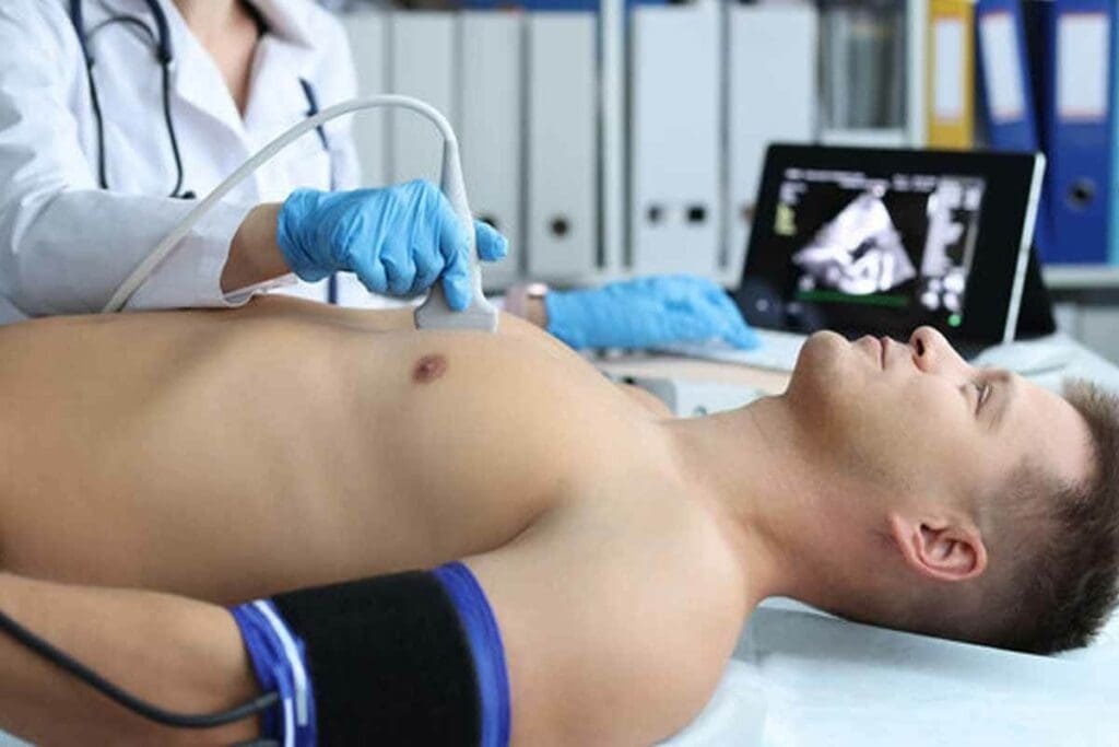
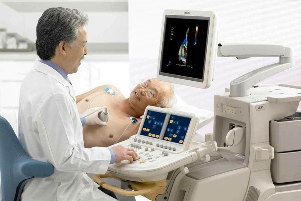
Echocardiograms are key in cardiology, using ultrasound to see the heart’s inside. This method is non-invasive and helps diagnose and track heart issues.
Types of Echocardiograms
There are many echocardiogram types, each for a different purpose. The most common ones are:
- Transthoracic Echocardiogram (TTE): This is the most common, where the probe is on the chest to get heart images.
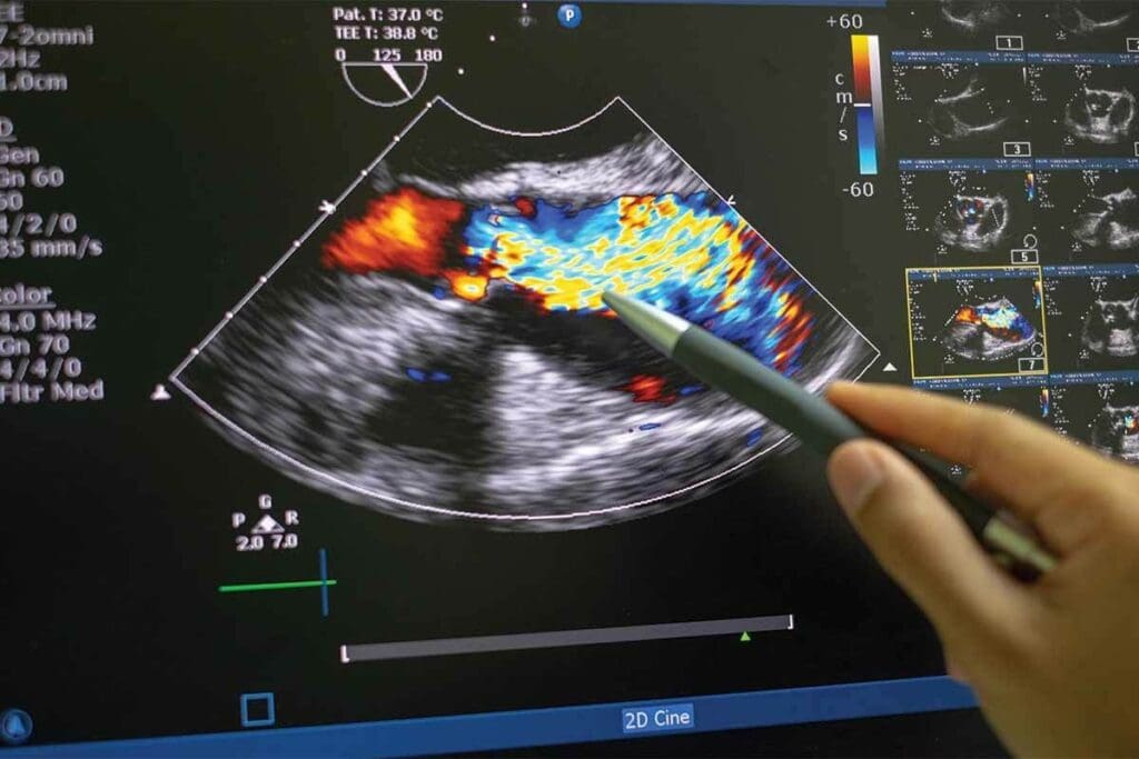
- Transesophageal Echocardiogram (TEE): The probe goes into the esophagus for closer heart images, great for the back of the heart.
- Stress Echocardiogram: Done before and after stress (like exercise or medicine) to see how the heart works under stress.
What an Echocardiogram Reveals About Heart Function
An echocardiogram shows a lot about the heart’s shape and how it works. It can find:
- The heart’s size and shape, helping spot problems like hypertrophic cardiomyopathy.
- How well the heart valves work, spotting issues like stenosis or regurgitation.
- The heart’s pumping ability, helping diagnose heart failure.
A leading cardiologist says, “Echocardiography is a window to the heart, giving insights for diagnosis and treatment.”
Advantages and Limitations of Ultrasound Imaging
Echocardiography is non-invasive, doesn’t use radiation, and shows images in real-time. But, it depends on the operator and can be hard to see in some cases, like obesity or lung disease.
When comparing echocardiograms to CT scans, it’s important to think about what you need to diagnose. CT scans show the coronary arteries well, but echocardiograms give real-time heart function info. The choice depends on the situation and what’s needed.
In summary, echocardiograms are essential in heart imaging, giving a lot of info on heart function and structure. Knowing about the different types and their uses helps doctors make better decisions for patients.
Cardiac CT Scan: Detailed 3D Visualization
The cardiac CT scan, also known as a heart scan, is a key tool for doctors. It gives detailed 3D images of the heart. These images help find blockages and calcium deposits in the heart’s arteries.
Calcium Scoring vs. Coronary CT Angiography
Cardiac CT scans have two main uses: calcium scoring and coronary CT angiography. Calcium scoring measures calcium plaque in the heart’s arteries. This is a sign of heart disease. Coronary CT angiography shows detailed images of the heart’s arteries. It helps doctors see blockages and check the heart’s health.
These tests have different roles. Calcium scoring is for early risk checks. Coronary CT angiography is for those at higher risk or with heart symptoms.
Detecting Coronary Artery Disease and Blockages
Cardiac CT scans are great for finding heart disease and blockages. They give clear images of the heart’s arteries. This lets doctors spot problems early. Research on PubMed Central shows these scans are key in diagnosing and managing heart disease.
Benefits and Radiation Considerations
Cardiac CT scans have many benefits but involve some radiation. New technology has made these scans safer. Modern scanners use less radiation to get high-quality images. It’s important to talk to your doctor about the risks and benefits.
Getting a cardiac imaging test can worry some patients. But, these scans are done in a safe place. Our team makes sure you’re comfortable and safe during the test.
Nuclear Cardiac Stress Tests: SPECT Imaging
Nuclear cardiac stress tests, like SPECT imaging, are key in finding heart problems. They check how well blood flows to the heart muscle. This helps doctors see how the heart works when it’s stressed and find issues that aren’t seen when it’s at rest.
How SPECT Imaging Works
SPECT imaging uses a tiny amount of radioactive tracer in the blood. This tracer sends out gamma rays that a camera catches. It makes detailed 3D pictures of the heart. Doctors can see how blood flows to the heart muscle, both when it’s resting and when it’s stressed.
Conditions Diagnosed with SPECT
SPECT imaging is great for finding problems like myocardial ischemia. This is when the heart muscle doesn’t get enough blood, causing pain or other symptoms. It also spots damaged heart tissue after a heart attack. By looking at blood flow during stress and rest, SPECT gives doctors a clear picture of the heart’s health.
Procedure and Patient Experience
For a SPECT imaging test, patients usually walk on a treadmill or bike to stress their heart. The tracer is given right when they’re exercising the hardest. Then, they go to the SPECT camera for pictures. The whole thing is watched over by doctors to keep everyone safe and comfortable. Most people can go back to their usual activities right after.
Understanding SPECT imaging helps patients see its importance. It’s a key part of checking the heart’s health. With SPECT, doctors can make better plans for treatment. This leads to better health and a better life for patients.
PET Scans: The Advanced Cardiac Imaging Test
PET scans are a big step forward in heart imaging. They give us detailed views of heart function and help spot heart diseases better.
PET vs. SPECT: Key Differences in Cardiac Imaging
PET and SPECT scans differ in how clear their images are. PET scans show more detailed pictures, helping us find heart problems more accurately. They give us a better look at how the heart works and blood flows.
When we look at PET and SPECT, we see they serve different needs. SPECT is more common and has been used for longer. But PET’s new tech often means more accurate results.
Clinical Applications of Cardiac PET
Cardiac PET scans are great for finding heart artery disease and checking if heart muscle is alive. They help us choose the best treatment for complex heart issues.
- Diagnosing coronary artery disease with high accuracy
- Assessing myocardial viability before revascularization procedures
- Evaluating the effectiveness of cardiac treatments over time
Advantages and Accessibility Factors
PET scans are good because they give exact numbers on blood flow to the heart. This helps us make treatment plans that fit each patient’s needs.
But, PET scans are not as common as other tests. This is because they cost more and need special gear. Even so, their benefits in heart diagnosis make them very useful in cardiology.
Cardiac MRI: High-Resolution Imaging Without Radiation
We use Cardiac MRI to see the heart’s details without radiation. This method is key in cardiology. It gives a full view of the heart’s shape and how it works.
Evaluating Heart Structure and Function
Cardiac MRI checks the heart’s structure and function closely. It shows the heart’s chambers, walls, and valves clearly. Doctors can then see how well the heart is working and spot any problems.
The benefits of Cardiac MRI include:
- High-resolution imaging without ionizing radiation
- Detailed images of the heart’s anatomy and function
- Ability to assess cardiac performance and detect abnormalities
Diagnosing Cardiomyopathies and Heart Damage
Cardiac MRI is great for finding cardiomyopathies and heart damage. It spots scar tissue, inflammation, and other issues that other tests can’t see.
| Condition | Cardiac MRI Findings | Clinical Implication |
| Hypertrophic Cardiomyopathy | Thickened heart muscle | Increased risk of heart failure and arrhythmias |
| Myocardial Infarction | Scar tissue in the heart muscle | Potential for heart failure and need for revascularization |
| Myocarditis | Inflammation of the heart muscle | Need for anti-inflammatory treatment and monitoring |
When Cardiac MRI is Recommended Over Other Tests
Cardiac MRI is chosen when other tests don’t give clear results. It’s best for detailed heart views, like in complex heart diseases or cardiac tumors.
Key scenarios where Cardiac MRI is preferred include:
- Complex congenital heart disease evaluation
- Assessment of cardiac tumors or masses
- Detailed evaluation of cardiomyopathies
Cardiac MRI offers detailed images without radiation. It’s a key tool in cardiology. It helps in checking the heart’s structure and function, diagnosing cardiomyopathies, and finding heart damage.
Chest X-rays: Basic Cardiac Assessment
Chest X-rays are often the first step in diagnosing cardiac issues. They give clinicians a broad overview of heart size and any abnormalities. This basic yet valuable diagnostic tool is key in the initial assessment of heart health.
What a Chest X-ray Can (and Cannot) Show About the Heart
A chest X-ray provides a quick snapshot of the heart’s size, shape, and position. It can show signs of heart failure, like fluid in the lungs. It can also indicate if the heart is enlarged. But, it lacks the detail needed to diagnose specific heart conditions or complex cardiac structures.
Emergency Uses in Cardiac Care
In emergency settings, chest X-rays are invaluable. They help quickly assess patients with suspected cardiac issues. They can identify acute conditions like pulmonary edema or cardiogenic shock. This quick insight allows healthcare providers to make swift decisions for patient care.
Limitations as a Cardiac Diagnostic Tool
While chest X-rays are useful for initial assessments, they have significant limitations. They cannot provide detailed images of the heart’s structure or function. Nor can they diagnose coronary artery disease or other complex cardiac conditions. For such detailed evaluations, other types of cardiovascular tests like echocardiograms or cardiac MRIs are necessary.
In conclusion, chest X-rays serve as a fundamental test of the heart. They offer a preliminary evaluation that can guide further diagnostic procedures. Understanding their capabilities and limitations is key for effective cardiac care.
Coronary Angography: Visualizing Coronary Arteries
Coronary angiography is a detailed heart imaging method. It lets doctors see the coronary arteries clearly. This is key for spotting and treating coronary artery disease, a major cause of heart attacks.
Catheterization Procedure Explained
The angiography process starts with a thin, flexible tube called a catheter. It’s inserted into a blood vessel in the leg or arm. The goal is to reach the coronary arteries. This is done under local anesthesia to make it less painful.
Next, a contrast dye is injected into the arteries. X-ray images are then taken to see how blood flows and spot any problems. The whole thing usually takes about 30 minutes to an hour, depending on the case.
Diagnostic and Therapeutic Applications
Coronary angiography is used for both checking and treating heart issues. It helps doctors see how bad the artery disease is and find blockages. This helps them decide the best treatment.
It can also be used with angioplasty to widen blocked arteries. Sometimes, a stent is placed to keep the artery open.
| Procedure | Description | Benefits |
| Coronary Angiography | Insertion of a catheter to visualize coronary arteries | Detailed imaging of coronary arteries, diagnosis of blockages |
| Angioplasty | Use of a balloon to widen narrowed arteries | Restores blood flow, relieves symptoms |
| Stent Placement | Insertion of a stent to keep the artery open | Prevents re-narrowing, improves long-term outcomes |
Risks and Recovery Considerations
Coronary angiography is usually safe, but there are risks. These include bleeding, infection, and allergic reactions to the dye. Though rare, serious issues like heart attack or stroke can happen.
After the procedure, patients are watched for hours. Most can go home the same day. Resting for a day or two is advised, and following the doctor’s recovery plan is important. Side effects like bruising or discomfort at the site are common but usually go away on their own.
Preparing for Your Cardiac Imaging Procedures
To make your cardiac imaging procedure go smoothly, follow specific preparation guidelines. We know that getting a cardiac imaging test can be stressful. But being prepared can make you feel less anxious and help the test go well.
General Preparation Guidelines
While the exact preparation steps may vary, there are some general tips. These include:
- Tell your doctor about any medicines you’re taking, including over-the-counter ones and supplements.
- Avoid eating or drinking for a certain time before the test, as your doctor will tell you.
- Take off any jewelry or metal objects that could get in the way of the imaging equipment.
- Wear loose, comfy clothes.
It’s very important to follow the specific instructions from your healthcare provider. They might have extra steps based on your health and the test you’re getting.
Test-Specific Preparations
Different cardiac imaging tests need different preparations. For example:
| Test Type | Preparation Requirements |
| Echocardiogram | Usually, no special preparation is needed, but you might need to take off your upper body clothes. |
| Cardiac CT Scan | You might need to avoid caffeine and certain medicines before the test. Wear loose clothes without metal. |
| Nuclear Stress Test | Avoid eating or drinking for a few hours before. You might also need to skip certain medicines. |
Knowing the specific preparation for your test helps you get ready properly. This ensures the test goes well.
Questions to Ask Your Cardiologist Before Testing
Before your cardiac imaging test, ask your cardiologist some questions. This helps you prepare well. You might ask:
- What are the risks of this test?
- How should I prepare for it?
- Are there any medicines or foods I should avoid before the test?
- What will the test results tell us about my heart?
- How long will it take to get my test results?
By asking these questions and following the preparation tips, you can make sure your test goes smoothly. You’ll also get accurate and helpful results.
Conclusion: Making Informed Decisions About Heart Imaging
Knowing about different cardiac imaging tests is key to good heart health. We’ve looked at many tests, like echocardiograms and cardiac CT scans. We also talked about nuclear stress tests, PET scans, and cardiac MRI.
Each test gives special views of the heart’s structure and function. This helps doctors diagnose and treat heart disease well. By picking the right test, you can understand your heart better. Then, you can work with your team to create a treatment plan just for you.
At our place, we aim to give top-notch healthcare with full support. We suggest talking to your cardiologist about your options. Ask about the good and bad of each test. This way, you can help manage your heart health and make smart choices about your care.
FAQ
What is a heart scan called?
Heart scans have different names. They can be called cardiac CT scans, echocardiograms, or nuclear cardiac stress tests.
What are the different types of cardiac imaging tests?
There are many cardiac imaging tests. These include echocardiograms, cardiac CT scans, and nuclear cardiac stress tests (SPECT). Other tests are PET scans, cardiac MRI, chest X-rays, and coronary angiography.
What is the difference between an echocardiogram and a CT scan?
An echocardiogram uses sound waves to see the heart. A CT scan uses X-rays to make detailed 3D images of the heart and blood vessels.
How does SPECT imaging work?
SPECT imaging uses a small amount of radioactive material. It’s injected into the bloodstream. Then, a camera detects it to show the heart’s blood flow.
What is cardiac MRI used for?
Cardiac MRI checks the heart’s structure and function. It helps diagnose heart damage and is used when other tests are unclear.
What can a chest X-ray show about the heart?
A chest X-ray shows the heart’s size and shape. It also checks for lung or tissue issues. But, it can’t pinpoint specific heart problems.
What is coronary angiography?
Coronary angiography uses a catheter to inject contrast material. It helps see the coronary arteries. This helps find blockages and guide treatment.
How do I prepare for a cardiac imaging test?
Preparing for cardiac tests varies. You might need to avoid certain foods and meds. Always follow your cardiologist’s instructions.
What are the benefits and risks of cardiac imaging tests?
These tests give valuable heart info. They help diagnose and manage heart disease. But, some tests use radiation or have risks. Talk to your cardiologist about these.
How do I choose the right cardiac imaging test?
Choosing a test depends on your condition and history. Talk to your cardiologist. They’ll help pick the best test for you, considering its benefits and limits.
References
- Anand, S. S. (2009). Clinical applications of PET and PET-CT. Clinical Radiology, 64(9), 860-869. https://www.sciencedirect.com/science/article/abs/pii/S0377123709800993


