
Computed Tomography (CT) scans have changed medical imaging a lot. They started in the 1970s. Godfrey Hounsfield and Allan Cormack created them. They won the Nobel Prize in 1979 for this. CT, also known as CAT CT, stands for computerized axial tomography. Both terms refer to the same imaging technique that uses X-rays and computer processing to create detailed cross-sectional images of the body. This technology remains a cornerstone of modern diagnostic medicine.
The history of CT scans shows a lot of progress. From the first scan in 1971 to today’s advanced ones.
CT scan technology has grown a lot. It has changed how we care for patients and diagnose diseases. Today’s scanners make very clear images. This helps doctors treat patients better.
At Liv Hospital, we use the latest CT scanners. We aim to give top-notch healthcare with the best technology.
CT scanning technology has changed medical diagnostics a lot. We’ll look at what CT scans are and how they’re different from X-rays. We’ll also clear up any confusion about this technology.
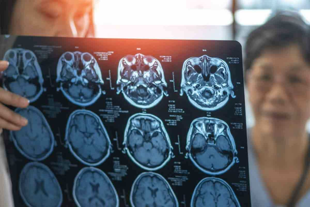
A CT scan uses computer-processed X-rays from different angles to show cross-sections of the body. Unlike X-rays, which are two-dimensional, CT scans give a detailed, three-dimensional view. This helps doctors diagnose and plan treatments better.
For example, CT scans can show injuries or diseases more clearly than X-rays. A study on the National Center for Biotechnology Information website says CT scans are key in modern medicine.
CT, CAT, and Computed Axial Tomography mean the same thing. “CT” is short for Computed Tomography. “CAT” comes from Computed Axial Tomography, which was used in early scanners. These scanners could only scan in one plane. Now, CT scanners can scan in many planes and make 3D images.
Here’s a table to show the differences and similarities between these terms:
| Term | Meaning | Usage |
| CT | Computed Tomography | General term for the technology |
| CAT | Computed Axial Tomography | Historically, specific to axial scanning |
| Computed Axial Tomography | Full form of CAT | Describes the original axial scanning technique |
Knowing these terms helps doctors and patients talk about medical tests clearly.
We thank Godfrey Hounsfield and Allan Cormack for the CT scan. Their work changed how we diagnose diseases.
Godfrey Hounsfield, an English engineer, and Allan Cormack, a physicist from South Africa, won the Nobel Prize in Physiology or Medicine in 1979. They invented the CT scanner. Hounsfield worked on X-ray technology at EMI Central Research Laboratories. Cormack focused on the math behind image reconstruction from X-rays.
Their work was key in changing medical diagnostics. Hounsfield made the first CT scanner that worked well. It was set up in 1971 at Atkinson Morley Hospital in London. Cormack’s theory helped Hounsfield’s practical work, creating a new technology.
The first CT scan was done on October 1, 1971. It was a big step in medical imaging. It showed how CT scans could give detailed images of the body, better than X-rays.
CT scans quickly became a key tool in hospitals. They help doctors diagnose and treat many diseases. Today, they are used all over the world.
Knowing about Hounsfield and Cormack helps us see how far medical imaging has come. Their work has saved many lives. It also keeps pushing the limits of new medical technologies.
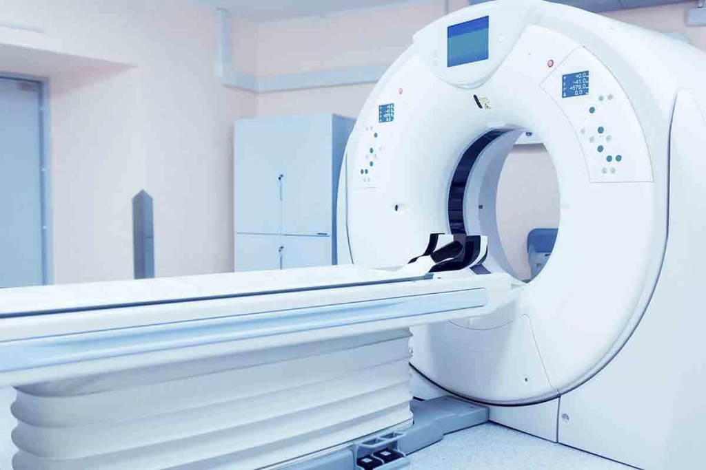
CT scanning combines advanced X-ray sources, detectors, and computer algorithms. This mix creates detailed, cross-sectional images of the body. It has greatly improved how we diagnose diseases.
Modern CT scanners use advanced X-ray sources and detectors. They capture the data needed for creating images. The X-ray source sends beams through the patient’s body. Detectors then catch the X-rays and turn them into electrical signals.
The X-ray tube is key, making a fan-shaped or cone-shaped beam. This beam rotates around the patient. The table also moves, gathering a continuous dataset.
The detectors’ raw data is processed by computers. Sophisticated algorithms turn this data into detailed images. These algorithms also remove artifacts and improve image quality.
Thanks to advanced computers, we can make multiplanar reconstructions (MPRs) and 3D images. These help doctors see the patient’s anatomy clearly. This is very useful for complex diagnoses and treatment plans.
The invention of CT scans was a big step forward in medical imaging. It has changed how doctors diagnose and treat diseases. Thanks to Computerized Axial Tomography (CAT), we can now see inside the body more clearly.
Before CT scans, doctors used X-rays, but they had their limits. CT technology brought cross-sectional imaging, giving a clearer view of the body’s inside. This has made diagnosing diseases faster and more accurate.
Some key changes include:
CT scans have many benefits over other imaging methods. These include:
Doctors say CT scans have been a huge change in medical diagnostics. They give us deep insights into the human body. This view is shared by many in the medical field, showing the big impact of the CT scan invention on patient care.
“CT scans have revolutionized the field of medical imaging, providing healthcare professionals with a powerful tool for diagnosis and treatment planning.” “ A radiologist
In conclusion, the impact of CT on medical diagnostics has been huge. It has changed how we diagnose and treat diseases. As technology keeps improving, we can look forward to even better diagnostic tools in the future.
CT scans are key in diagnosing and managing many medical conditions. They give detailed images of the body’s inside. This helps improve patient care and treatment results.
CT scans are widely used in oncology for finding tumors and staging cancer. They show the size, location, and spread of tumors. This is vital for choosing the right treatment.
They also help doctors see if treatments are working. This helps make better decisions for patient care.
In emergency medicine, CT scans are key for trauma patients. They quickly show internal injuries like bleeding or organ damage. This allows for quick and right actions.
The speed and accuracy of CT scans are very important. Every minute is critical in emergencies.
CT scans are also key for vascular diseases like aneurysms and stenosis. They show detailed images of blood vessels. This helps doctors understand the disease’s severity and plan treatments.
This is important for preventing complications and improving patient outcomes.
In summary, CT scans are a versatile tool in modern medicine. They provide detailed images of the body’s inside. This makes them essential for diagnosing cancer, emergency care, and managing vascular diseases.
Computed tomography has seen big changes, making medical imaging better. These updates have improved image quality and made scans safer. They’ve also opened up new uses for CT scans in medicine.
CT technology has come a long way. The biggest leap was from single-slice to multi-detector CT (MDCT) scanners. MDCT scanners have made scans faster and images clearer. This is a big plus in emergencies where fast and accurate diagnosis is key.
MDCT scanners can scan bigger areas in one go. This cuts down the time needed for detailed images. It also helps with complex scans like cardiac CT angiography, which needs fast and clear images to see the heart’s movement.
As CT tech has improved, so has the effort to lower radiation for patients. Dose reduction technologies are now a big focus in making CT scanners. This is to cut down radiation risks while keeping image quality high.
Many methods have been used to lower doses. For example, automated exposure control systems adjust doses based on patient size and the scanned area. Also, new techniques like iterative reconstruction help make images clear at lower doses. This means scans can be accurate even with less radiation.
These steps forward in reducing doses have made CT scans safer. This is good for patients who need many scans or are more at risk from radiation, like kids and young adults.
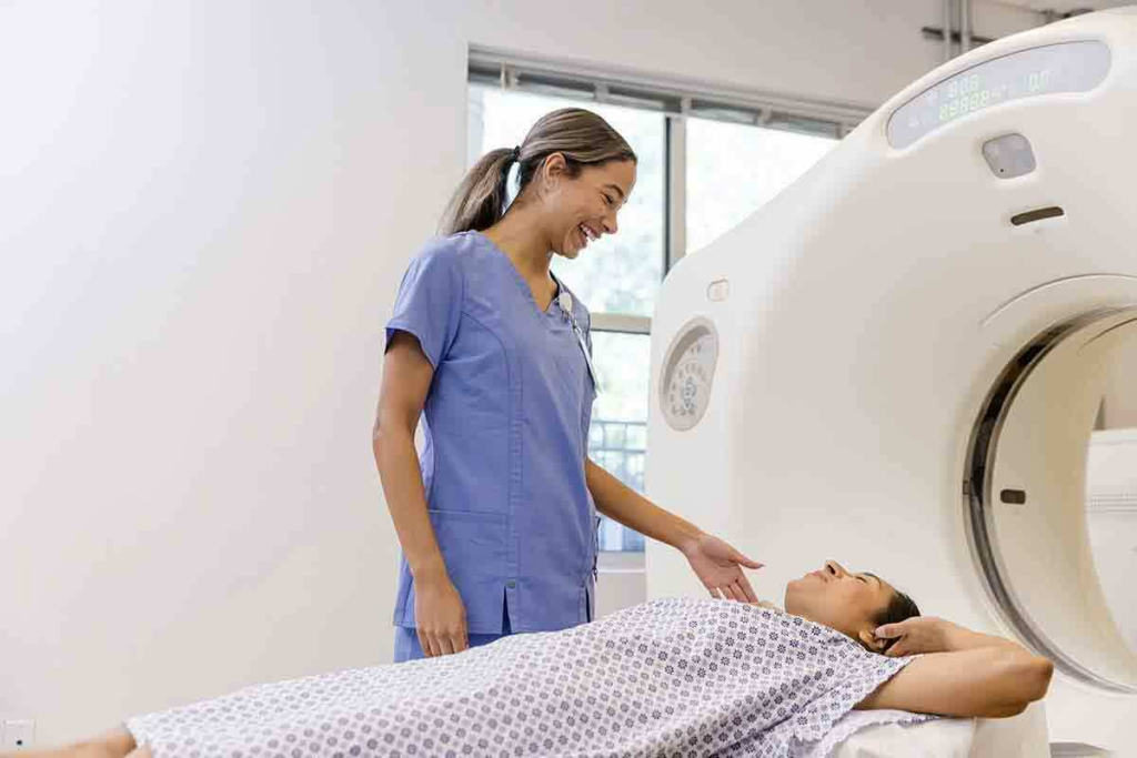
Knowing what to expect during a CT scan can really help you feel more at ease. We get that medical tests can be scary. That’s why we’re here to help you understand what’s going to happen.
How you prepare for a CT scan depends on the type. You might need to fast or avoid certain medicines. It’s very important to listen to your doctor’s instructions to make sure the scan goes smoothly.
If you’re getting a CT scan with contrast, you might need to arrive early. This could be an injection or something you drink, depending on the scan. We’ll tell you what you need to do ahead of time.
During the scan, you’ll lie on a table that moves through a big machine. The scan itself is usually very quick, taking just a few minutes. You might need to hold your breath for a bit to get clear pictures.
Contrast media helps show important details in your body. If you get contrast, you might feel a slight pinch when it’s given. Our team is trained to make this as comfortable as possible.
Understanding the CT scan process can really help reduce your stress. If you have any questions or worries, just ask your doctor. We’re here to support you every step of the way.
CT technology has changed medical diagnostics worldwide. But, its reach is not the same everywhere. This leads to differences in healthcare access.
CT scanners are more common in rich countries. Developed nations have a higher density of CT scanners. This means more people can get advanced tests. But, poor countries struggle to get and keep CT tech because of money and infrastructure issues.
There are efforts to help. International groups and projects are working to bring more medical imaging to areas that need it most.
Buying and running CT scanners costs a lot. Healthcare policies play a big role in who gets CT scans. How much money is available and how it’s spent affects CT use.
Leaders need to find a balance. They must make sure CT tech is used well and fairly, considering the costs of healthcare.
Looking back, CT scanning technology has made a big impact on healthcare. It’s set to keep growing with new research and tech. We’re looking forward to better image quality, lower doses, and more accurate diagnoses.
These improvements will help doctors find and treat diseases sooner and more accurately. As CT scans get cheaper and more available, they’ll help more people around the world. We’re excited for a future where CT scans are even more precise, safe, and helpful.
CT scanning will play an even bigger role in medicine as it evolves. It will help in many medical fields, making it a key part of modern healthcare. As we progress, we must keep focusing on safety and care for patients. This way, CT scans will always be a valuable tool in healthcare.
A CT (Computed Tomography) scan is a medical test. It makes detailed images of the body’s inside. It uses computer-processed X-rays.
Godfrey Hounsfield and Allan Cormack invented the CT scanner. They won the Nobel Prize in 1979 for their work.
CT scans show detailed 3D images. Traditional X-rays give 2D images. CT scans use many X-rays to create detailed images.
CT scans help find tumors, check for injuries, and diagnose vascular diseases. They also guide medical procedures.
A CT scan uses an X-ray source and detector. A computer then makes detailed images of the body’s inside.
A single-slice scanner takes one image at a time. Multi-detector scanners take many images at once. This makes scans faster and clearer.
A CT scan’s time varies. It depends on the scan type and area being checked. Most scans take a few minutes.
Preparation for a CT scan varies. Some scans need contrast media or fasting. Always follow the doctor’s instructions.
Contrast media make certain body parts or lesions more visible. This helps doctors make more accurate diagnoses.
CT scans use X-rays, but they are generally safe. The benefits of getting a clear diagnosis are worth it. New technologies also reduce radiation.
CT technology has grown a lot. From single-slice to multi-detector scanners, it’s faster and safer now. New technologies also help reduce radiation.
Computed axial tomography (CAT) is another name for CT scanning. It uses computer processing to create images from X-rays.
The first clinical CT scan was in 1971. It was a big step in medical imaging technology.