
Discover which chamber of heart receives oxygenated blood and its role in circulation.
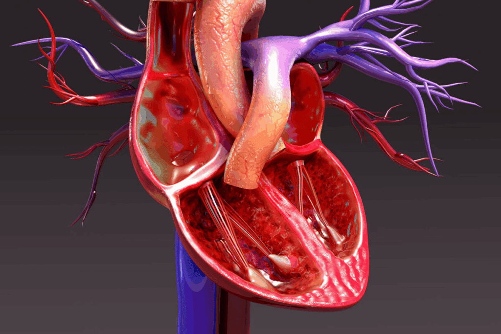
The human heart is a complex organ that plays a vital role in maintaining life. It has four chambers: two atria and two ventricles. Knowing how these chambers work together is key to understanding life-sustaining circulation.
The left atrium is one of the heart’s chambers. Its main job is to get oxygen-rich blood from the lungs through the pulmonary veins. This blood then goes to the left ventricle, which pumps it to the rest of the body.
At Liv Hospital, we help you learn about the left atrium’s role in the circulatory system. Our team of experts is committed to providing top-notch healthcare. We offer full support for international patients.
Understanding the heart’s structure is key to knowing how it keeps us healthy. The heart is a complex organ that is vital for our circulatory system. Its anatomy helps us see how it functions.
The heart has four chambers: the right and left atria, and the right and left ventricles. These chambers work together to make sure blood flows right through the heart and out to the body. The atria receive blood returning to the heart, while the ventricles pump blood out.
The four chambers of the heart work together to help blood flow. Here’s how they function:
This detailed process makes sure oxygenated blood gets to the body’s tissues. It also sends deoxygenated blood to the lungs for oxygen.

Learning how blood gets oxygen is essential to understanding the heart and blood system. Blood gets oxygen in the lungs. There, it picks up oxygen and drops off carbon dioxide through gas exchange.
The lungs make deoxygenated blood oxygen-rich. This starts when deoxygenated blood, full of carbon dioxide, comes from the right ventricle to the lungs. In the lungs, gas exchange happens.
Here, carbon dioxide leaves, and oxygen goes into the blood. This oxygen-rich blood then goes back to the heart through the pulmonary veins into the left atrium.
The alveoli’s special structure helps with gas exchange. They have lots of blood capillaries close to the air. This makes gas exchange very efficient.
Oxygen-rich blood goes from the lungs to the heart and then to the body. On the other hand, deoxygenated blood, full of carbon dioxide, goes back to the heart to get oxygen. The left ventricle sends oxygen-rich blood to the body.
The right ventricle sends deoxygenated blood to the lungs. It’s important to know the difference to understand how the heart works. The heart’s chambers keep oxygen-rich and deoxygenated blood separate.
The left atrium is the heart chamber that gets oxygen-rich blood from the lungs. This is key for keeping the body’s oxygen levels up. We’ll dive into its anatomy and role in more detail.
The left atrium is one of the heart’s four chambers, found on the upper left. It has a muscular pouch called the left auricle, which helps it hold more blood. The left atrium gets oxygen-rich blood from the lungs through four pulmonary veins.
The left atrium’s walls are thinner than the ventricles. This is because it doesn’t need to pump blood as hard. Yet, they are strong enough for efficient blood transfer.
The left atrium gets oxygen-rich blood from the lungs via the pulmonary veins. There are four pulmonary veins, two from each lung, that empty into it. This blood then moves to the left ventricle through the mitral valve.
The process can be summarized in the following table:
| Structure | Function |
| Pulmonary Veins | Carry oxygenated blood from the lungs to the left atrium |
| Left Atrium | Receives oxygenated blood from the pulmonary veins and pumps it into the left ventricle |
| Mitral Valve | Allows blood to flow from the left atrium into the left ventricle while preventing backflow |
Understanding the left atrium’s role in getting oxygenated blood is key. It’s a vital part of the circulatory process. The left atrium’s function ensures oxygen-rich blood is spread throughout the body.
Oxygenated blood returns to the heart in a specific path. This path is key for delivering oxygen to the body’s tissues and organs.
Oxygen-rich blood comes back from the lungs through the pulmonary veins to the left atrium. This is the first step in the heart’s journey. The left atrium holds the blood until it’s ready to move on.
The left atrium sends oxygenated blood to the left ventricle through the mitral valve. The left ventricle is strong and muscular. It’s made to pump blood to the body.
Key steps in this process include:
The left ventricle pumps blood into the aorta, the biggest artery. The aorta sends it to the rest of the body. This step is vital for oxygen to reach everywhere.
We can summarize the path of oxygenated blood as follows:
The pulmonary veins carry oxygen-rich blood from the lungs to the heart. They play a special role in the circulatory system. This is different from other veins that carry blood without oxygen.
The pulmonary veins are built for efficient blood flow back to the heart. There are four of them, two from each lung, that send oxygenated blood to the left atrium. Their job is key to keeping the body’s tissues oxygenated.
The anatomy of pulmonary veins is designed for their task. Their walls are thinner than other veins but strong enough for the job.
Pulmonary veins are the only veins carrying oxygenated blood. This makes them unique in the circulatory system. Unlike most veins, which carry blood without oxygen, pulmonary veins carry blood that’s been oxygenated in the lungs.
This special function is essential for blood circulation. Without pulmonary veins, oxygenated blood wouldn’t return to the heart. This would stop oxygen from reaching the body’s tissues and organs.
The heart’s right side deals with deoxygenated blood from the body. This is key for keeping the body’s oxygen levels up. We’ll look at how the right side of the heart processes this blood.
The right atrium is the first stop for deoxygenated blood when it returns to the heart. It gets blood from the body through the superior vena cava and the inferior vena cava. Then, it pumps this blood into the right ventricle through the tricuspid valve.
The chamber on the right bottom side of the heart is the right ventricle. It’s a muscular chamber that sends deoxygenated blood to the lungs. The right ventricle gets blood from the right atrium and then sends it through the pulmonary valve into the pulmonary artery, which takes it to the lungs.
The right ventricle is built to handle the pressure needed to pump blood to the lungs. Though important, it needs less pressure than the left ventricle, which pumps blood to the whole body.
Deoxygenated blood goes from the right atrium to the right ventricle, then to the lungs via the pulmonary artery. In the lungs, it picks up oxygen and releases carbon dioxide through gas exchange. The now-oxygenated blood returns to the heart through the pulmonary veins into the left atrium, ready to be sent to the body.
The journey of deoxygenated blood to the lungs is essential for the circulatory system. It ensures blood is oxygenated and ready for the body. The right side of the heart, mainly the right ventricle, is key in this process.
The left and right chambers of the heart are different in structure and function. This difference is mainly because of the oxygen content in the blood they handle. Knowing these differences helps us understand how the heart works and its role in our body.
The left and right chambers of the heart have unique features. The left ventricle, for example, has a thicker wall than the right ventricle. This is because it needs to pump blood with oxygen to the whole body, which requires more strength.
Key structural differences include:
The left and right chambers of the heart have different jobs. The left chambers get oxygen-rich blood from the lungs and send it to the body. The right chambers get oxygen-poor blood from the body and send it to the lungs to get oxygen.
| Chamber | Function | Blood Oxygen Content |
| Left Atrium | Receives oxygenated blood from lungs | Oxygenated |
| Left Ventricle | Pumps oxygenated blood to body | Oxygenated |
| Right Atrium | Receives deoxygenated blood from body | Deoxygenated |
| Right Ventricle | Pumps deoxygenated blood to lungs | Deoxygenated |
The oxygen content in the blood changes between the left and right chambers of the heart. The left chambers have oxygen-rich blood, while the right chambers have oxygen-poor blood. This difference is key to the heart’s efficiency in circulating blood.
The left ventricle gets oxygen-rich blood from the left atrium and sends it to the body. The right ventricle gets oxygen-poor blood from the right atrium and sends it to the lungs. This shows how the heart keeps blood flowing efficiently through the body.
The heart is key in the circulatory loop, moving blood between the lungs and the body. This process is vital for delivering oxygen to tissues and bringing deoxygenated blood back to the lungs.
Pulmonary circulation is the path blood takes from the heart to the lungs and back. The right ventricle sends deoxygenated blood to the lungs through the pulmonary arteries. In the lungs, blood gets oxygen and loses carbon dioxide through gas exchange.
The oxygen-rich blood then returns to the heart via the pulmonary veins, entering the left atrium.
Systemic circulation is the path of oxygenated blood from the heart to the body and back. The left ventricle is key, pumping oxygenated blood to the body through the aorta and its branches. As this blood reaches the body’s tissues, it releases oxygen and picks up carbon dioxide, becoming deoxygenated.
This deoxygenated blood then returns to the heart through the venous system, entering the right atrium.
The integration of pulmonary and systemic circulation ensures a continuous supply of oxygenated blood to the body’s tissues. We rely on this dual circulation system to maintain the delicate balance of oxygen and carbon dioxide in the body. The heart, with its four chambers, works tirelessly to facilitate this process, making it a vital organ for sustaining life.
In summary, the complete circulatory loop is a vital process that involves the coordinated effort of the heart, lungs, and blood vessels. Understanding this complex system helps us appreciate the intricacies that keep us alive and functioning.
The health of heart chambers is key to heart health. At Liv Hospital, we focus on the best global medical practices. We understand the heart’s structure and function are vital for well-being.
Disorders in the heart chambers can greatly affect health. This is why we pay close attention to them.
The left atrium is important for getting oxygen-rich blood from the lungs. Problems like atrial fibrillation can harm its function. Atrial fibrillation causes an irregular and fast heart rate, making it hard for the left atrium to pump blood.
We know treating atrial fibrillation is key to prevent complications. It helps improve patient health.
Left atrial dysfunction can affect blood oxygen levels. When the left atrium doesn’t pump well, less oxygen is delivered to the body. This can harm many bodily functions and overall health.
At Liv Hospital, we focus on diagnosing and treating left atrial dysfunction. This helps keep blood oxygen levels up and improves heart health.
Accurate diagnosis is key for checking heart chamber function. Tools like echocardiography and cardiac MRI are very helpful. They let doctors see the heart’s structure and function, spotting problems and assessing their severity.
We use the latest diagnostic technologies for our patients. This ensures they get the right treatment for their conditions.
Understanding heart chamber function and using advanced diagnostics can greatly improve patient outcomes. It helps keep the heart healthy.
We often get things wrong about how the heart works, like the flow of blood. Knowing how the heart is built and works is key to fixing these mistakes.
Many think oxygenated and deoxygenated blood mix in the heart. But, the heart’s wall keeps them apart. This wall, called the septum, stops the mixing of these two blood types.
The left side of the heart, including both the left atrium and left ventricle, contains oxygenated blood. On the other hand, the right side, with the right atrium and right ventricle, holds deoxygenated blood. This separation is key for delivering oxygen to the body’s tissues.
Some think the right side of the heart has oxygenated blood. But, it’s actually the left side. The left atrium gets oxygen-rich blood from the lungs. This blood then goes to the left ventricle, which pumps it to the body.
To show how it works, let’s look at a table:
| Heart Chamber | Type of Blood | Source/ Destination |
| Left Atrium | Oxygenated | Receives from lungs via pulmonary veins |
| Left Ventricle | Oxygenated | Pumps to the body |
| Right Atrium | Deoxygenated | Receives from the body via superior and inferior vena cava |
| Right Ventricle | Deoxygenated | Pumps to the lungs |
The table shows the left side deals with oxygenated blood, and the right side with deoxygenated blood. This division is vital for the heart’s job and our health.
“The heart’s ability to separate oxygenated and deoxygenated blood is key for our health.”
— A Cardiologist
In summary, knowing how the heart works helps clear up blood flow myths. By understanding the left side has oxygenated blood and the heart’s wall keeps blood types separate, we see how complex and efficient our circulatory system is.
Knowing how the heart works is key to understanding the circulatory system. The left atrium is the heart chamber that gets oxygen-rich blood from the lungs. At Liv Hospital, we focus on top-notch healthcare for international patients. We make sure everyone gets the best care for their heart health.
The heart has four chambers that work together to move blood. The left atrium is important because it gets the oxygen-rich blood. Learning about this helps us see why heart health is so critical.
We’re dedicated to giving the best care to patients worldwide. We make sure each person gets care that fits their needs. Our goal is to improve lives with our advanced medical skills and caring approach.
The left atrium gets oxygen-rich blood from the lungs through the pulmonary veins.
The left atrium is key. It takes in oxygen-rich blood from the lungs and sends it to the left ventricle.
The right ventricle holds deoxygenated blood. It pumps this blood to the lungs for oxygen.
The pulmonary veins carry oxygen-rich blood from the lungs to the heart. They go straight to the left atrium.
The left atrium gets oxygen-rich blood from the lungs through the pulmonary veins.
The right ventricle sends deoxygenated blood to the lungs for oxygenation.
The left side of the heart, including the left atrium and ventricle, holds oxygenated blood.
Pulmonary veins carry oxygen-rich blood from the lungs to the heart. They send it to the left atrium.
Oxygenated blood comes back from the lungs to the left atrium via the pulmonary veins. It then goes to the left ventricle and is pumped to the aorta. From there, it spreads to the body.
Oxygenated blood is full of oxygen and goes from the lungs to the body’s tissues. Deoxygenated blood, on the other hand, is low in oxygen and high in carbon dioxide. It returns from the body to the lungs for oxygen.
The left atrium gets blood returning from the lungs through the pulmonary veins.
Whiteman, S., Saker, E., Courant, V., Salandy, S., Gielecki, J., & Loukas, M. (2019). An anatomical review of the left atrium. Translational Research in Anatomy, 17, Article 100052. https://www.sciencedirect.com/science/article/pii/S2214854X19300512
Jain, V. (2023). Physiology, pulmonary circulatory system. In NCBI Bookshelf. National Center for Biotechnology Information. https://www.ncbi.nlm.nih.gov/books/NBK525948/