
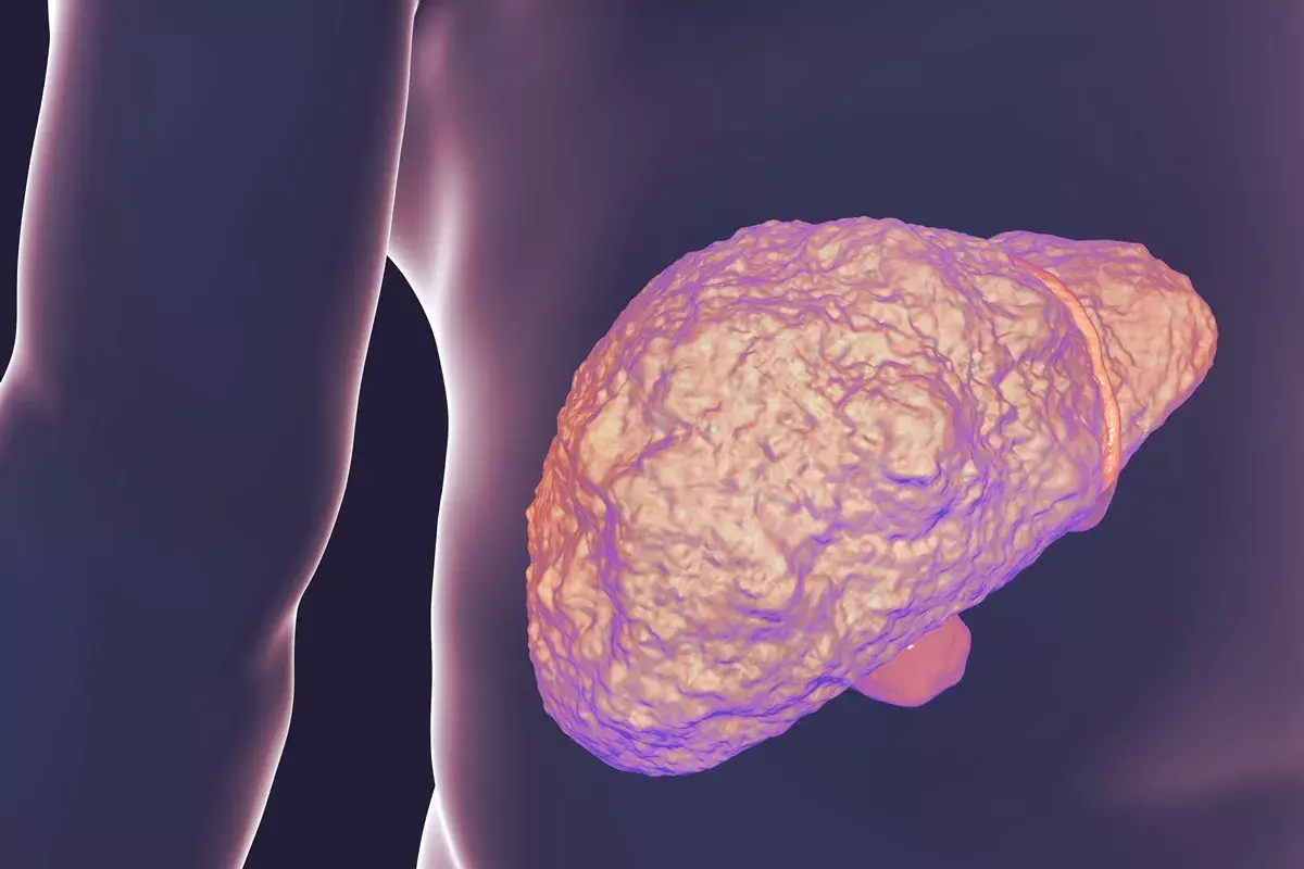
Knowing about the colon anatomy is key for doctors and patients. The colon is a big part of our digestive system. It’s about 5 to 6 feet long and helps absorb water and salts from food.
The colon has four main parts: the ascending, transverse, descending, and sigmoid colon. Pictures, diagrams, and images help us understand its complex structure and role.
At Liv Hospital, we offer reliable medical info and expert insights. We provide top-notch imaging and help with diagnosis.
Key Takeaways
- The colon is approximately 5 to 6 feet long.
- It is divided into four main anatomical sections.
- Understanding colon anatomy is vital for diagnosing gastrointestinal disorders.
- Visual guides and images aid in comprehending the colon’s structure.
- Liv Hospital provides high-quality imaging and diagnostic support.
The Colon in the Digestive System

It’s important to know how the colon works in our body. The colon is part of the large intestine. It helps absorb water and salts from what we can’t digest. It also stores waste until we can get rid of it.
Position Within the Gastrointestinal Tract
The colon is a key part of our digestive system. It goes from the cecum to the rectum. It sits in the belly, around the small intestine. This spot helps it do its job well.
Relationship to Surrounding Organs
The colon is close to other important organs. For example, the ascending colon is on the right side, near the liver. The transverse colon is in the upper belly, near the stomach and pancreas. Knowing these connections helps doctors find and treat problems.
Section of Colon | Location | Adjacent Organs |
Ascending Colon | Right side of abdomen | Liver |
Transverse Colon | Upper abdomen | Stomach, Pancreas |
Descending Colon | Left side of abdomen | Left kidney, Spleen |
Sigmoid Colon | Left lower quadrant | Rectum |
Understanding where the colon is and its connections to other organs helps us see its role. This knowledge is key for doctors and anyone wanting to know more about their health.
Understanding Colon Anatomy

The human colon is a key part of our digestive system, about 5 feet long. It’s a big part of the large intestine and is vital for our health.
Size and Dimensions
The colon is roughly 5 feet (150 cm) long and has five main parts: cecum, ascending colon, transverse colon, descending colon, and sigmoid colon. The colon’s size and dimensions are critical to its function, allowing it to perform its various roles in the digestive process.
On average, the colon is 5 to 6 feet long. This length helps it absorb water and electrolytes from waste.
General Structure and U-shaped Configuration
The colon’s shape is U-shaped, encircling the small intestine in the belly. This shape helps it absorb nutrients and move waste.
Medical images like colonoscopy and CT colonography show the colon’s U-shaped form.
- The colon’s U-shaped configuration allows it to frame the small intestine.
- This configuration is key for absorbing water and electrolytes.
- The colon’s structure fits its role in digestion.
Knowing about the colon’s anatomy is important for doctors and those interested in digestive health.
The Four Main Sections of the Colon
Knowing the four main parts of the colon is key to understanding its role. The colon is a vital part of our digestive system. It’s divided into sections, with the main ones being the ascending, transverse, descending, and sigmoid colon. Each part has its own role in helping the colon function properly.
Ascending Colon
The ascending colon is the first part of the colon. It’s on the right side of the abdomen. It’s about 15-20 cm long. This part of the colon is important for absorbing water and salts from the small intestine.
Transverse Colon
The transverse colon is the second part. It crosses the upper abdomen from right to left. It’s over 18 inches long. This section absorbs water and salts and is home to many types of bacteria.
Descending Colon
The descending colon is on the left side of the abdomen. It’s about 25-30 cm long. It helps make the stool more solid by absorbing more water and salts.
Sigmoid Colon
The sigmoid colon is the last part before the rectum. It has an S-shaped configuration in the left lower quadrant. It’s about 25-40 cm long. This part stores stool until it’s ready to be eliminated.
In summary, the four main sections of the colon work together to help digest food. Understanding these sections is important for diagnosing and treating colon-related issues.
Detailed Examination of the Ascending Colon
The ascending colon is a key part of the large intestine. It moves fecal matter upwards towards the transverse colon. It plays a vital role in the digestive process.
Anatomical Position on the Right Side
The ascending colon is on the right side of the abdomen. It goes from the cecum to the hepatic flexure. This position helps it move fecal matter upwards.
Connection to the Cecum and Ileocecal Valve
The ascending colon connects directly to the cecum, the start of the large intestine. The ileocecal valve controls the flow from the small intestine to the cecum. This valve is key to keeping the digestive process smooth.
Hepatic Flexure Junction Point
The hepatic flexure is where the ascending colon meets the transverse colon. It’s near the liver and is a key bend in the colon. This spot marks the transition from the ascending to the transverse colon.
The ascending colon’s main features are:
- Located on the right side of the abdomen
- Connected to the cecum and ileocecal valve
- Transports fecal matter upwards to the transverse colon
- Junctions with the transverse colon at the hepatic flexure
Knowing these details about the ascending colon is essential for understanding colon anatomy and its importance.
The Transverse Colon: Horizontal Pathway
The transverse colon is unique because it runs horizontally. It’s a key part of the digestive system. It helps move waste from the right to the left side of the abdomen.
Path Across the Upper Abdomen
The transverse colon runs horizontally across the upper abdomen. It’s held by the transverse mesocolon, which lets it move freely. This is important for the body’s movements and changes.
As it carries feces transversely from right to left, it’s near many important organs. Its location means it can be affected by or affect these organs.
Relationship to the Stomach and Pancreas
The transverse colon is near the stomach and pancreas. It’s below the stomach and sometimes touches it. The pancreas, behind the stomach, is also close, near the splenic flexure.
This close relationship is key to understanding some diseases. Problems with the stomach or pancreas can affect the transverse colon, and vice versa.
Splenic Flexure Junction Point3>
The transverse colon ends at the splenic flexure, a sharp bend. This is where it meets the descending colon. It’s near the spleen and is a key landmark. The splenic flexure is a common place for colon problems like diverticula and tumors.
Knowing the anatomy of the transverse colon is vital for diagnosing and treating gut issues.
- The transverse colon is a key component of the large intestine.
- It runs horizontally across the upper abdomen.
- Its position is closely related to the stomach and pancreas.
- The splenic flexure marks the end of the transverse colon.
The Descending Colon: Left-Side Anatomy
The descending colon is a key part of the large intestine. It runs down the left side of the abdomen. It helps move feces to the sigmoid colon and rectum.
Vertical Path Down the Left Abdomen
The descending colon starts near the spleen and goes down the left side of the abdomen. This path is important for moving feces to the sigmoid colon.
Relationship to the Left Kidney and Spleen
The descending colon is close to the left kidney and spleen. The splenic flexure, where the transverse colon meets the descending colon, is near the spleen. This is important for understanding the colon’s position in the abdomen.
“The colon’s anatomy is complex, with various sections working together to facilitate digestion and waste elimination.”
— Medical Anatomy Guide
Junction with the Sigmoid Colon
The descending colon meets the sigmoid colon, an S-shaped part of the colon, in the left lower quadrant. This is a key point as it marks the start of the sigmoid colon’s journey to the rectum.
Colon Section | Length | Key Features |
Descending Colon | 25-30 cm | Vertical path down the left abdomen, related to the left kidney and spleen |
Sigmoid Colon | 25-40 cm | S-shaped, located in the left lower quadrant |
Knowing about the descending colon’s anatomy is key to understanding the colon’s function. Its connection to other organs and its role in digestion make it a vital area of study in gut health.
The Sigmoid Colon’s S-Shaped Structure
The sigmoid colon is a key part of the large intestine. It has a unique S-shaped structure. This part of the colon stores feces until they are ready to leave the body. We will look at its anatomy and function.
Distinctive S-Shape in the Left Lower Quadrant
The sigmoid colon is in the left lower part of the abdomen. Its S-shape helps it store feces well. This shape is a key part of its anatomy, helping it do its job in digestion.
Mobility and Variation in Position
The sigmoid colon is quite mobile and can change its position. This flexibility is important for its job. It allows for the movement and storage of feces. Its mobility also affects its role in health issues.
Rectosigmoid Junction
The rectosigmoid junction is where the sigmoid colon meets the rectum. It’s a key area for understanding the health of the colon. This junction is often where health problems occur. Knowing about this area is vital for diagnosing and treating colon issues.
The sigmoid colon’s anatomy includes:
- S-shaped structure in the left lower quadrant
- Mobility and variation in position
- Transition to the rectum at the rectosigmoid junction
Understanding the sigmoid colon’s S-shape and its features helps us see its role in digestion. Pictures and images of the sigmoid colon can help us visualize its complex anatomy and function.
Histological Structure of the Colon Wall
Knowing how the colon wall is structured is key to understanding its role in digestion. The colon wall has several layers, each with its own job. These jobs help keep the digestive system healthy.
Mucosa Layer with Simple Columnar Epithelium
The mucosa is the innermost layer of the colon. It touches the contents inside the colon. This layer is covered in simple columnar epithelium, made up of colonocytes and goblet cells.
Colonocytes help absorb water and salts. Goblet cells make mucus, which helps feces move smoothly and protects the lining.
The mucosa also has intestinal crypts. These are like tubes that go into the layer below. They are filled with stem cells that keep the lining of the mucosa fresh.
Submucosa Layer
Under the mucosa is the submucosa. It’s a layer of loose tissue that helps the colon stay flexible. It has lots of blood vessels, lymphatic vessels, and nerves. These help the mucosa work well.
Muscularis Layer
The muscularis layer has two types of smooth muscle. The inner layer goes around in a circle, and the outer layer stretches out. When they work together, they help move things through the colon.
This layer is key for mixing and pushing feces along.
Serosa Layer
The serosa is the outermost layer. It’s a thin layer of cells that covers the outside of the colon. It makes a fluid that helps the colon move smoothly.
In summary, the colon wall is made up of many layers, each with its own important job. Knowing about these layers helps us understand how the colon works. It also helps us diagnose and treat digestive problems.
Blood Supply and Innervation of the Colon
It’s important to know how the colon gets its blood and nerves. This helps it do its job of absorbing water and getting rid of waste. The colon’s blood and nerve systems are key to its work.
Arterial Supply: Superior and Inferior Mesenteric Arteries
The colon gets its blood from two main arteries. The superior mesenteric artery feeds the right side, like the cecum and ascending colon. The inferior mesenteric artery takes care of the left side, including the sigmoid colon and rectum.
The superior mesenteric artery has branches for different parts of the colon. The inferior mesenteric artery also branches, supplying the descending and sigmoid colon.
Venous Drainage System
The colon’s veins work in a similar way to its arteries. The right side’s veins go to the superior mesenteric vein. The left side’s veins go to the inferior mesenteric vein. These veins then join to form the portal vein, which goes to the liver.
Lymphatic Drainage
The lymph system is important for the colon’s health. It helps fight off infections. Lymph from the colon goes to epicolic and paracolic lymph nodes. Then it moves to intermediate and principal lymph nodes and ends at preaortic lymph nodes near the aorta.
Autonomic Nerve Supply
The colon has nerves from both the sympathetic and parasympathetic systems. The sympathetic innervation comes from the lumbar spine. The parasympathetic innervation comes from the vagus nerve for the right side and the pelvic splanchnic nerves for the left. These nerves help control the colon’s movements and blood flow.
In summary, the blood and nerve systems of the colon are complex and essential. Knowing about them helps us understand and treat colon problems.
Clinical Significance of Colon Anatomy
The importance of colon anatomy in healthcare is huge. It helps us diagnose and treat colon problems better. Knowing the colon’s details is key for doctors to care for patients with gut issues.
Common Pathologies and Their Anatomical Basis
Diseases like inflammatory bowel disease (IBD) and colon cancer affect the intestines. The colon’s shape and structure are important in how these diseases develop and grow.
The ascending colon is often where colon cancer starts. Knowing its anatomy helps doctors find and treat it early. The sigmoid colon‘s shape makes it more likely to get diseases like diverticulitis.
Pathology | Anatomical Location | Clinical Significance |
Colon Cancer | Ascending Colon, Sigmoid Colon | Early detection is key for treatment |
Diverticulitis | Sigmoid Colon | Proper management is important to avoid complications |
Inflammatory Bowel Disease (IBD) | Various sections of the Colon | Needs ongoing management due to chronic nature |
Surgical Considerations Based on Anatomy
When it comes to surgery for colon problems, knowing the colon’s anatomy is vital. Surgeons must understand how different parts of the colon and nearby structures relate. This helps them plan surgeries well.
“A thorough understanding of colon anatomy is essential for surgeons to navigate the complexities of colon surgery and achieve optimal outcomes.”
— Surgical Gastroenterology Expert
For example, during a colectomy, surgeons must find and keep the blood vessels that feed the colon. This is to avoid surgery problems.
Knowing how colon anatomy affects health is critical for treating gut diseases. By understanding the anatomy of common diseases and the surgery needed, doctors can give better care to patients.
Visualizing the Colon: Modern Imaging Techniques
Modern imaging techniques have changed how we see the colon. They help us make more accurate diagnoses and treatments. These new tools let us spot problems and understand the colon’s complex structure better.
Colonoscopy: Direct Visualization
Colonoscopy lets us see inside the colon directly. A flexible tube with a camera and light is inserted into the rectum. This method finds polyps, cancer, and other issues. It also takes tissue samples for biopsies.
CT Colonography (Virtual Colonoscopy)
CT colonography, or virtual colonoscopy, uses X-rays and computers to show the colon’s details. It’s good for those who can’t have a regular colonoscopy. It spots polyps and other growths well.
MRI and Other Advanced Imaging Methods
Magnetic Resonance Imaging (MRI) is another way to see the colon. It shows the colon’s structure and the tissues around it. MRI helps diagnose many conditions. Other methods like ultrasound and PET scans are used too, for specific cases.
Normal vs. Abnormal Findings in Images
It’s important to know the difference between normal and abnormal colon images. Normal images show a smooth colon lining. Abnormal images might show polyps, inflammation, or other issues. Experts need to understand these images well to make accurate diagnoses.
Imaging Technique | Key Features | Clinical Applications |
Colonoscopy | Direct visualization, ability to biopsy | Polyp detection, cancer screening, diagnosis of gastrointestinal symptoms |
CT Colonography | Non-invasive, detailed 3D images | Polyp detection, cancer screening, for those who can’t have traditional colonoscopy |
MRI | Detailed soft tissue imaging, no radiation | Assessment of the colon and surrounding structures, diagnosis of various gastrointestinal conditions |
Conclusion
Knowing about colon anatomy is key for spotting and treating stomach problems. We’ve looked closely at the colon’s structure, its layers, and why it matters in health.
The colon is a big part of our digestive system. Its shape and layers are important for doctors and patients to know. We’ve covered the main parts of the colon: the ascending, transverse, descending, and sigmoid colon.
Understanding the colon helps doctors make better diagnoses and treatments. As we wrap up our look at colon anatomy, it’s clear that knowing it well is essential for better health care.
FAQ
What is the colon’s role in the digestive system?
The colon, or large intestine, absorbs water and electrolytes. It’s key to the digestive system.
What are the four main sections of the colon?
The colon has four main parts: the ascending, transverse, descending, and sigmoid colon. Each part has its own role.
Where is the ascending colon located?
The ascending colon is on the right side of the abdomen. It connects to the cecum and ileocecal valve.
What is the function of the transverse colon?
The transverse colon crosses the upper abdomen. It’s linked to the stomach and pancreas.
What is the sigmoid colon’s distinctive feature?
The sigmoid colon is S-shaped in the left lower quadrant. It’s mobile and can vary in position.
What is the histological structure of the colon wall?
The colon wall has layers like the mucosa, submucosa, muscularis, and serosa. Each layer is important for the colon’s function.
What is the blood supply to the colon?
The colon gets blood from the superior and inferior mesenteric arteries.
Why is understanding colon anatomy important?
Knowing colon anatomy helps diagnose and treat diseases like inflammatory bowel disease and colon cancer.
What imaging techniques are used to visualize the colon?
Colonoscopy, CT colonography, and MRI are used to see the colon. They help find problems and diagnose conditions.
What is the significance of the rectosigmoid junction?
The rectosigmoid junction is where the sigmoid colon meets the rectum. It’s key for understanding colorectal health.
Can you show a picture of the human colon and intestines?
We have diagrams and images of the colon and intestines. They help show its anatomy and structure.
What does the colon look like in the human body?
The colon is part of the large intestine, about 5 to 6 feet long. It forms a U-shape around the small intestine in the abdomen.
Are there any resources available to understand colon anatomy better?
We have detailed diagrams, pictures, and explanations. They help you understand colon anatomy and its role in digestion.
References:
National Health Service (NHS). Colon Anatomy: Structure and Function. Retrieved from https://www.nhs.uk/conditions/digestive-system/large-intestine/


