
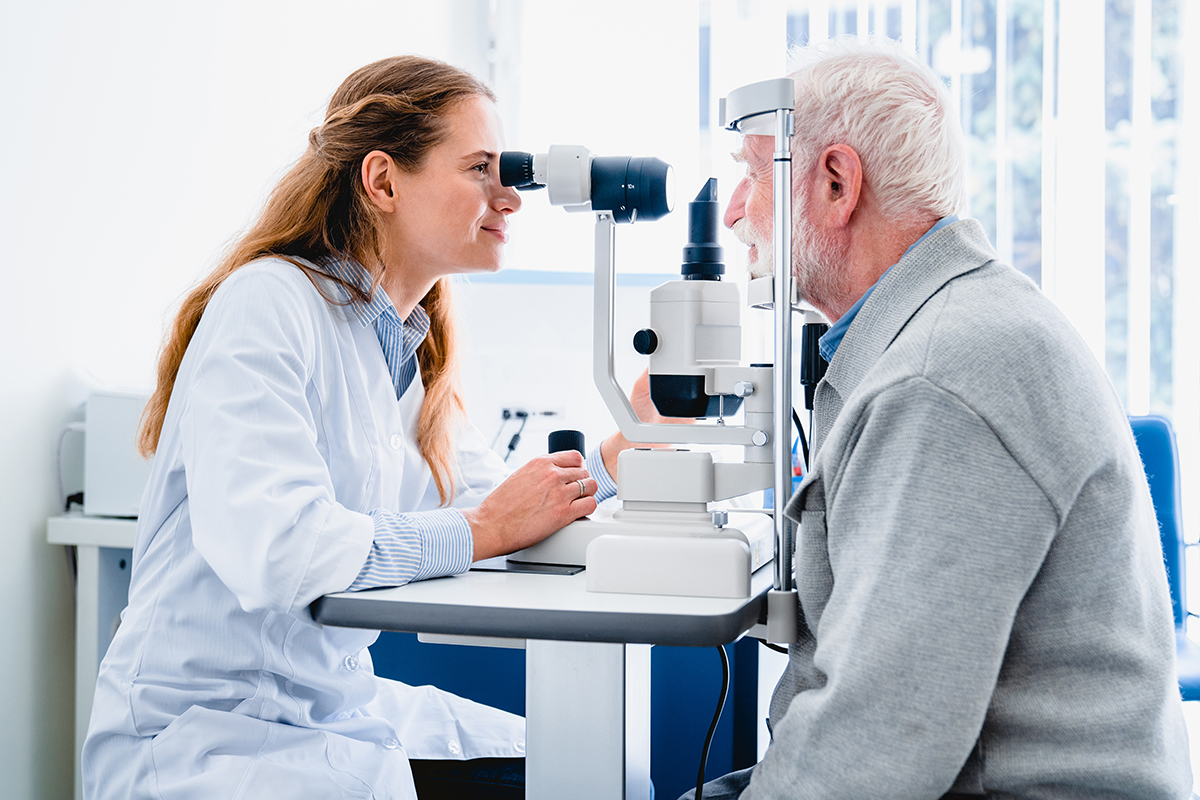
Testing cranial nerves 3, 4, and 6 is key for checking how well the nerves work. These nerves help us move our eyes. If they don’t work right, it could mean there’s a problem in the brain or along the nerve path. Learn the essential cranial nerve 3 test. This guide details how to assess the oculomotor nerve, pupil response, and eye movements.
We will look at why testing these nerves is important. We’ll also give a detailed guide on how to do these tests. The oculomotor, trochlear, and abducens nerves control our eye muscles and movement. This is why they are important for finding and treating neurological problems.
Knowing how to test these nerves helps doctors find and treat problems early. It’s important to check these nerves well to catch serious health issues early.
Key Takeaways
- Testing cranial nerves 3, 4, and 6 is essential for neurological examination.
- These nerves control extraocular muscles and eye movement.
- Abnormalities in their function can indicate specific pathologies.
- A systematic clinical examination is necessary for accurate assessment.
- Understanding the proper testing methodology is important for healthcare professionals.
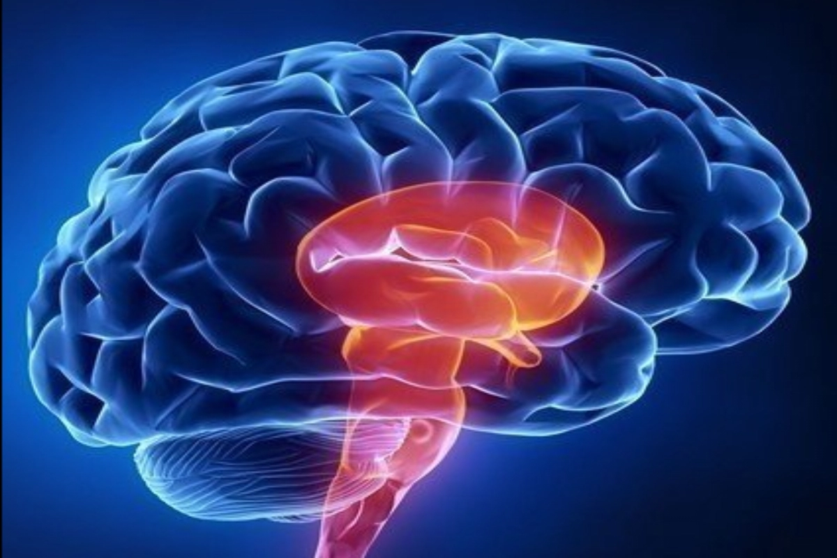
Understanding Cranial Nerves 3, 4, and 6

Testing cranial nerves 3, 4, and 6 is key in checking eye movements. These nerves, or oculomotor, trochlear, and abducens, help our eyes move in many ways. Knowing how they work helps doctors find and treat brain problems.
The Role of Ocular Cranial Nerves in Neurological Assessment
The oculomotor (CN III), trochlear (CN IV), and abducens (CN VI) nerves control eye muscles. The oculomotor nerve helps most eye movements. The trochlear nerve controls eye tilt and depression. The abducens nerve helps eyes move outward.
Clinical Significance of Proper Testing
Testing these nerves is important for finding brain problems. Eye movement issues can show signs of stroke, multiple sclerosis, or brain tumors. Doctors use these tests to find where problems are and what they are.
Experts say, “Checking cranial nerves III, IV, and VI is very important. It tells us a lot about the brain and its connections.”
Anatomy and Function of the Oculomotor Nerve (CN3)
The oculomotor nerve, or CN3, is key in controlling eye movements and other important functions. It is one of the three cranial nerves that move the eyeball.
Muscles Innervated by CN3
The oculomotor nerve controls several muscles. These include the superior rectus, medial rectus, inferior rectus, and inferior oblique muscles. Together, they help the eyes move in many ways.
Additional Functions: Pupillary Constriction and Eyelid Elevation
The oculomotor nerve also controls pupillary constriction through parasympathetic fibers. It also helps the levator palpebrae superioris muscle lift the eyelid. These roles are key in checking if CN3 is working right.
Testing the oculomotor nerve looks at its motor and parasympathetic functions. This includes eye movement, eyelid elevation, and how pupils react to light.
Pathway of the Oculomotor Nerve
The oculomotor nerve starts in the oculomotor nucleus of the midbrain. It goes through the interpeduncular fossa. Then, it passes between the posterior cerebral artery and the superior cerebellar artery. It enters the cavernous sinus and reaches the superior orbital fissure. Knowing this path helps find where problems might be.
Damage to the oculomotor nerve can cause symptoms like ptosis, dilated pupils, and trouble moving the eyes. So, knowing its anatomy and function is important for correct diagnosis and treatment.
Anatomy and Function of the Trochlear Nerve (CN4)
The trochlear nerve is the fourth cranial nerve. It controls a specific muscle that helps move the eyes. This nerve is unique and plays a key role in eye movement.
The Superior Oblique Muscle
The trochlear nerve connects to the superior oblique muscle. This muscle is one of six that move the eyes. It helps rotate the eye, moving it downward and inward.
Eye Movement Controlled by CN4
The superior oblique muscle, controlled by the trochlear nerve, is vital for eye movement. It helps us look down and inward, important for reading or walking stairs. This muscle ensures our eyes move smoothly together.
Unique Features of the Trochlear Nerve
The trochlear nerve has special features. It’s the only nerve that comes out of the back of the brainstem. It also has the longest path inside the skull. Plus, it’s the smallest nerve by the number of axons it has. Knowing these facts helps in diagnosing and treating nerve problems.
To test the trochlear nerve, we check how well the superior oblique muscle works. We look at how well the patient can move their eyes down and inward. Tests like the double Maddox rod test help find any issues with this muscle.
Anatomy and Function of the Abducens Nerve (CN6)
Learning about the Abducens nerve is key for diagnosing and treating neurological issues. The Abducens nerve, or CN6, is the sixth cranial nerve. It controls eye movements. Knowing about CN6’s anatomy and function helps us understand its importance in medical practice.
The Lateral Rectus Muscle
The Abducens nerve controls the lateral rectus muscle. This muscle is one of six that move the eyes. The lateral rectus muscle is unique because it’s only controlled by CN6. It’s vital for eye movement tests.
The muscle’s main job is to move the eye outward. This is away from the face’s midline.
Lateral Gaze Function
The Abducens nerve is key for looking sideways. When it works right, our eyes move smoothly. This is important for daily tasks like driving or reading.
A leading neurologist said, “The Abducens nerve’s role in lateral gaze is fundamental to our ability to interact with the world around us.” This shows how important this nerve is in our lives.
Anatomical Course and Vulnerability
The Abducens nerve’s long path inside the brain makes it prone to injury. Its vulnerability is a big worry in neurological checks. Damage can cause trouble moving the eye outward.
Healthcare pros need to know about the Abducens nerve’s path and risks. This helps them diagnose and treat CN6-related issues well. It ensures the best care for patients.
Preparation for Cranial Nerve Testing
Getting ready is key for a good cranial nerve test. We must make sure we have all the needed items. Also, the patient should be well-prepared.
Required Equipment
The tools for testing cranial nerves 3, 4, and 6 are simple but important. The main items are:
- A penlight or flashlight for checking the pupils
- An occluder to block one eye during the test
- A target for the patient to track with their eyes
Having the right tools ready makes the test go smoothly.
Patient Positioning and Instructions
It’s important to position the patient correctly for accurate results. They should be comfortable and able to move their eyes easily.
It’s also key to give clear instructions. We should explain the test in simple terms. Tell them to follow the target with their eyes without moving their head.
Documentation Preparation
Good documentation is vital for recording the test results. We should have a form that includes:
Test Component | Normal Findings | Abnormal Findings |
Pupillary Response | Equal and reactive to light | Unequal or non-reactive |
Extraocular Movements | Full and coordinated movements | Limited or uncoordinated movements |
Eyelid Function | Normal elevation and closure | Ptosis or weakness |
Accurate records help track patient progress. They also guide decisions on further tests or treatments.
Cranial Nerve 3 Test: Complete Assessment Protocol
Testing the oculomotor nerve, or cranial nerve 3, is key in neurological exams. It checks eye movements and functions. The nerve controls muscles for eye movement, pupil size, and eyelid opening.
Assessing Pupillary Response
Checking how pupils react is vital for nerve 3 tests. First, we look at the pupils’ size, shape, and if they’re even. Then, we shine a light in one eye to see if the pupil gets smaller or bigger.
This test shows if the nerve works right. It checks if the nerve can make the pupil smaller.
Evaluating Eyelid Function and Ptosis
Looking at eyelid function is also important. We check for ptosis, or eyelids that droop. The nerve that controls eyelid lifting is part of this check.
If this muscle is weak, the eyelid might droop. This could mean the nerve is not working right.
Testing Extraocular Movements
Testing how eyes move is a big part of the exam. We have the patient follow a target with their eyes. This checks if the eyes move smoothly and fully.
The nerve controls muscles that help eyes move. If these muscles don’t work right, we might see problems in eye movement.
This detailed test helps doctors find and treat nerve problems. It ensures patients get the best care.
Testing the Trochlear Cranial Nerve (CN4)
CN4, the trochlear nerve, controls the superior oblique muscle. Its testing requires specific methods. This nerve is key for eye movements, like looking down and in. Testing it helps diagnose and treat related issues.
Evaluating Downward and Inward Gaze
To check the trochlear nerve, we look at eye movement down and in. The superior oblique muscle, controlled by CN4, handles this. We ask patients to follow a moving target, like a penlight. Trouble with this movement suggests nerve problems.
Key observation points include: eye movement smoothness, keeping focus, and any double vision or discomfort.
The Double Maddox Rod Test
The Double Maddox Rod Test helps check the trochlear nerve. It uses a Maddox rod with two parallel lines for each eye. Patients look at a light, and we observe the lines. If the lines seem off due to nerve issues, it’s a sign of trouble.
Detecting Superior Oblique Dysfunction
Spotting superior oblique dysfunction is key for diagnosing trochlear nerve problems. We look for signs like eye deviation up or double vision from image rotation. The Bielschowsky head tilt test also helps, showing eye deviation changes with head tilt.
Common Challenges in Trochlear Testing
Testing the trochlear nerve can be tricky. Patients, often children or those with cognitive issues, may struggle to follow instructions. It’s also hard to tell if the nerve is the problem or if it’s something else. A thorough exam and sometimes extra tests are needed for a correct diagnosis.
How to Test the Abducens Nerve (CN6)
We will explore how to test the abducens nerve, a key part of checking the nervous system. The abducens nerve, or cranial nerve VI, helps us look to the side. It controls the lateral rectus muscle. Testing this nerve is important for checking eye movement and finding any problems.
Lateral Gaze Assessment Techniques
To test the abducens nerve, we check if the patient can look sideways. We ask them to look away from the middle of their body. We watch both eyes to see if they move properly. If they can’t move their eyes well, it might mean the nerve is not working right.
Lateral gaze assessment is done by asking the patient to follow a moving target. This test helps us see if the abducens nerve and the muscle it controls are working well.
Identifying Abducens Palsy
Abducens palsy means the lateral rectus muscle is weak or paralyzed. This makes it hard to look to the side. People with this problem might see double when looking to the side. We check their eye movements to see if they can move their eyes outward.
We also look for signs like compensatory head movements. These are when patients turn their head to help their eyes move better.
Distinguishing True from Pseudo-Abducens Palsy
It’s important to tell true abducens palsy from pseudo-abducens palsy. Pseudo-abducens palsy looks like true abducens palsy but has different causes. A detailed check-up and tests might be needed to figure out the real cause.
- True abducens palsy means the nerve is damaged.
- Pseudo-abducens palsy is caused by other issues that look like nerve problems.
Getting the right diagnosis is key for good treatment. Knowing how to test the abducens nerve helps doctors take better care of their patients.
The H-Pattern Testing Method for CN 3, 4, and 6
The H-pattern test checks the health of cranial nerves 3, 4, and 6. It uses eye movements to do this. This method helps doctors see how well these nerves work.
Step-by-Step Procedure
To do the H-pattern test, follow these steps:
- Put the patient in a comfy position and make sure they know what to do.
- Hold a target, like a penlight or finger, about 12-18 inches from the patient’s face.
- Move the target in an H-pattern. Start at the center, then go laterally, down and up diagonally, and back to the center.
- Tell the patient to follow the target with their eyes without moving their head.
Watch the patient’s eye movements for any odd signs, like nystagmus or trouble following the target.
Interpreting Eye Movements
Understanding the H-pattern test results is key. It shows how well the extraocular muscles work. The test can spot problems like:
- Eye movement limits in certain directions.
- Nystagmus or saccadic movements.
- Diplopia or double vision.
Cranial Nerve | Primary Muscle | Action |
CN 3 (Oculomotor) | Medial Rectus | Adduction |
CN 4 (Trochlear) | Superior Oblique | Intorsion, Abduction |
CN 6 (Abducens) | Lateral Rectus | Abduction |
Common Errors in H-Pattern Testing
Common mistakes include moving the target too fast or not keeping a steady distance. To avoid these, practice the H-pattern test. This will help you move smoothly and control the motion.
Documentation of Findings
It’s important to document accurately. Write down if the patient can follow the target, any nystagmus, and double vision complaints. Also, note any eye movement limits and where they happen.
Recognizing Abnormal Findings and Pathologies
Cranial nerves 3, 4, and 6 are key for eye function. Their problems can mean serious health issues. It’s important to spot and understand these nerve problems for the right diagnosis and care.
Oculomotor Nerve Palsy Presentation
Oculomotor nerve palsy, linked to nerve 3, shows as droopy eyelids, big pupils, and trouble moving the eyes. Clinical assessment checks how bad these symptoms are and how they affect vision.
The seriousness of oculomotor nerve palsy can differ. Finding out the cause, like a blood clot or injury, is key.
Trochlear Nerve Dysfunction Signs
Trochlear nerve problems, linked to nerve 4, make it hard to look down and in. This can cause diplopia, or double vision.
People might tilt their head to help with these symptoms. This is something to watch for in the trochlear cranial nerve test.
Abducens Nerve Palsy Characteristics
Abducens nerve palsy, affecting nerve 6, makes it hard to look sideways. This leads to esotropia, or eyes turning inward.
The abducens test is key for finding this problem. It can be caused by things like high pressure in the brain or blood issues.
Differential Diagnoses to Consider
When looking at problems with nerves 3, 4, and 6, it’s important to think about other possible causes. These can be other brain or nerve issues that might look like or mix with these nerve problems.
A full check-up, including a detailed medical history and more tests, is often needed. This helps get the right diagnosis and treatment.
Conclusion
Testing cranial nerves 3, 4, and 6 is key in a full neurological check-up. Knowing how these nerves work helps doctors spot and treat many health issues.
Checking these nerves is vital for finding problems with eye movements and how pupils react. By carefully testing, we can find signs of serious health problems.
We’ve looked at how to test these nerves, like the H-pattern method and checking pupil reactions. Using these methods helps doctors give better care and improve patient results.
To do cranial nerve tests well, doctors need to know a lot about the nerves. As we get better at these exams, we can make more accurate diagnoses and treatments.
FAQ
How do you test cranial nerves 3, 4, and 6?
We check these nerves by looking at eye movements, how pupils react, and eyelid function. We use the H-pattern testing method to see how well these nerves work.
What is the function of the oculomotor nerve?
The oculomotor nerve helps control most eye movements. It also controls how pupils get smaller and how eyelids move up.
How do you test the trochlear nerve?
We test the trochlear nerve by checking how eyes move down and in. We also use the Double Maddox Rod Test to find problems with the superior oblique muscle.
What is the role of the abducens nerve in eye movement?
The abducens nerve helps the eye move sideways. We check this by seeing how well the eye moves laterally.
How do you distinguish between true and pseudo-abducens palsy?
To tell true from pseudo-abducens palsy, we look closely at symptoms and medical history.
What is the H-pattern testing method?
The H-pattern testing method checks how well nerves 3, 4, and 6 work. It looks at eye movements in an H shape.
How do you assess pupillary response?
We check pupillary response by shining a light in the eyes. Then we watch how pupils change size.
What are the signs of oculomotor nerve palsy?
Signs of oculomotor nerve palsy include droopy eyelids, big pupils, and limited eye movement.
How do you detect superior oblique dysfunction?
We find superior oblique dysfunction with the Double Maddox Rod Test. We also check how eyes move down and in.
What are the characteristics of abducens nerve palsy?
Abducens nerve palsy makes it hard to move eyes sideways. It also causes eyes to turn inward.
What are the common challenges in trochlear testing?
Challenges in trochlear testing include finding small problems with the superior oblique muscle. It’s hard to tell these problems from other causes of double vision.
How do you document findings from cranial nerve testing?
We write down what we find from cranial nerve tests. This includes any eye movement problems or other issues.
References
National Center for Biotechnology Information. Evidence-Based Medical Guidance. Retrieved from https://www.ncbi.nlm.nih.gov/books/NBK386/

