
We often overlook the complex movements of our head, neck, and shoulders. But these actions are thanks to the spinal accessory nerve, also known as cranial nerve 11 or CN XI.
This nerve is a pure motor nerve. It helps control two major muscles: the sternocleidomastoid and trapezius. These muscles let us rotate our head, flex our neck, and lift our shoulders.
The spinal accessory nerve is key for these movements. Damage to it during neck surgery can cause permanent shoulder drooping and muscle weakness.
Key Takeaways
- The spinal accessory nerve is a pure motor nerve that controls head, neck, and shoulder movements.
- It innervates the sternocleidomastoid and trapezius muscles.
- Damage to the nerve can result in permanent shoulder drooping and muscle weakness.
- Understanding the function of cranial nerve 11 is essential for patients undergoing head and neck procedures.
- The nerve plays a vital role in keeping our shoulders in the right position.
The Fundamental Role of Cranial Nerve Eleven

Cranial nerve eleven controls muscles in the neck and shoulder. It’s a pure motor nerve, known as the spinal accessory nerve. It’s key for head, neck, and shoulder movements.
The spinal accessory nerve has a complex history. It was once thought to have both spinal and cranial parts. Now, its cranial part is seen as part of the vagus nerve.
Definition as a Pure Motor Nerve
Cranial nerve eleven is a pure motor nerve. It sends motor signals to muscles, not sensory information. This lets it control muscles like the sternocleidomastoid and trapezius with precision.
This nerve is vital for many movements. It helps with head rotation, neck flexion, and shoulder elevation. Its fibers come from the spinal cord and brainstem, showing its unique origins.
Historical Context and Naming
The spinal accessory nerve has fascinated anatomists for centuries. It was once believed to have both cranial and spinal roots. This led to its complex naming.
The name “accessory” comes from its perceived role. It was thought to help other nerves, like the vagus nerve, in their tasks.
Historical Term | Modern Understanding | Significance |
Cranial Root | Now considered part of the vagus nerve | Reclassification reflects improved understanding of nerve functions |
Spinal Root | Remains associated with cranial nerve eleven | Critical for motor functions of the neck and shoulder |
Learning about the spinal accessory nerve’s history helps us appreciate its role in anatomy. As we study cranial nerve eleven, we understand its importance in health and disease.
Anatomical Origins and Unique Structure

The spinal accessory nerve is special because it has a dual origin system. It comes from both the spinal and cranial roots. This unique feature is key to understanding its complex structure and function.
The Dual Origin System
This nerve’s dual origin system makes it different from other cranial nerves. It has a spinal root and a cranial root. Each part has its own origins and roles.
Spinal Root from Cervical Segments C1-C5
The spinal root comes from the upper cervical spinal cord, from C1 to C5. These nerve fibers ascend through the foramen magnum into the cranial cavity. There, they join the cranial root.
- The spinal root mainly controls the sternocleidomastoid and trapezius muscles.
- Nerve fibers from C1-C5 form the spinal root.
Cranial Root Components
The cranial root of the spinal accessory nerve comes from the cranial root components. These are from the brainstem. Even though the cranial root is smaller, it’s part of the vagus nerve. It plays a role in the nerve’s function.
Knowing the anatomical origins and structure of the spinal accessory nerve is vital. It helps us understand its role in controlling important head and neck movements.
Nerve Pathway and Anatomical Course
It’s important to know how cranial nerve 11 works. The spinal accessory nerve has a special path. This path helps it control important muscles in the neck and shoulder.
Ascension Through the Foramen Magnum
The spinal accessory nerve goes up through the foramen magnum. This is a big hole in the back of the skull. The foramen magnum is key because it lets the nerve move from its spinal roots to its cranial path.
It’s like a doorway for the nerve to enter the skull. Then, it goes through the posterior cranial fossa.
Exit via the Jugular Foramen
After going through the posterior cranial fossa, the nerve leaves the skull through the jugular foramen. This foramen is important because it’s where several cranial nerves exit. The nerve’s exit through the jugular foramen is a key part of its journey to the muscles it needs to control.
Medical experts say, “The jugular foramen is a complex structure. It lets vital nerves and vessels pass through.”
This shows how complex the paths cranial nerves take are to reach their destinations.
Distribution to Target Muscles
After leaving the skull, the spinal accessory nerve goes to its target muscles. These are the sternocleidomastoid and trapezius muscles. The nerve’s path is designed for precise control. This allows for movements like head rotation, lateral flexion, and shoulder elevation.
The spinal accessory nerve’s path is key to its role in movement. Knowing this path helps us understand its importance in controlling important movements.
Primary Functions of the Spinal Accessory Nerve
The spinal accessory nerve controls the sternocleidomastoid and trapezius muscles. It helps with head, neck, and shoulder movements. This nerve is key for many important actions.
Motor Control Capabilities
The spinal accessory nerve controls the muscles it connects to. It sends signals for precise movements. This control is vital for simple head turns and complex shoulder actions.
- Control of sternocleidomastoid muscle for head rotation and lateral flexion
- Innervation of trapezius muscle for shoulder elevation and scapular rotation
- Coordination of movements between the head, neck, and shoulders
Critical Movements of Head and Neck
The nerve helps with head and neck movements through the sternocleidomastoid muscle. This muscle lets us rotate our head and tilt it laterally. These actions are key for daily tasks and keeping good posture.
Some important movements include:
- Head rotation, allowing us to turn our head from side to side
- Lateral flexion, enabling us to tilt our head towards our shoulders
Shoulder Movement Coordination
The trapezius muscle, innervated by the spinal accessory nerve, is vital for shoulder movements. It helps lift the shoulders and rotate the scapula. These actions are important for arm movement and upper body function.
“The trapezius muscle is essential for movements such as shrugging and lifting the arms above the head, highlighting the importance of the spinal accessory nerve in everyday activities.”
Understanding the spinal accessory nerve’s functions shows its role in our daily lives. It’s important to keep it healthy.
Sternocleidomastoid Muscle: Structure and Function
The sternocleidomastoid muscle is key for moving the head and neck. It’s located in the neck and helps with side-to-side movements and head turns. This muscle gets its nerve supply from the spinal accessory nerve.
Anatomical Positioning and Attachments
This muscle runs diagonally in the neck. It has two parts (sternal and clavicular) that come together. They attach to the mastoid part of the temporal bone and the top part of the occipital bone.
Here’s where it attaches:
- The sternal head starts from the manubrium of the sternum.
- The clavicular head starts from the middle third of the clavicle.
- It inserts into the mastoid process of the temporal bone and the lateral half of the superior nuchal line of the occipital bone.
Lateral Flexion Mechanisms
The sternocleidomastoid muscle helps tilt the neck to one side. When it contracts on one side, it tilts the head to that side. This brings the ear closer to the shoulder.
Head Rotation Toward Opposite Side
This muscle also helps turn the head to the opposite side. When it contracts on one side, it rotates the head to the other side. This is important for turning the head.
To better understand the sternocleidomastoid muscle, let’s look at a table:
Function | Description | Action |
Lateral Flexion | Tilts the head to the same side | Brings ear towards shoulder |
Head Rotation | Rotates head to opposite side | Turns head contralateral |
Knowing how the sternocleidomastoid muscle works is important. It helps diagnose and treat problems with the spinal accessory nerve and neck movements.
Trapezius Muscle: Neural Control and Movement
The trapezius muscle is a key player in the shoulder area. It helps with shrugging and lifting the arms. It gets its instructions from the spinal accessory nerve, which is vital for its actions.
Origins from Skull Base and Thoracic Vertebrae
The trapezius muscle starts from the skull base to the thoracic vertebrae. This wide starting point lets it move the scapula and shoulder joint in many ways.
Specifically, the trapezius muscle starts from:
- The external occipital protuberance and the superior nuchal line of the occipital bone
- The spinous processes of the seventh cervical vertebra (C7) to the third thoracic vertebra (T3)
Functional Divisions and Actions
The trapezius muscle is divided into three parts: descending, transverse, and ascending. Each part moves the scapula in different ways:
Part | Action |
Descending | Elevates the scapula |
Transverse | Retracts the scapula |
Ascending | Depresses the scapula |
Shoulder Elevation and Shrugging Biomechanics
The descending part of the trapezius muscle is key for lifting the shoulder and shrugging. When we shrug, the trapezius muscle tightens. This lifts the scapula and the shoulder.
Shrugging biomechanics involve:
- Contraction of the trapezius muscle
- Elevation of the scapula
- Movement of the shoulder girdle upwards
The way the trapezius muscle and the spinal accessory nerve work together is key for controlling the shoulder. Knowing this is important for diagnosing and treating shoulder issues.
Cranio-Caudal Organization of Nerve Fibers
The spinal accessory nerve’s anatomy is shaped by its cranio-caudal organization. This means how nerve fibers are arranged from head to tail. It’s key for the nerve’s function. The nerve, with roots from both the head and spine, has a unique fiber arrangement.
Segmental Arrangement Patterns
Nerve fibers in the spinal accessory nerve follow a segmental pattern. This segmental arrangement helps us see how the nerve controls muscles and movements. It ensures precise muscle control.
Studies show the fibers are organized based on their function. For example, fibers for the sternocleidomastoid and trapezius muscles are arranged differently. This reflects their unique roles.
Lower Cervical Control of Trapezius
The trapezius muscle is mainly controlled by the lower cervical segments of the spinal accessory nerve. The lower cervical control of the trapezius muscle is key for shoulder movements. It allows for elevation and rotation of the shoulder.
- The lower cervical segments (C3-C4) contribute to the innervation of the trapezius muscle.
- This innervation pattern allows for the precise control of shoulder movements.
- The trapezius muscle plays a critical role in movements like shrugging and arm elevation.
Upper Cervical Control of Sternocleidomastoid
The sternocleidomastoid muscle, on the other hand, is controlled by the upper cervical segments. The upper cervical control enables the sternocleidomastoid to perform head rotation and neck lateral flexion.
“The sternocleidomastoid muscle is innervated by the upper cervical segments, which allows for its unique functions in head and neck movements.”
The different control patterns for the trapezius and sternocleidomastoid muscles show the nerve’s complexity. Knowing these patterns is vital for diagnosing and treating nerve disorders.
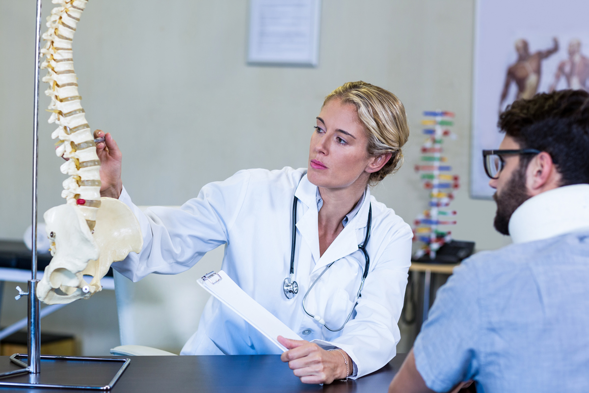
Clinical Assessment and Diagnostic Approaches
Assessing the spinal accessory nerve involves physical exams, electromyography, and imaging. These methods help us check how well cranial nerve 11 works and find any problems.
Physical Examination Techniques
A detailed physical exam is key to checking the trapezius and sternocleidomastoid muscles. Manual muscle testing helps us see how strong these muscles are. It shows if there’s weakness or if they’re not even.
We watch how well patients can shrug their shoulders and turn their heads. Any unusual movements or muscle weakness can mean nerve trouble.
Electromyography and Nerve Conduction Studies
Electromyography (EMG) is a tool for checking muscle electrical activity. We use fine electrodes in the muscles to see how they work. This gives us clues about nerve and muscle health.
Nerve conduction studies (NCS) help us see if the nerve is working right. They help find where nerve problems are and how bad they are.
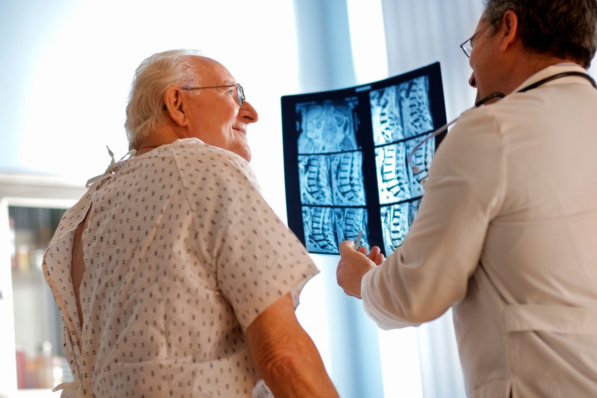
Imaging Modalities for Evaluation
We also use imaging to look at the spinal accessory nerve. Magnetic Resonance Imaging (MRI) is great for finding things like tumors or injuries that might hurt the nerve.
Computed Tomography (CT) scans help us see the neck and shoulder area. They give us important info about nerve problems or damage.
Pathologies and Disorders of Cranial Nerve 11
Understanding the pathologies and disorders of Cranial Nerve 11 is key for effective management and rehabilitation. The spinal accessory nerve faces various conditions that can greatly affect a person’s quality of life.
These pathologies include traumatic and iatrogenic injuries, neurological conditions, and congenital abnormalities. Each category presents unique challenges in diagnosis and treatment.
Traumatic and Iatrogenic Injuries
Traumatic injuries to the spinal accessory nerve can happen due to accidents, falls, or other trauma. Iatrogenic injuries, on the other hand, are caused by medical procedures in the neck area.
These injuries can cause significant problems, like muscle weakness or paralysis. This can make neck movements and shoulder elevation hard.
Neurological Conditions Affecting Function
Neurological conditions can also impact the spinal accessory nerve. This includes neuropathies, nerve compressions, and systemic neurological diseases.
These conditions can weaken muscles, cause atrophy, or change muscle tone. Accurate diagnosis is key for proper management.
Congenital Abnormalities
Congenital abnormalities of the spinal accessory nerve are rare but can have big implications for the affected individual.
These abnormalities may be linked to other congenital conditions or syndromes. A thorough evaluation is needed to address all health aspects.
Pathology | Effects on Cranial Nerve 11 | Clinical Presentation |
Traumatic Injuries | Damage to nerve fibers | Weakness or paralysis of sternocleidomastoid and trapezius muscles |
Iatrogenic Injuries | Nerve damage during surgery | Neck movement difficulties, shoulder elevation problems |
Neurological Conditions | Impaired nerve function | Muscle weakness, atrophy, or altered tone |
Congenital Abnormalities | Abnormal nerve development | Variable, depending on the nature of the abnormality |
Therapeutic Interventions and Rehabilitation
Managing spinal accessory nerve dysfunction needs a mix of treatments and rehab. Every patient is different, so we tailor our plans to fit their needs.
Conservative Management Approaches
First, we try conservative methods. This includes pain management with meds, physical therapy to keep muscles moving, and lifestyle changes to ease muscle strain.
We also suggest alternative therapies like acupuncture and massage. These can help with pain and make life better.
Surgical Repair Techniques
If other methods don’t work, surgery might be needed. For nerve injuries, we use nerve grafting and nerve transfer. These surgeries aim to fix nerve damage and boost muscle power.
Choosing the right surgery depends on the nerve damage’s extent and where it is. We team up with neurosurgeons to pick the best surgery for each patient.
Physical Therapy Protocols
Physical therapy is key in rehab for spinal accessory nerve issues. Our therapy plans aim to strengthen muscles, improve movement, and help patients get back to normal.
Our therapy might include exercises for the sternocleidomastoid and trapezius muscles. We also do scapular stabilization exercises to better shoulder function. Plus, we use functional training to help with daily tasks.
We focus on educating and empowering patients during rehab. Knowing their condition and being part of their treatment plan helps them get better and live better.
Conclusion
Understanding the spinal accessory nerve, or cranial nerve 11, is key. It controls important movements of the head, neck, and shoulders. This nerve has a special dual origin, making it unique.
It connects to muscles like the sternocleidomastoid and trapezius. These muscles help us move our head, neck, and shoulders. This is why the spinal accessory nerve is so important.
Damage to this nerve can cause big problems. Doctors use tests like physical exams and imaging to find issues. They then treat it with different methods, like surgery or physical therapy.
In short, the spinal accessory nerve is vital for our movements. Knowing about it helps doctors give better care. This improves patients’ lives and outcomes.
FAQ
What does the accessory nerve do?
The accessory nerve, also known as cranial nerve 11 or CN XI, controls two main muscles. These are the sternocleidomastoid and the trapezius. It helps with head rotation, lateral flexion, and shoulder elevation.
What is the function of cranial nerve 11?
Cranial nerve 11, or the spinal accessory nerve, is key for head, neck, and shoulder movements. It does this by controlling the sternocleidomastoid and trapezius muscles.
Which cranial nerve innervates the sternocleidomastoid and trapezius?
The spinal accessory nerve, or cranial nerve 11, innervates the sternocleidomastoid and trapezius muscles. This allows for head rotation, lateral flexion, and shoulder elevation.
What is the spinal accessory nerve?
The spinal accessory nerve is a pure motor nerve. It has both spinal and cranial origins. It controls the sternocleidomastoid and trapezius muscles.
What are the primary functions of the spinal accessory nerve?
The spinal accessory nerve’s main functions are motor control. It enables critical movements of the head, neck, and shoulders. This includes head rotation, lateral flexion, and shoulder elevation.
How is the spinal accessory nerve assessed clinically?
Clinically, the spinal accessory nerve is assessed through physical exams, electromyography, and imaging. These methods help evaluate nerve function and identify issues.
What pathologies can affect the spinal accessory nerve?
Pathologies like traumatic and iatrogenic injuries, neurological conditions, and congenital abnormalities can affect the spinal accessory nerve. These can impact nerve function and patient outcomes.
What therapeutic interventions are available for managing spinal accessory nerve dysfunction?
Therapeutic interventions include conservative management, surgical repair, and physical therapy. These aim to restore nerve function and improve patient outcomes.
What is the role of the spinal accessory nerve in shoulder movement?
The spinal accessory nerve is vital for shoulder movement. It innervates the trapezius muscle, enabling shoulder elevation and shrugging.
What is cranial nerve 11 responsible for?
Cranial nerve 11, or the spinal accessory nerve, controls the sternocleidomastoid and trapezius muscles. It enables various movements, including head rotation, lateral flexion, and shoulder elevation.
