
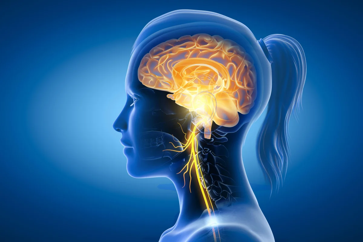
The cranial nerve system is complex. It has twelve paired nerves that start from the brain and brainstem. Knowing where these nerves come from and how they connect is key for doctors to diagnose and treat.cranial nerves tableRecalling the Start: Your First leukemia symptoms in a Child
The brainstem is where ten of these nerves begin. The other two, the olfactory and optic nerves, start in the cerebrum. Learning about these nerves helps doctors find problems and give the best care.
Key Takeaways
- The cranial nerves control vital functions including vision, taste, hearing, facial movement, and autonomic regulation.
- Ten of the twelve cranial nerves emerge from the brainstem.
- Understanding cranial nerve anatomy is essential for diagnosing and treating related disorders.
- The brainstem is a critical area for the origin of cranial nerves.
- Accurate knowledge of cranial nerve innervation patterns is critical for clinical neurological assessment.
Anatomy and Organization of Cranial Nerves

It’s important to know about the anatomy and organization of cranial nerves. They are a complex set of nerves that come directly from the brain. These nerves control many bodily functions.
Definition and Basic Structure
Cranial nerves start from the brain, including the brainstem. There are 12 pairs, labeled I through XII. Each has its own functions and areas it covers. A cranial nerve has nerve fibers, which can be sensory, motor, or both.
The cranial nerve origin and cranial nerve innervation are key to their anatomy. Some nerves are sensory, motor, or both. Knowing this helps doctors diagnose and treat.
Brainstem Nuclei Organization
The brainstem, made up of the midbrain, pons, and medulla oblongata, is where most cranial nerves start. The brainstem’s nuclei are arranged in a specific way. Sensory nuclei are usually found more to the back and sides. Motor nuclei are more to the front.
Rostral to Caudal Arrangement
Cranial nerves come out of the brainstem in order from I to XII. The midbrain is where nerves III and IV start. The pons is home to nerves V, VI, VII, and VIII. The medulla oblongata is where nerves IX, X, XI, and XII begin. This order helps us understand their spatial and functional roles.
Brainstem Level | Cranial Nerves | Primary Function |
Midbrain | III, IV | Oculomotor, Trochlear |
Pons | V, VI, VII, VIII | Trigeminal, Abducens, Facial, Vestibulocochlear |
Medulla Oblongata | IX, X, XI, XII | Glossopharyngeal, Vagus, Accessory, Hypoglossal |
In conclusion, the anatomy and organization of cranial nerves are complex. Knowing about the cranial nerve origin and cranial nerve innervation is key. It’s also important to understand their organization in the brainstem and cranial nerves for neurological assessment and treatment.
Classification of Cranial Nerves

The cranial nerves are divided into sensory, motor, and mixed types. This helps us understand their roles in our body. It’s important for both learning and medical purposes.
Sensory Nerves
Sensory nerves send information to our brain. Nerves I, II, and VIII are all sensory. Cranial Nerve I (Olfactory Nerve) helps us smell. Cranial Nerve II (Optic Nerve) lets us see. Cranial Nerve VIII (Vestibulocochlear Nerve) is key for hearing and balance.
“The senses are the instruments by which we perceive the world around us.” – Aristotle
Motor Nerves
Motor nerves control our muscles. Nerves III, IV, VI, XI, and XII are all motor. Cranial Nerve III (Oculomotor Nerve), Cranial Nerve IV (Trochlear Nerve), and Cranial Nerve VI (Abducens Nerve) help our eyes move. Cranial Nerve XI (Accessory Nerve) and Cranial Nerve XII (Hypoglossal Nerve) control our neck and tongue.
Mixed Nerves
Mixed nerves have both sensory and motor fibers. Cranial Nerve V (Trigeminal Nerve), Cranial Nerve VII (Facial Nerve), Cranial Nerve IX (Glossopharyngeal Nerve), and Cranial Nerve X (Vagus Nerve) are examples. They handle facial sensation, facial muscle control, swallowing, and more.
Nerve Type | Cranial Nerves | Primary Functions |
Sensory | I, II, VIII | Smell, Vision, Hearing/Balance |
Motor | III, IV, VI, XI, XII | Eye movements, Neck/Tongue movements |
Mixed | V, VII, IX, X | Facial sensation, Motor control, Swallowing, Visceral innervation |
Comprehensive Cranial Nerves Table: Origins and Innervation
We have created a detailed table to help you understand cranial nerves better. It shows where they start, how they exit, and what they do. This table is great for both doctors and students, making it easy to learn about cranial nerves.
Table Structure and Key Components
The table is easy to use, with clear columns and rows. It lists the origins, exit points, and functional classifications of each nerve. You’ll find important info like the nerve’s name, where it starts, how it exits the brainstem, and its main tasks.
Nuclei Origins and Exit Points
Knowing where cranial nerves start and exit is key to understanding them. The table makes it easy to see how these nerves come out of the brainstem and leave the skull. It shows their paths clearly.
Functional Classifications
Cranial nerves are grouped by what they do. The table sorts them into sensory, motor, and mixed nerves. This helps you see their role in sensing, moving, or doing both.
Clinical Applications of the Table
The table is very useful in medical settings. It helps doctors quickly check cranial nerves during exams. It’s also a great tool for students to learn about cranial nerves.
This table gives a clear view of cranial nerves. It makes learning about their complex anatomy easier. It also helps in checking their function in a clinical setting.
Cerebrum-Originating Cranial Nerves
The olfactory and optic nerves start in the cerebrum. They are key to our sense of smell and sight. These nerves help us understand and connect with our world.
Olfactory Nerve (Cranial Nerve I)
The olfactory nerve carries smell information from the nose to the brain. It’s the first cranial nerve and comes from the nasal cavity. It’s special because it connects directly to the cerebrum.
Smelling works when odor molecules meet olfactory sensory neurons. These neurons send signals to the olfactory bulb. There, the brain processes this information further.
Optic Nerve (Cranial Nerve II)
The optic nerve sends visual info from the retina to the brain. It’s made of axons from ganglion cells in the retina. It’s vital for seeing.
Signals from light hitting the retina go through the optic nerve. They cross over at the optic chiasm. Then, they go to the visual cortex for processing. Damage can cause vision problems, including blindness.
Knowing how the olfactory and optic nerves work is key for diagnosing and treating problems. Doctors test our senses of smell and sight to find issues.
Midbrain Cranial Nerves
The midbrain is a key part of the brainstem. It’s where important cranial nerves start. These nerves help with hearing, seeing, and eye movements. We’ll look at the nerves from this area, their structure, and why they matter.
Oculomotor Nerve
The oculomotor nerve, or Cranial Nerve III, comes from the midbrain. It controls many eye muscles. The oculomotor nerve works with the superior, medial, inferior rectus, and inferior oblique muscles, plus the levator palpebrae superioris muscle. This lets our eyes move in many ways.
This nerve also helps our pupils get smaller and our lenses focus. If it gets damaged, we might see our eyelid droop, our eyes turn outward, and our pupils get bigger.
- Controls extraocular muscles (superior rectus, medial rectus, inferior rectus, inferior oblique)
- Innervates levator palpebrae superioris muscle
- Carries parasympathetic fibers for pupil constriction and lens accommodation
Trochlear Nerve
The trochlear nerve, or Cranial Nerve IV, also starts in the midbrain. It’s the thinnest and longest cranial nerve. The trochlear nerve is special because it comes out of the back of the brainstem. It mainly works with the superior oblique muscle.
This muscle helps our eyes turn inward, move down, and out. If the trochlear nerve is hurt, we might see double vision.
In short, the midbrain is home to two vital cranial nerves: the oculomotor and trochlear nerves. Knowing about them helps us understand and treat brain disorders.
Pons Cranial Nerves
The pons is a key part of the brainstem. It is where many important cranial nerves start. These nerves help with facial expressions, sending sensory info, and eye movements.
Trigeminal Nerve
The trigeminal nerve does two main things. It feels sensations on the face and helps chew food.
- It senses pain, temperature, touch, and how our body is positioned.
- It also controls the muscles we use to chew.
Abducens Nerve
The abducens nerve makes the eye move outwards. It’s a motor nerve.
Key Function:It lets the eye move to the side, away from the body’s center.
Facial Nerve
The facial nerve has two roles. It makes facial muscles move and carries taste from the tongue.
Vestibulocochlear Nerve
The vestibulocochlear nerve is all about sound and balance. It’s a sensory nerve.
- The cochlear part helps us hear.
- The vestibular part helps with balance and knowing where we are in space.
Medulla Oblongata Cranial Nerves
The medulla oblongata is home to four key cranial nerves. These nerves handle important tasks like swallowing, speaking, and tongue movements.
Glossopharyngeal Nerve
The glossopharyngeal nerve (IX) helps with swallowing and making saliva. It also sends signals from the back third of the tongue. Plus, it controls the stylopharyngeus muscle.
- Functions: swallowing, salivation, taste sensation
- Innervation: stylopharyngeus muscle, parotid gland
Vagus Nerve
The vagus nerve (X) is essential for many body functions. It controls the heart rate, digestion, and breathing.
- Regulation of heart rate
- Control of smooth muscle in the gastrointestinal tract
- Modulation of respiration
Accessory Nerve
The accessory nerve (XI) has two parts. The cranial root joins the vagus nerve. The spinal root helps move the neck and shoulders by innervating the sternocleidomastoid and trapezius muscles.
- Functions: neck and shoulder movement
- Innervation: sternocleidomastoid and trapezius muscles
Hypoglossal Nerve
The hypoglossal nerve (XII) controls the tongue’s movements. It helps the tongue stick out, pull back, and change shape.
Damage to this nerve can cause tongue weakness or paralysis. This makes it hard to speak and swallow.
Functional Systems of Cranial Nerves
Understanding the cranial nerves’ roles is key to grasping their importance in our bodies. These nerves are part of several systems. They include special sensory, branchial motor, visceral motor, and somatic motor systems.
Special Sensory Systems
Special sensory systems handle sensory info like vision, hearing, taste, and smell. The nerves involved in these functions are vital for how we interact with the world.
The olfactory nerve (CN I) and optic nerve (CN II) are key players in these systems. The vestibulocochlear nerve (CN VIII) also plays a role, handling sound and balance info.
Branchial Motor System
The branchial motor system manages muscles from the branchial arches. It’s important for actions like chewing, facial expressions, and swallowing.
The trigeminal nerve (CN V), facial nerve (CN VII), glossopharyngeal nerve (CN IX), and vagus nerve (CN X) all contribute to this system. They control muscles essential for many bodily functions.
Visceral Motor System
The visceral motor system, or autonomic nervous system, controls organs’ involuntary actions. Cranial nerves are key in this system, focusing on parasympathetic functions.
The vagus nerve (CN X) is a major player, providing parasympathetic signals to organs in the chest and belly. The oculomotor nerve (CN III) also has a role, controlling the eye’s smooth muscles.
Somatic Motor System
The somatic motor system handles voluntary movements through skeletal muscles. Several cranial nerves are involved, enabling various motor actions.
The oculomotor nerve (CN III), trochlear nerve (CN IV), and abducens nerve (CN VI) manage the eye muscles. The hypoglossal nerve (CN XII) controls the tongue muscles. These nerves are vital for precise movement.
Functional System | Cranial Nerves Involved | Primary Functions |
Special Sensory | I, II, VIII | Smell, vision, hearing, balance |
Branchial Motor | V, VII, IX, X | Mastication, facial expression, swallowing |
Visceral Motor | III, VII, IX, X | Parasympathetic control of organs |
Somatic Motor | III, IV, VI, XII | Eye movements, tongue movements |
Clinical Assessment of Cranial Nerves
Checking cranial nerves is key for doctors to spot and treat brain problems. They look at each nerve carefully. This helps find out if there’s damage.
Systematic Examination Approach
Checking cranial nerves needs a careful plan. Doctors start with the olfactory nerve (I) to see if smells are recognized. They then check eyesight and how eyes move.
They also check how well the face feels and moves. The facial nerve (VII) is tested by looking at facial expressions and taste. The eighth nerve is checked for hearing and balance.
Lower nerves are tested too. The ninth and tenth nerves help with swallowing. The eleventh nerve is checked by seeing if the shoulders and head move right. The twelfth nerve is tested by watching the tongue move.
Cranial Nerve | Function | Assessment Method |
I (Olfactory) | Smell | Smell identification tests |
II (Optic) | Vision | Visual acuity and field tests |
III, IV, VI (Oculomotor, Trochlear, Abducens) | Eye movements | Eye movement examination |
Common Findings in Cranial Nerve Dysfunction
Problems with cranial nerves can show up in many ways. You might see blurry vision or double vision. Facial weakness, hearing loss, and trouble speaking or swallowing are common too.
“Accurate diagnosis of cranial nerve dysfunction requires a complete understanding of their anatomy and function.”
— Medical Expert, Neurologist
Diagnostic Techniques
Doctors use MRI and CT scans to see the nerves. They also use tests like EMG and nerve conduction studies. These help figure out how well nerves work.
By using these methods, doctors can find and treat problems with cranial nerves.
Pathologies Affecting Cranial Nerves at the Brainstem
Pathologies at the brainstem can greatly affect cranial nerve function. The brainstem connects the cerebrum to the spinal cord. It houses the nuclei and tracts of the cranial nerves, making it key for many neurological functions.
Vascular Disorders
Vascular disorders are a big worry for brainstem health. Ischemic strokes and hemorrhages can harm cranial nerve nuclei or their tracts in the brainstem. We’ll look at how issues like vertebral artery dissection or basilar artery thrombosis can cause problems with cranial nerves.
The symptoms of vascular disorders affecting cranial nerves at the brainstem vary. For example, a lesion in the midbrain can damage the oculomotor nerve (CN III). This can cause ptosis and ophthalmoplegia.
Inflammatory and Demyelinating Conditions
Inflammatory and demyelinating conditions, like Multiple Sclerosis (MS), can harm cranial nerves at the brainstem. MS plaques in the brainstem can affect cranial nerve nuclei or their tracts. This can lead to a variety of symptoms.
We’ll talk about how MS can cause symptoms like diplopia, facial weakness, or dysphagia. These symptoms can occur due to damage to nerves like the abducens (CN VI), facial (CN VII), or glossopharyngeal (CN IX) nerves.
Neoplastic Processes
Neoplastic processes, including both benign and malignant tumors, can affect cranial nerves at the brainstem. Tumors like schwannomas, meningiomas, and gliomas can grow within or near the brainstem. They can compress or infiltrate cranial nerve structures.
The impact of these tumors depends on their location, size, and growth rate. For instance, a schwannoma of the trigeminal nerve can cause facial pain and numbness.
Traumatic Injuries
Traumatic injuries to the brainstem can result from head trauma. These injuries can affect cranial nerves directly or indirectly. The severity and location of the trauma determine the extent of cranial nerve involvement.
We’ll explore how traumatic brain injuries can lead to cranial nerve dysfunction. This includes conditions like cranial nerve palsies or complex neurological syndromes resulting from brainstem damage.
Conclusion
Understanding the complex anatomy and function of cranial nerves is key for diagnosing and managing neurological disorders. The brainstem, where most cranial nerves start or pass through, is vital for controlling our body’s functions. Our detailed look at the 12 cranial nerves has shown their origins, how they connect, and their roles.
This article’s cranial nerves summary is a great tool for healthcare workers and those wanting to learn about the nervous system. Knowing how the brainstem and cranial nerves work together helps us understand the nervous system’s complexities.
In summary, the cranial nerves are a vital part of the nervous system. Studying them helps us learn more about neurology. As we dive deeper into the brain and nervous system, we can find better ways to diagnose and treat neurological disorders.
FAQ
What are cranial nerves and what is their significance?
Cranial nerves are a complex system that controls many bodily functions. This includes movement, sensation, and bodily functions. Knowing how they work is key to diagnosing and treating disorders.
How are cranial nerves classified?
Cranial nerves are divided into sensory, motor, and mixed types. Each type has its own functions and characteristics. This helps us understand their roles and importance in health.
What is the brainstem and how are cranial nerves organized within it?
The brainstem connects the cerebrum to the spinal cord. It’s where many cranial nerves start. Knowing how they’re arranged helps us understand their functions and importance.
What are the cranial nerves that originate from the cerebrum?
The olfactory and optic nerves start in the cerebrum. They carry information about smell and vision.
Which cranial nerves emerge from the midbrain?
The oculomotor and trochlear nerves come from the midbrain. They control eye movements and other functions.
What is the function of the trigeminal nerve?
The trigeminal nerve is a mixed nerve. It sends sensory information from the face and controls chewing muscles.
How are cranial nerves assessed clinically?
Doctors check cranial nerves through a detailed examination. They use imaging and electrophysiological tests to do this.
What are some common pathologies that can affect cranial nerves at the brainstem?
Vascular disorders, inflammation, demyelination, tumors, and injuries can harm cranial nerves at the brainstem.
What is the significance of understanding cranial nerve origins and innervation?
Knowing where cranial nerves come from and how they connect is vital. It helps in diagnosing and treating disorders and understanding their importance.
Where can I find a complete table of cranial nerves?
You can find a detailed table of cranial nerves online. It shows their origins, exit points, and types. This helps in understanding their roles and significance.
What is the role of the brainstem in cranial nerve function?
The brainstem is key to cranial nerve function. Many cranial nerves start here and are organized within its nuclei.
References
National Center for Biotechnology Information. Evidence-Based Medical Guidance. Retrieved from https://www.ncbi.nlm.nih.gov/books/NBK544297/


