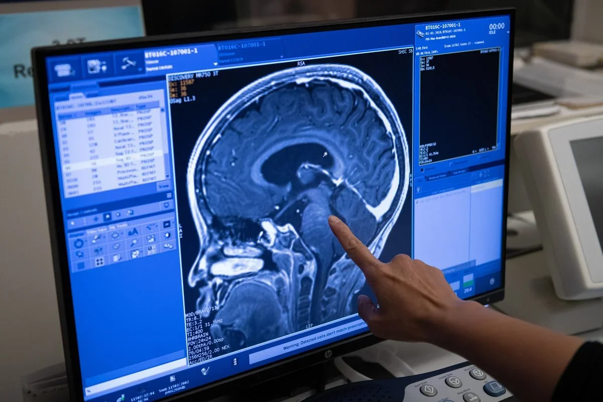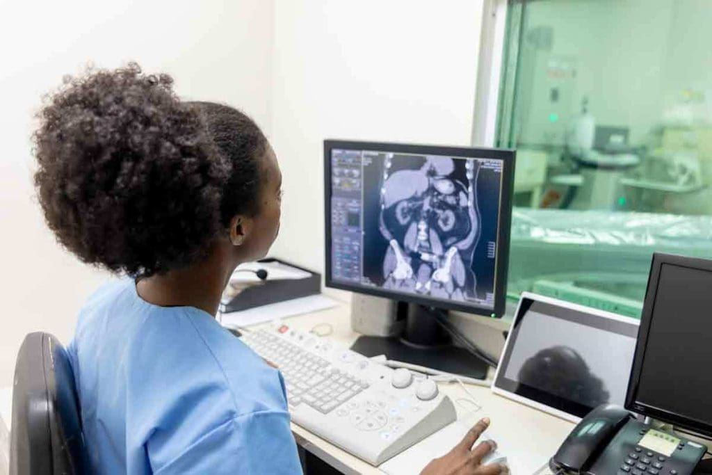
Accurate diagnosis is key for liver health. A CT liver scan takes many images from different angles. This gives a full view of what’s inside. It’s vital for spotting liver problems like tumors and cysts.
Using a ct scan for liver with contrast helps doctors find liver issues better. At Liv Hospital, we focus on our patients. We use top standards for early detection and diagnosis. We want to give patients the facts about ct of liver with contrast and new ways to see the liver.
Key Takeaways
- CT liver scans provide detailed cross-sectional images of the liver, aiding in accurate diagnosis.
- The use of contrast agents in CT scans improves detection rates for various liver conditions.
- Liv Hospital employs a patient-centered, internationally recognized approach for diagnosis.
- Advanced CT scans can detect tumors, cysts, and other liver abnormalities.
- Early detection through CT liver scans is key for effective treatment planning.
Understanding CT Liver Scan Technology
It’s important to know how CT liver scans work. They help us see the liver clearly and find problems. This helps us plan the best treatment.
Definition and Basic Principles of Liver CT
A CT liver scan uses X-rays and computers to show the liver’s inside. It lets us see details that other scans can’t. This is key for finding and fixing liver issues.
CT scans work by moving an X-ray source around the body. It takes pictures from many angles. Then, a computer makes these pictures into clear images.
Key parts of CT liver scan tech are:
- X-ray tube and detectors
- Computer processing unit
- Image reconstruction algorithms
Evolution of CT Scanning for Liver Assessment
CT scanning has gotten better over time. Now, we can see more clearly and quickly. We can even spot tiny problems.
Some big changes in CT scanning include:
- Multidetector CT (MDCT): Scans faster and shows more detail.
- Dual-Energy CT: Helps tell different tissues apart better.
- Advanced image reconstruction techniques: Makes pictures clearer and smoother.
These updates have made CT liver scans very useful. They help doctors diagnose and treat liver problems accurately.
Key Thing #1: CT Liver Scan Offers Detailed Cross-Sectional Imaging
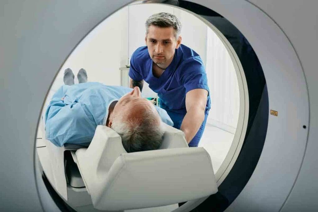
CT liver scans give detailed views of the liver. They help doctors see the liver from many angles. This helps them understand its structure and find any problems.
How CT Creates Comprehensive Liver Visualization
CT liver scans use X-rays to make detailed liver images. They take X-ray shots from different angles. Then, these shots are turned into detailed images.
Key benefits of CT liver scan cross-sectional imaging include:
- Detailed visualization of liver anatomy
- Accurate detection of lesions and abnormalities
- Guidance for biopsies and other interventional procedures
Comparing Healthy Liver CT Scan vs. Abnormal Findings
A healthy liver CT scan looks uniform. But, an abnormal scan might show changes. These could be signs of fatty liver disease or tumors.
For example, a CT scan can tell the difference between a simple cyst and a complex lesion. This is key for choosing the right treatment. By comparing scans, doctors can understand a patient’s condition better. They can then plan the best course of action.
Abnormal findings on a CT liver scan may include:
- Hypodense lesions suggestive of cysts or tumors
- Hyperdense areas indicating possible hemorrhage or calcification
- Diffuse changes in liver texture, such as those seen in steatosis
Key Thing #2: CT of Liver with Contrast Dramatically Improves Detection
Using contrast agents in liver CT scans makes liver structures much clearer. This is key for spotting liver problems accurately.
How Contrast Agents Enhance Liver Visualization
Contrast agents are essential for better liver CT scans. They make different liver tissues stand out. This makes finding problems easier.
Contrast-enhanced CT scans give a clearer view of the liver’s anatomy and issues. This is vital for spotting tumors and other problems that might not show up without contrast.
The 97% Sensitivity Rate for Malignant Tumor Detection
Research shows that CT scans of the liver with contrast are very good at finding cancer. A study in the Journal of Immunopathology found a 97% detection rate for cancerous liver tumors. This high rate is key for early treatment.
The high detection rate comes from contrast agents making tumors stand out. This is critical for knowing how serious the cancer is and how to treat it.
Using contrast-enhanced CT liver scans helps doctors make more accurate diagnoses. These detailed images help plan better treatments and keep track of progress.
Key Thing #3: Wide Range of Detectable Liver Conditions
We use CT liver scans to find many liver problems. They are key in today’s liver health care.
Detecting Various Liver Abnormalities
CT liver scans can spot many liver issues, like tumors and cysts. This is important for correct diagnosis and treatment plans.
Primary and Metastatic Tumor Identification
CT liver scans are great for finding primary and metastatic tumors. They show where and how big tumors are. This helps doctors know how serious the cancer is and what treatment to use.
Primary liver tumors start in the liver. Metastatic tumors come from other places. Finding these tumors right is key to managing them well.
Steatotic Liver Disease Detection
CT liver scans can also spot steatotic liver disease. This is when liver cells get too much fat. Catching it early can stop it from getting worse.
CT scans can measure liver fat. This helps doctors see how bad the disease is and if treatments are working.
Cyst and Lesion Characterization
CT liver scans can also look at cysts and lesions in the liver. These might be simple cysts or more complex ones. Knowing what they are helps doctors decide the best course of action.
With detailed images, CT scans help doctors tell if a lesion is harmless or could be cancer. This guides how to treat it.
Key Thing #4: Advanced CT Technologies Improve Diagnostic Accuracy
Advanced CT technologies have changed the game in diagnostic imaging. They offer unmatched accuracy in liver assessments. This has greatly improved our ability to spot and diagnose liver issues. It leads to better care for patients.
Dual-Energy CT for Enhanced Tissue Differentiation
Dual-energy CT scanners are a big leap in CT technology. They use two X-ray energy levels to take images. This makes it easier to tell different tissues apart. A study in PMC12428841 shows dual-energy CT boosts accuracy in liver diagnosis.
Dual-energy CT helps spot liver lesions better. It also makes liver tissue and blood vessels clearer. This could mean fewer extra tests for patients.
Achieving 98% Diagnostic Accuracy with Optimized Parameters
Getting CT scanning right is key for accurate diagnoses. Tweaking the settings can make CT images much clearer. This leads to more accurate diagnoses. With the right settings, accuracy can hit 98% in some cases.
The table below shows how tweaking CT settings can boost accuracy:
| Scanning Parameter | Standard Setting | Optimized Setting | Diagnostic Accuracy |
| kVp | 120 | 100/140 (dual-energy) | 95% |
| mAs | 200 | 250 | 96% |
| Slice Thickness | 5mm | 1mm | 98% |
Using advanced CT tech like dual-energy CT and fine-tuning settings can greatly improve accuracy. This leads to better care for patients.
Key Thing #5: AI-Powered Analysis Enhances CT Liver Scan Results
We are now using AI to make CT liver scans better. Artificial intelligence (AI) and machine learning algorithms are changing radiology. They are making CT liver scans more accurate.
AI analysis is not just a help; it’s essential for better diagnosis. It uses advanced algorithms to improve CT liver scan results. This makes them more reliable and accurate.
Machine Learning Algorithms for Liver Segmentation
AI is key in CT liver scans for liver segmentation. Machine learning algorithms can spot the liver and separate it from other parts. This is important for:
- Accurate liver volume measurement
- Finding lesions and abnormalities
- Planning surgeries
AI makes liver segmentation faster and more consistent. It helps radiologists work less and improves scan quality.
Improved Detection Reliability with AI Assistance
AI also makes finding problems in CT liver scans better. AI looks at huge amounts of data to spot liver issues. This includes:
- Malignant tumors
- Cysts and benign lesions
- Fatty liver disease
AI helps radiologists make more accurate diagnoses. It reduces mistakes. Studies show AI can lead to quicker diagnoses, helping patients.
In summary, AI in CT liver scans is a big step forward. It makes liver segmentation and detection more reliable. This leads to better and faster diagnosis of liver problems.
The Complete CT Liver Scan Procedure
Knowing about the CT liver scan procedure helps patients feel ready and calm. We walk our patients through each step, making sure they know what to expect.
What to Expect During Your Liver Cat Scan
When you get to the CT liver scan, our skilled radiology team will welcome you. They’ll explain everything and answer your questions. The scan is quick, usually taking just a few minutes.
You’ll lie on a comfy table that slides into the CT scanner, a big, doughnut-shaped machine. You might need to hold your breath for a bit to get clear images. Our team will talk to you through an intercom, helping you through it. If contrast material is used, it goes through an IV, which might feel a bit warm.
Duration and Patient Experience
The whole CT liver scan procedure takes about 15 to 30 minutes. But the actual scanning is much quicker. Most of the time is spent getting you ready and in the right spot.
Most people find the experience pretty comfortable and easy. The table moves smoothly into the scanner, and the machine helps with claustrophobia. Our staff works hard to keep you comfortable during the whole process.
| Procedure Step | Duration | Patient Experience |
| Preparation | 5-10 minutes | Changing into a gown, removing jewelry, and IV placement if needed |
| Scanning | 2-5 minutes | Lying on the table, being moved into the scanner, and holding breath when instructed |
| Total Time | 15-30 minutes | From arrival to completion, including preparation and scanning |
By knowing the CT liver scan procedure, patients can prepare better. This reduces anxiety and makes the diagnostic process smoother.
Key Thing #6: Preparation Requirements for Optimal CT Liver Scan Results
Getting ready for a CT liver scan is key to getting good results. Patients must follow specific steps to get accurate data. Their healthcare provider will give them detailed instructions.
Pre-Scan Instructions and Guidelines
Before the scan, patients get clear instructions. It’s important to follow these steps carefully to avoid problems. These steps include:
- Removing any metal objects or jewelry that could interfere with the scan
- Wearing comfortable, loose-fitting clothing
- Arriving early to complete any necessary paperwork and preparation
Talking clearly with your healthcare provider is vital. Make sure you understand all the pre-scan requirements. If you have questions, ask them before the scan.
Dietary and Medication Considerations
What you eat and take as medicine is important for the scan. Patients might need to fast or follow a special diet before the scan. This helps ensure the scan images are clear.
Also, tell your healthcare provider about all your medicines. Some medicines might need to be changed or stopped before the scan. This prevents any bad reactions or effects on the scan results.
By following the instructions, dietary rules, and medicine guidelines, patients help get the best scan images. These images are essential for accurate diagnosis and treatment planning.
Key Thing #7: Benefits of CT Scan for Liver Evaluation
CT scans have changed how we check the liver. They are quick, don’t hurt, and very accurate. This helps doctors care for patients better and plan treatments well.
Non-Invasive Nature and Quick Procedure Time
CT scans are great because they don’t hurt. They let doctors check the liver without surgery. This makes it safer and more comfortable for patients.
Also, CT scans are fast. The scan itself only takes a few minutes. But getting ready and settled in takes longer. This is good for patients who are in pain.
Supporting Early Intervention and Treatment Planning
CT scans help find problems early. They show detailed images of the liver. This helps doctors catch issues like liver cancer sooner.
These scans also help plan treatments. Doctors can see how bad the damage is. They can plan surgeries or check if treatments are working. This helps make treatment plans that fit each patient’s needs.
In short, CT scans are very helpful for liver checks. They are safe, quick, and accurate. They help doctors find problems early and plan treatments well.
Key Thing #8: Limitations and Considerations of Liver CT Scans
CT scans are useful for diagnosing, but they have their limits. It’s key to know the downsides when using them for liver checks.
Radiation Exposure Considerations
One big issue with CT scans is the radiation they use. Too much radiation can increase cancer risk. We aim to keep doses low while keeping images clear.
A typical abdominal CT scan gives 5 to 7 millisieverts (mSv) of radiation. This is more than the yearly background radiation of 3 mSv. We follow the ALARA principle to keep doses as low as possible.
Contrast Agent Risks and Contraindications
Contrast agents are another thing to think about. They’re usually safe but can cause allergic reactions or more serious issues in rare cases.
For people with kidney problems, contrast agents can be risky. We check kidney health before using them and take steps to reduce risks.
| Contrast Agent Risk Factors | Precautions |
| Pre-existing kidney disease | Assess kidney function before contrast administration |
| History of allergic reactions | Administer pre-medication or use alternative contrast agents |
| Diabetes | Monitor kidney function and consider alternative imaging |
Cases Where Alternative Imaging May Be Preferred
In some cases, other imaging methods might be better than CT scans for liver checks. MRI is often chosen for those with kidney issues or at high risk for kidney problems from contrast agents.
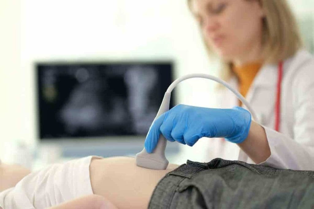
Ultrasound is another option, great for initial checks or when avoiding radiation is key. We pick the best imaging method based on each patient’s needs and health history.
In summary, CT scans are valuable for liver exams but have their limits. Knowing these helps us choose the best imaging for each patient’s care.
Comparing CT Liver Scan with Alternative Diagnostic Methods
CT liver scans are used to check liver health, but how do they compare to other methods? It’s key to know the good and bad of different imaging ways.
MRI vs CT for Liver Assessment
MRI and CT scans are both important for liver checks, but they’re used in different ways. CT scans give detailed images fast, great for emergencies or trauma checks. MRI shows soft tissue better, helping spot some liver issues.
Choosing MRI or CT depends on the liver problem, patient needs, and the doctor’s question. MRI is better for the biliary system and some liver spots that CT can’t see.
Ultrasound and Other Complementary Techniques
Ultrasound is a key tool for liver checks, being non-invasive and affordable. It’s not as detailed as CT or MRI but great for fatty liver and cirrhosis. It’s often the first choice because it’s easy to get and doesn’t use radiation.
PET scans are used too, for liver function and cancer detection. The right imaging choice depends on the situation and what info is needed.
In summary, CT scans are just one part of many diagnostic tools like MRI, ultrasound, and more. Knowing each method’s strengths helps doctors choose the best for each patient. This leads to better diagnoses and treatments.
Conclusion: The Future of CT Liver Scanning Technology
CT liver scanning has changed how we diagnose and find liver problems. It uses detailed images and special contrast agents. This makes CT scans very important in medicine.
We expect big changes in CT technology soon. These will make finding diseases better and safer for patients. We’ll see clearer images, less radiation, and more use of AI to help doctors.
These updates will help us catch liver diseases early. They will also lead to better treatment plans. As CT tech gets better, we’ll see even better care for liver patients.
FAQ
What is a CT liver scan, and how does it work?
A CT liver scan uses X-rays and computer tech to show detailed liver images. It works by moving an X-ray machine around the body. This captures images from different angles, making detailed liver pictures.
What is the role of contrast agents in CT liver scans?
Contrast agents, or dyes, make liver structures and lesions more visible during a CT scan. They are given through an IV. This helps doctors see tumors and cysts better.
How does a CT liver scan compare to other imaging modalities like MRI and ultrasound?
CT liver scans give detailed images and are great for finding liver lesions and tumors. MRI shows soft tissues well and is used to check liver lesions. Ultrasound is cheap and non-invasive but doesn’t show as much detail as CT or MRI.
What are the benefits of using a CT liver scan for liver evaluation?
CT liver scans are quick, non-invasive, and show detailed images. They help doctors diagnose accurately. This leads to better treatment plans and outcomes for patients.
Are there any limitations or risks associated with CT liver scans?
Yes, CT scans use radiation and contrast agents can cause allergic reactions or kidney damage. In some cases, like pregnancy or severe kidney disease, other imaging methods might be better.
How do I prepare for a CT liver scan?
To prepare for a CT liver scan, you might need to fast and stop certain medications. Your doctor will give you specific instructions to get the best scan results.
What can I expect during a CT liver scan procedure?
During a CT liver scan, you’ll lie on a table that moves into a scanner. The scan is fast, lasting 10-30 minutes, and is painless. You might need to hold your breath briefly for clear images.
Can a CT liver scan detect all types of liver conditions?
CT liver scans can find many liver conditions, like tumors and cysts. But, how well it detects them depends on the scan quality and contrast agents used.
How has AI and machine learning impacted CT liver scan analysis?
AI and machine learning have made CT liver scans better. They help with liver segmentation and detection. These technologies can lower false positives and make diagnoses more accurate.
What advancements can we expect in CT liver scanning technology?
Future CT liver scanning will have better hardware, advanced algorithms, and more AI. These changes will improve how well scans diagnose and help patients.
Reference
Diagnostic accuracy of triphasic CT scan in differentiating malignant from benign focal liver lesions


