Last Updated on November 27, 2025 by Bilal Hasdemir

It’s important to know the differences in medical imaging radiation for patient safety and to make informed decisions. A CT radiation dose chart helps compare radiation from various imaging tests.
RadiologyInfo.org says a CT scan of the abdomen and pelvis has an effective dose of about 7.7 mSv. This is similar to 2.6 years of natural background radiation. This info helps patients make better choices about their medical tests.
Key Takeaways
- Understanding radiation doses from medical imaging procedures is key to patient safety.
- A CT radiation dose chart is useful for comparing radiation from different tests.
- The effective dose from a CT scan of the abdomen and pelvis is about 7.7 mSv.
- This dose is like 2.6 years of natural background radiation.
- With a CT radiation dose chart, patients can make better choices about their tests.
Understanding Medical Radiation Exposure
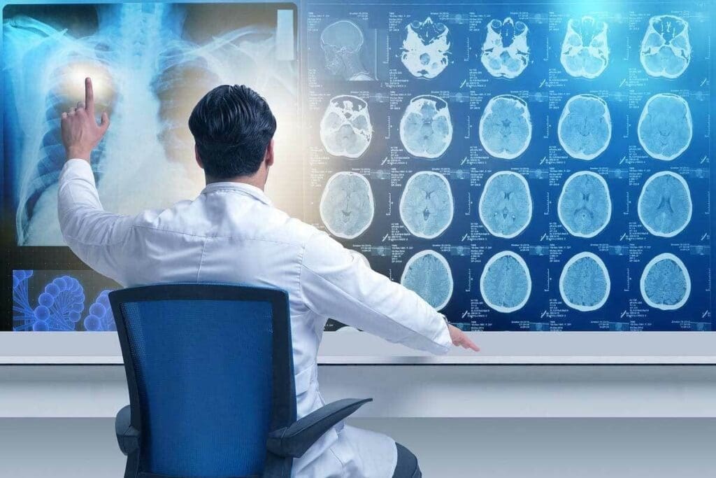
Medical imaging technology is getting better, and knowing about radiation risks is key. Techniques like X-rays and CT scans use ionizing radiation to create images for doctors.
Types of Medical Imaging That Use Radiation
Many medical imaging methods use radiation to help diagnose and treat diseases. These include:
- X-rays
- Computed Tomography (CT) scans
- Fluoroscopy
- Nuclear medicine procedures
Each method has its own use and level of radiation exposure.
Why Radiation Dose Measurement Matters
Measuring radiation doses is vital for checking patient safety. It helps keep radiation exposure low. The dose is usually measured in millisieverts (mSv).
| Imaging Procedure | Typical Radiation Dose (mSv) |
| Chest X-ray | 0.1 |
| CT Abdomen | 10 |
| Fluoroscopy (e.g., barium swallow) | 1-10 |
Knowing the radiation doses for different tests helps doctors and patients make better choices about imaging.
Radiation Measurement Units Explained
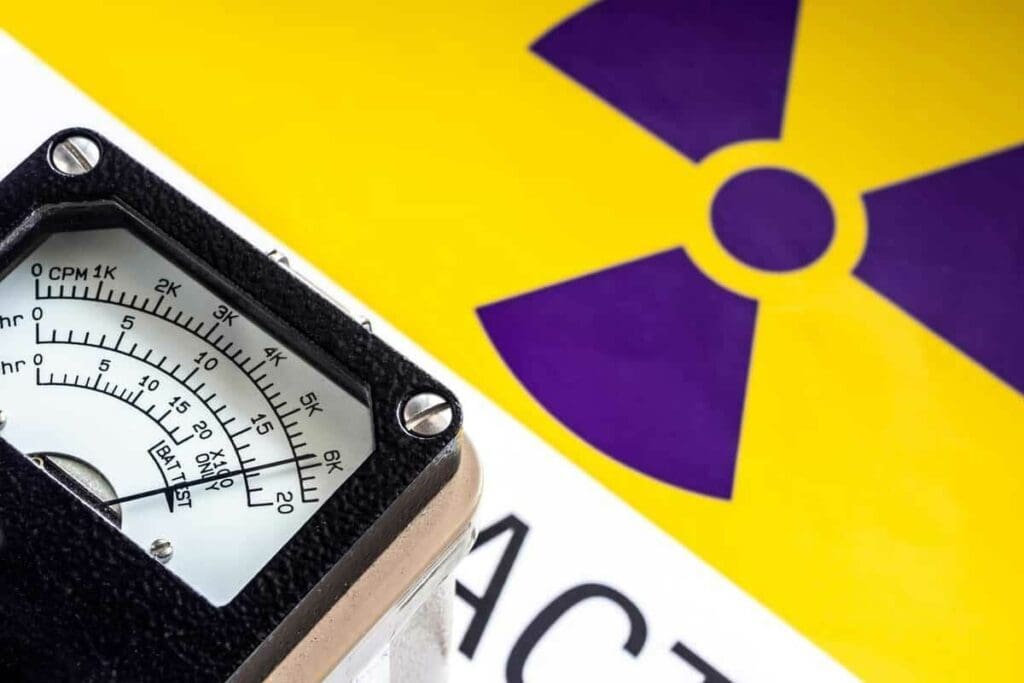
It’s important to know about radiation measurement units when comparing medical imaging procedures. The dose from these procedures is usually measured in millisieverts (mSv) or millirem (mrem).
Millisieverts (mSv) vs. Millirem (mrem)
The millisievert (mSv) is the standard unit for measuring effective dose in the International System of Units (SI). It shows the biological effect of radiation. In contrast, millirem (mrem) is used in the United States and equals one-thousandth of a rem. The conversion is simple: 1 mSv = 100 mrem.
Converting Between Different Radiation Units
To switch between mSv and mrem, use these conversion factors:
- To convert mSv to mrem, multiply by 100.
- To convert mrem to mSv, divide by 100.
For instance, a CT scan with a dose of 10 mSv is the same as 1000 mrem. Knowing these units and how to convert them is key to comparing radiation doses from various medical imaging procedures.
CT Radiation Dose Chart: A Comprehensive Overview
It’s important for doctors and patients to know about CT scan radiation doses. A CT radiation dose chart shows the radiation levels for different CT scan types.
Common CT Scan Procedures and Their Typical Doses
CT scans give out different amounts of radiation. For example, an abdomen and pelvis CT scan has a dose of about 7.7 mSv. Here are doses for other common CT scans:
- Head CT: approximately 2 mSv
- Chest CT: around 7 mSv
- CT angiography (heart): about 12 mSv
The dose can change based on the scanner, patient size, and the scan’s details.
How CT Doses Compare to Natural Background Radiation
To understand these doses better, compare them to natural background radiation. In the U.S., people get about 3 mSv of background radiation each year. So, a typical abdominal CT scan (7.7 mSv) is like getting 2.6 years of background radiation.
This comparison helps patients and doctors decide when to use CT scans. It balances the benefits of diagnosis with the risks of radiation.
X-Ray Radiation Exposure Levels
It’s important for patients to know about X-ray radiation exposure. X-rays help doctors see inside the body but involve radiation. This is why understanding this is key.
Typical Radiation Doses for Different X-Ray Procedures
The dose of radiation from X-rays changes with each procedure. For example, a chest X-ray gives about 0.1 mSv. This is like 10 days of natural background radiation. More detailed scans, like fluoroscopy, can have higher doses.
Here’s a look at typical doses for common X-ray tests:
- Chest X-ray: 0.1 mSv
- Extremity X-ray (e.g., arm or leg): 0.001-0.01 mSv
- Spinal X-ray: 0.5-1.5 mSv
Factors That Affect X-Ray Radiation Exposure
Many things can change how much radiation you get from an X-ray. The X-ray machine, the type of scan, and your size matter. For example, bigger patients might need more radiation for clear images.
A radiology expert says, “Using the right X-ray techniques and modern machines can lower radiation while keeping images clear.”
CT Abdomen Radiation Dose: What Patients Should Know
Knowing about the radiation from abdominal CT scans is key to making smart choices. These scans are vital for doctors to see inside the body. They help find and treat many health issues.
Typical Dose Range for Abdominal CT Scans
Abdominal CT scans usually give off 6 to 20 millisieverts (mSv) of radiation. This amount changes based on the scan type and the person’s size. For comparison, we all get about 3 mSv of natural radiation each year.
- A standard abdominal CT scan can give a dose like 2-6 years of natural background radiation.
- The exact dose can change based on the scanner and the patient’s size.
Risk-Benefit Analysis for Abdominal Imaging
Though there are risks from radiation, the benefits of CT scans often outweigh them. It’s important to weigh the good against the bad.
- Diagnostic Accuracy: CT scans give detailed images for better diagnoses.
- Risk Assessment: Doctors decide if a scan is needed and look for other options.
- Dose Optimization: New CT scanners aim for the lowest dose for clear images.
By knowing about the CT abdomen radiation dose and what affects it, patients can make better choices about their scans.
Chest X-Ray Radiation Exposure: Facts and Figures
Chest X-rays give off a small amount of radiation. Knowing this can ease worries for patients. A standard chest X-ray is fast and doesn’t hurt. It’s used to find many chest problems.
How Much Radiation Is in a Standard Chest X-Ray
A standard chest X-ray has about 0.1 mSv of radiation. This is like 10 days of natural background radiation. Here’s how it compares:
- A flight across the country exposes you to about 0.035 mSv of cosmic radiation.
- In the United States, you get about 0.01 mSv of background radiation every day.
Cumulative Effects of Multiple Chest X-Rays
Even though one chest X-ray has a low dose, many can add up. Doctors need to keep track of how many X-rays you’ve had. This helps avoid too much radiation. Talk to your doctor about your X-ray history to understand your total radiation dose.
Important things to remember:
- The risk from radiation builds up over your whole life.
- You might need more chest X-rays to check and keep track of some health issues.
- Doctors always try to use the least amount of radiation needed for good images.
How Much Radiation in CT Scan vs X-Ray: The Key Differences
Understanding the radiation difference between CT scans and X-rays is key for patients and doctors. Both are used a lot, but they have different uses and radiation levels.
CT scans give detailed images of the body, helping with complex diagnoses. X-rays are quicker and simpler for bones and some soft tissues. The radiation levels are quite different between these two.
Why CT Scans Deliver Higher Radiation Doses
CT scans have higher radiation because they’re more complex. They use a computer to combine X-ray images from different angles. This means more radiation overall.
An abdominal CT scan can give 6 to 20 millisieverts (mSv) of radiation. A chest X-ray is much less, about 0.1 mSv. This big difference is because CT scans give more detailed images.
Balancing Diagnostic Benefits Against Radiation Risks
Even with more radiation, CT scans are often worth it, like in emergencies. Doctors must weigh the need for accurate diagnoses against radiation risks.
Doctors aim to use the least radiation needed for good images. This helps lower risks while keeping images useful. It’s all about finding the right balance.
Knowing the radiation differences helps patients make better choices. By understanding the benefits and risks, patients and doctors can choose the best imaging options together.
Radiation Risk Assessment in Medical Imaging
Medical imaging technologies that use radiation need a detailed risk assessment. These tools are vital for diagnosis, but we must know the risks of radiation exposure.
Understanding Relative Risk from Medical Radiation
The risk from medical radiation is low but not zero. The benefits of imaging often outweigh the risks, like in emergency cases. Knowing the risk helps us make better choices about using radiation in imaging.
Several factors affect the risk, including:
- The dose of radiation received
- The age and health status of the patient
- The type of imaging procedure performed
Patient Populations at Higher Risk from Radiation Exposure
Some patients face higher risks from radiation. These include:
- Children and young adults: Their growing bodies are more vulnerable to radiation.
- Pregnant women: There’s a risk of harm to the developing fetus.
- Patients needing repeated imaging: The total radiation dose can increase risks.
For these groups, it’s key to balance the benefits of imaging against the risks. We should also look for non-radiation imaging options when possible.
Strategies to Reduce Radiation Exposure in Medical Imaging
There’s a growing need to cut down on radiation in medical imaging. This field is vital for diagnosis, but we must use it wisely to protect patients.
Dose optimization is key to lowering radiation exposure. It means adjusting settings to use the least amount of radiation needed for clear images.
Dose Optimization Techniques
Techniques like automated exposure control adjust doses based on patient size and body part. Iterative reconstruction also boosts image quality, allowing for lower doses.
| Technique | Description | Benefits |
| Automated Exposure Control | Adjusts radiation dose based on patient size and body part | Reduces radiation exposure, improves image quality |
| Iterative Reconstruction | Improves image quality, allows for lower doses | Enhances diagnostic accuracy, reduces radiation exposure |
When to Consider Alternative Non-Radiation Imaging
In some cases, non-radiation imaging like MRI or ultrasound is better. These methods offer valuable insights without the risks of radiation.
Choosing the Right Imaging Modality: Doctors should think about the patient’s condition and the diagnostic needs. They should weigh the risks and benefits of each imaging option.
By using dose optimization and non-radiation imaging, doctors can reduce radiation risks. This way, they keep diagnostic accuracy high while protecting patients.
Tracking Cumulative Radiation Exposure
It’s key to track how much radiation patients get from many medical imaging tests. This is very important for people who have had lots of CT scans or other tests that use radiation.
The Importance of Medical Imaging History
Keeping a detailed medical imaging history is very important. It should list all tests that use radiation, like CT scans and X-rays. This way, patients and doctors can see how much radiation they’ve had over time.
A study on the National Center for Biotechnology Information website shows that keeping accurate records is key. It helps figure out how much radiation someone has had.
Tools for Monitoring Lifetime Radiation Exposure
There are many tools and technologies to track radiation exposure over a lifetime. These include:
- Radiation dose tracking software
- Electronic health records (EHRs) that include radiation exposure data
- Patient registries that track radiation exposure from medical imaging
Using these tools helps doctors make smart choices about future tests. They can weigh the benefits of tests against the risks of radiation.
Advances in Low-Dose Imaging Technology
CT technology has made big strides, leading to lower radiation doses for patients. This progress comes from new scanner parts and better image-making algorithms.
New CT Technologies That Reduce Radiation Exposure
In recent years, new CT tech has emerged to cut down radiation. These include:
- Iterative Reconstruction (IR): IR boosts image quality while cutting down needed doses.
- High-Pitch CT Scanners: These scanners scan faster, lowering radiation exposure.
- Spectral CT: This tech can tell different materials apart, possibly cutting down on scan numbers.
A study on the National Center for Biotechnology Information shows these advances have greatly helped reduce CT doses.
Future Directions in Radiation Dose Reduction
Future tech will likely bring even more dose cuts. This includes better image-making methods and AI in imaging. AI could adjust scans in real-time to lower radiation even more.
| Technology | Description | Potential Benefit |
| Iterative Reconstruction | Improves image quality at lower doses | Reduced radiation exposure |
| High-Pitch CT | Faster scanning times | Lower overall dose |
| Spectral CT | Material differentiation based on atomic number | Potential reduction in multiple scans |
The push for low-dose imaging shows a big commitment to safer medical scans. As these techs get better, they’ll be key in the future of medical imaging.
Conclusion: Making Informed Decisions About Medical Imaging
It’s important for patients to know about the radiation doses from medical imaging. Different tests, like CT scans and X-rays, have different levels of radiation. This knowledge helps patients make better choices about their health.
Patients should talk to their doctors about the benefits and risks of these tests. Knowing this information helps them decide what’s best for them. This way, they can balance the need for diagnosis with the possible risks.
When choosing medical imaging, patients should think about several things. These include the type of test, the radiation dose, and other options. By being involved in their care, patients can get the right information without too much radiation.
Being well-informed about medical imaging helps patients feel more confident in their healthcare. It ensures they get the best care that fits their needs.
FAQ
What is the typical radiation dose for a CT scan of the abdomen?
An abdominal CT scan usually has a radiation dose of 6 to 20 millisieverts (mSv). This depends on the procedure and the patient’s size.
How much radiation is in a standard chest X-ray?
Chest X-rays expose patients to about 0.1 millisieverts (mSv) of radiation.
What is the difference between millisieverts (mSv) and millirem (mrem)?
Millisieverts (mSv) and millirem (mrem) measure radiation dose. One millisievert equals 100 millirem.
Why do CT scans deliver higher radiation doses compared to X-rays?
CT scans use more radiation because they need higher energy to create detailed images. This is different from X-rays.
How can I minimize radiation exposure during medical imaging procedures?
To reduce radiation, ask your doctor about dose-saving methods. Consider non-radiation imaging options. Keep a record of your imaging history.
What are the possible risks from radiation in medical imaging?
Radiation exposure can increase cancer risk, genetic mutations, and other health issues. This is more concerning for those who have many scans.
How can I track my cumulative radiation exposure from medical imaging?
Keep a record of your imaging history. Use tools and software to monitor your lifetime radiation exposure.
Are there any new technologies that reduce radiation exposure in medical imaging?
Yes, new CT technologies and imaging methods aim to lower radiation. Examples include low-dose CT scanners and advanced image techniques.
How do I balance the diagnostic benefits of medical imaging against the risks of radiation exposure?
Talk to your doctor about the risks and benefits. Consider the need for the scan and other diagnostic options.
What patient populations are at higher risk from radiation exposure?
Children, pregnant women, and those with a radiation history are more at risk. They are more vulnerable to radiation effects.
Can I get a record of my past medical imaging procedures and radiation exposure?
Yes, you can ask for a record of your past imaging. This helps track your radiation exposure and guides future scans.
Reference
- National Center for Biotechnology Information. (2000, March 3). Radiation doses in computed tomography. https://pmc.ncbi.nlm.nih.gov/articles/PMC1117635/


