Last Updated on November 27, 2025 by Bilal Hasdemir
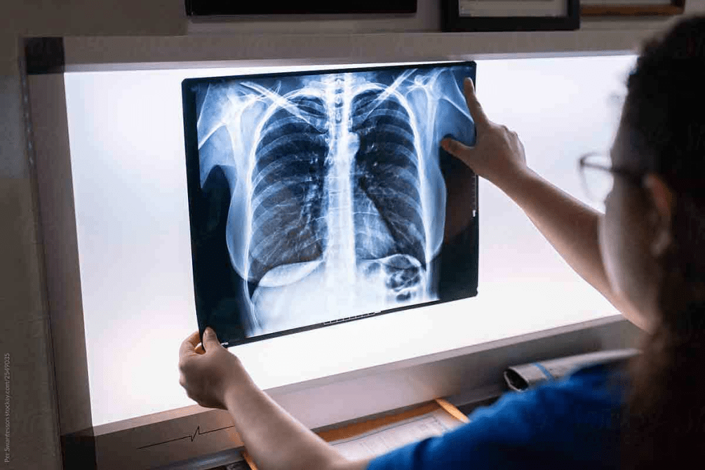
ChCT Scan Chest With Contrast: Essential Findings & Comparisonoosing between a chest CT scan with contrast and one without is key to diagnosing diseases. At Liv Hospital, we pick the best imaging method for each patient. This helps ensure safer and more accurate diagnoses. A CT scan uses X-rays to take pictures of the body’s organs, bones, and tissues.
A CT scan chest with contrast is great for spotting blood vessels, tumors, and organs in the chest. It makes it easier to find vascular problems and cancers. CT scan chest with contrast: What does it show? Get essential facts on how contrast enhances images compared to a non-contrast scan.
Key Takeaways
- CT scans with contrast make blood vessels and veins stand out.
- Non-contrast CT scans are used to check the lungs for infections or diseases.
- The choice between a CT scan with or without contrast depends on the medical condition.
- Contrast agents help show specific areas of the body being examined.
- CT scans are a painless, noninvasive way to find and diagnose medical issues.
The Basics of Chest CT Imaging

A CT scan of the thorax is a high-tech tool that uses X-rays to show detailed images of the chest. It’s great for looking at the lungs, heart, and big blood vessels. Let’s dive into how CT scans work and why they’re so detailed.
What is a CT Scan of the Thorax?
A CT scan of the thorax, or chest CT, is a test that shows what’s inside the chest. It helps doctors find and track many health issues in the lungs, heart, and nearby areas.
How CT Technology Creates Detailed Images
CT technology uses X-rays to take pictures of the body’s inside from different sides. A computer then puts these images together to show detailed cross-sections of the body. The CT scanner moves around the patient, taking X-ray measurements to make these images.
CT scans have changed how we look at the chest. They help doctors find problems early and accurately. The clear images from CT scans are key to planning treatments.
| Key Features | Description |
| Cross-sectional Imaging | CT scans provide detailed images of the body’s internal structures in cross-section. |
| Use of X-rays | CT scans use X-rays to capture images from multiple angles. |
| Computer Reconstruction | A computer reconstructs the X-ray images into detailed cross-sectional images. |
CT Scan Chest With Contrast: An Overview
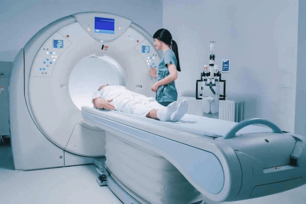
Contrast-enhanced CT scans of the chest are key for checking vascular and tumoral conditions. They help us see the chest’s inside better. The contrast agent, given through an IV, makes certain areas stand out. This makes diagnosing and treating chest issues easier.
Definition and Purpose
A CT scan chest with contrast uses a contrast agent to show certain areas in the chest better. The main goal is to see different tissues and find problems like tumors or vascular diseases.
Types of Contrast Media Used
For chest CT scans, iodine-based contrast is most often used. Iodine-based contrast agents are best because they make blood vessels and some organs show up well.
The right contrast agent depends on the patient’s health and the issue being checked.
How Contrast Agents Enhance Visibility
Contrast agents change how X-rays interact with body tissues. When given, they make certain areas show up more during the CT scan. This helps doctors spot chest problems like tumors or blood vessel issues more accurately.
| Type of Contrast Agent | Primary Use | Characteristics |
| Iodine-based | Vascular structures, tumors | Effective for making blood vessels and some organs visible |
| Barium-based | Gastrointestinal tract | Used for the GI tract, not usually for chest CT scans |
| Gadolinium-based | MRI contrast, sometimes used in CT for specific cases | Less common for CT scans but used in some cases |
CT Chest Without Contrast: When and Why
Choosing a CT chest scan without contrast depends on the patient’s health. We often suggest these scans for quick and accurate diagnoses. This is true when certain medical conditions are suspected.
Common Indications for Non-Contrast Studies
Non-contrast CT scans of the chest help check lung health and find lung nodules. They also look at bone and soft tissue. These scans are great for lung diseases like COPD or pneumonia.
Other reasons for non-contrast CT chest scans include:
- Detecting lung nodules or masses
- Assessing bone metastases or fractures
- Evaluating soft tissue abnormalities
- Guiding interventional procedures, such as biopsies
Benefits of CT Thorax Without Contrast
One big plus of CT thorax without contrast is the lower risk of allergic reactions. This is good for patients with allergies or at risk of kidney problems from contrast.
Also, non-contrast scans are quicker and simpler than those with contrast. This is helpful in emergencies where time is critical.
Key advantages of non-contrast CT chest scans include:
- Reduced risk of allergic reactions
- Faster scan times
- Lower risk of contrast-induced nephropathy
- Cost-effective
We carefully choose when to use non-contrast CT chest scans. This helps us give accurate diagnoses and effective treatments to our patients.
What Does a Chest CT Scan With Contrast Show?
Contrast-enhanced chest CT scans give a detailed look at blood vessels and possible issues. They use a contrast agent to show detailed images. These images help doctors diagnose many conditions.
Enhanced Visualization of Vascular Structures
The contrast agent makes blood vessels stand out in chest CT scans. This makes it easier to spot any problems. “The use of intravenous contrast material improves the detection of vascular abnormalities, such as aneurysms or dissections”. This is key to finding issues with blood vessels in the chest.
Tumor and Mass Characterization
Contrast-enhanced CT scans are great for looking at tumors and masses in the chest. The contrast agent helps tell different tissues apart. This gives doctors important information for treatment plans.
Key benefits of contrast in tumor characterization include:
- Improved delineation of tumor boundaries
- Better assessment of tumor vascularity
- Enhanced detection of tumor necrosis or calcification
Pleural Disease Assessment
Pleural diseases, which affect the lining around the lungs, can be checked with contrast-enhanced chest CT scans. The contrast agent helps spot pleural thickening, effusions, and other issues. Accurate diagnosis is key for managing diseases like pleurisy or pleural mesothelioma.
Medical professionals use contrast-enhanced CT scans to make informed decisions. These scans help see blood vessels, tumors, and pleural diseases clearly. They are essential in today’s diagnostic medicine.
What CT Chest Without Contrast Reveals
A CT chest scan without contrast is a key tool for diagnosing chest issues. It helps us see different chest health aspects, even when contrast isn’t needed or could be harmful.
Lung Parenchyma Evaluation
Non-contrast CT chest scans are mainly used to check the lung tissue. They help spot diseases like emphysema, fibrosis, or pneumonia. These scans show details that regular X-rays can’t.
Lung parenchyma evaluation is key for chronic lung disease diagnosis and tracking. It shows how much lung damage there is and helps decide treatment.
Pulmonary Nodule Detection
Non-contrast CT scans are great at finding lung nodules, which are abnormal lung growths. They can spot nodules as small as a few millimeters. Most nodules are harmless, but CT scans watch their size and shape to check for cancer.
Finding nodules early is vital for lung cancer management. Low-dose CT scans are suggested for lung cancer screening in people at high risk.
Bone and Soft Tissue Assessment
A CT chest without contrast also checks bone and soft tissue issues. It spots fractures, bone metastases, or other bone problems. It also looks at soft tissue masses or lesions outside the lungs.
This detailed check helps find conditions not directly linked to the lungs but are important for overall health.
Key Differences in Diagnostic Capabilities
CT chest scans with and without contrast have different uses. They help doctors see the chest in different ways. This affects how well they can diagnose chest problems.
Image Quality Comparison
Scans with contrast show more detail in blood vessels and tumors. This is because the contrast agent makes these areas stand out more. Contrast-enhanced CT scans are great for spotting and studying lesions, looking at blood vessels, and finding chest issues.
Scans without contrast are better for looking at lung tissue and finding calcifications. They are good enough to spot lung nodules and other lung problems.
Sensitivity and Specificity Differences
The ability to find problems varies with contrast use. Contrast-enhanced scans are better at finding vascular issues and tumors. This is because they show these areas more clearly.
- CT chest with contrast is more sensitive for:
- Detecting vascular abnormalities
- Characterizing tumors and masses
- Assessing pleural disease
Non-contrast scans are safer because they don’t use contrast. They are better for looking at lung tissue and finding calcifications.
Limitations of Each Technique
Both types of scans have downsides. Contrast scans can cause allergic reactions and kidney problems, mainly in those with kidney issues.
Non-contrast scans are safer but might not show all details. They are not as good at finding vascular problems or tumors.
| Feature | CT Chest with Contrast | CT Chest Without Contrast |
| Image Quality for Vascular Structures | High | Low |
| Sensitivity for Tumor Detection | High | Moderate |
| Risk of Contrast-Induced Nephropathy | Present | Absent |
In conclusion, choosing between CT chest scans with or without contrast depends on the situation. It’s about what the doctor needs to see and the patient’s health. Knowing the differences helps doctors make the best choice for their patients.
Clinical Applications: When to Choose Contrast vs. Non-Contrast
Choosing between CT chest scans with or without contrast depends on the patient’s condition. It’s key for good care and accurate diagnosis to know when to use contrast.
Conditions Best Evaluated With Contrast
For vascular diseases, a CT chest with contrast is best. It shows blood vessels clearly, helping spot issues like pulmonary embolism. Tumors and masses also benefit from contrast, as it highlights their blood supply and size.
Mediastinal abnormalities and some infections are also better seen with contrast. It helps distinguish between different structures, making diagnosis easier.
Conditions Best Evaluated Without Contrast
Some conditions don’t need contrast. For example, lung tissue and nodule detection work well without it. Non-contrast scans are also good for looking at bones and soft tissues.
Patients with kidney issues or contrast allergies usually get non-contrast scans. This is safer for them.
| Condition | CT Chest With Contrast | CT Chest Without Contrast |
| Vascular Diseases | Recommended for diagnosis | Limited utility |
| Tumors and Masses | Enhances characterization | May not fully assess extent |
| Lung Parenchyma Evaluation | Not necessary | Effective for evaluation |
| Pulmonary Nodule Detection | Not required | Sufficient for detection |
Knowing when to use contrast in CT chest scans helps doctors choose the best imaging for each patient. This ensures accurate diagnosis and care.
Prep for CT Scan of Chest: Patient Guidelines
Understanding what you need to do before a CT scan of the chest is key. This includes knowing what to do for contrast-enhanced and non-contrast scans. Getting ready right helps make sure the scan goes well and gives accurate results.
Preparation for Contrast-Enhanced Studies
For scans that use contrast, you need to follow some special steps. These steps help avoid risks and make sure the contrast works right.
- Fasting Requirements: You might need to not eat for a while before the scan. This helps avoid choking and makes sure the contrast is absorbed well.
- Allergy Assessment: Tell your doctor if you’re allergic to iodine or contrast agents. This is important to avoid allergic reactions.
- Medication Disclosure: Tell your doctor about all the medicines you take. Some might need to be changed or stopped before the scan.
- Hydration: Drinking plenty of water is often suggested. It helps your kidneys process the contrast agent.
Preparation for Non-Contrast Studies
For non-contrast chest CT scans, the prep is simpler. But there are some things to keep in mind.
- Clothing: Wear loose, comfy clothes without metal parts. You might get a gown to wear during the scan.
- Remove Metal Objects: Take off all metal things, like jewelry, glasses, and dental work, before the scan.
- Informing Your Healthcare Provider: Tell your doctor about any health issues, like claustrophobia or trouble lying down. They can talk about possible help.
By following these steps, you can help make sure your CT scan of the chest is safe and effective. This will give you the best possible results.
Safety Considerations and Possible Risks
CT scans are very useful for finding health problems. But, they also have some safety issues and risks. It’s important for patients to know about these to stay safe and make good choices.
Contrast Allergy and Adverse Reactions
One big safety issue with CT scans is the chance of allergic reactions to contrast media. Contrast agents can cause mild to severe reactions. We check patients for allergies before using contrast to lower this risk.
Common side effects include hives, itching, and a stuffy nose. Rarely, severe reactions like anaphylaxis can happen. We’re ready to deal with emergencies with the right medical help.
Kidney Function and Contrast-Induced Nephropathy
Another important safety point is how contrast media affects the kidneys. Contrast-induced nephropathy (CIN) is a risk, mainly for those with kidney problems or dehydration. We check kidney health before the scan by looking at creatinine levels and making sure patients are hydrated.
For those at higher risk, we might choose other imaging options or use hydration to lower CIN risk.
Radiation Exposure Considerations
CT scans use ionizing radiation, which slightly increases cancer risk. We aim to keep radiation doses as low as possible (ALARA) while getting good images.
We use the latest CT scanners to scan at lower doses. We also plan each scan carefully to keep radiation exposure low for the needed diagnostic information.
By knowing and dealing with these safety issues and risks, we make sure CT scans are safe and useful. This way, we get important health information while keeping patients safe.
How Radiologists Interpret Different CT Chest Studies
Radiologists are key in reading CT chest scans. They use their skills to look at images with and without contrast. This helps them spot many chest problems, like blood vessel issues and lung nodules.
Reading and Analyzing Contrast-Enhanced Images
For contrast-enhanced CT images, radiologists search for clear views of blood vessels and any problems. The contrast agent makes these areas stand out. This makes it easier to find issues like pulmonary embolism or aortic dissection.
They check the enhanced images for:
- Oddities in blood vessels
- Details about tumors and whether they’ve spread
- How far have diseases in the pleura spread
Evaluating Non-Contrast CT Findings
Non-contrast CT scans are looked at for natural differences in tissue density. Radiologists study the lung to find signs of pneumonia or lung fibrosis. They also check bones and soft tissues for any issues, like fractures or tumors.
The main things they look for in non-contrast CT scans are:
- Checking the lungs for diseases
- Finding and understanding lung nodules
- Looking at bones and soft tissues for problems
By studying both types of CT images, radiologists can make accurate diagnoses. This helps doctors decide on the best treatment.
Conclusion
Choosing between a CT scan chest with contrast and one without is key for diagnosing chest issues. We’ve looked at the differences between these two methods. We’ve seen their strengths and when to use them.
A CT scan with contrast is great for checking blood vessels, tumors, and pleural diseases. But a CT scan without contrast is better for lung issues, finding nodules, and looking at bones and soft tissues.
Whether to use contrast or not depends on the question being asked. Knowing the good and bad of each helps doctors make the best choice for their patients.
In short, both CT scans with and without contrast are essential for chest imaging. Picking the right one helps get accurate diagnoses and the best treatment plans.
FAQ
What is a chest CT scan?
A chest CT scan is a test that uses X-rays and computer technology to show detailed images of the chest. It looks at the lungs, heart, and blood vessels.
What is the difference between a CT scan with contrast and without contrast?
A CT scan with contrast uses dye to highlight areas like blood vessels and tumors. Without contrast, it’s used to see lung details and find nodules.
What is contrast media used for in a CT scan?
Contrast media make certain body parts, like blood vessels and tumors, more visible. This helps doctors diagnose many conditions.
How is the contrast agent administered during a CT scan?
The contrast agent is given through an IV line. This lets the dye spread throughout the body before the scan.
What are the benefits of a non-contrast CT chest scan?
Non-contrast CT scans are good for checking lung details and finding nodules. They also look at bones and soft tissue without the risk of allergic reactions.
What does a chest CT scan with contrast show?
A chest CT scan with contrast shows more about blood vessels, tumors, and pleural disease. It helps diagnose conditions like blood clots and cancer.
How do I prepare for a CT scan of the chest with contrast?
To get ready for a CT scan with contrast, you might need to fast for a few hours. Tell your doctor about any allergies or health issues. Arrive early to get the contrast agent.
What are the risks associated with CT scans?
CT scans can expose you to radiation and cause allergic reactions to contrast agents. They can also harm the kidneys, mainly in people with kidney disease.
How do radiologists interpret CT chest studies?
Radiologists look at CT chest images for signs of tumors, nodules, and vascular diseases. They check how well the contrast agent works.
What is the role of CT scans in diagnosing lung diseases?
CT scans are key in finding lung diseases like cancer and COPD. They give detailed images of the lungs and surrounding tissues.
Can I undergo a CT scan if I have kidney disease?
If you have kidney disease, you need to be careful before a CT scan with contrast. The dye can make kidney function worse.
How long does a CT scan of the chest take?
A CT scan of the chest takes a few minutes. But getting ready and processing the images can take longer.
References
- American Academy of Family Physicians. (2013, August 31). When to order contrast-enhanced CT. American Family Physician, 88(5), 312-314. Retrieved October 2025, from https://www.aafp.org/pubs/afp/issues/2013/0901/p312.html
- Jeon, Y. J., et al. (2025). Efficacy of contrast versus non-contrast CT surveillance for pulmonary nodules: A comparative study. Scientific Reports. Retrieved October 2025, from https://www.nature.com/articles/s41598-025-90124-x


