Last Updated on November 27, 2025 by Bilal Hasdemir

Choosing between a CT ultrasound scan and an abdominal ultrasound is key for diagnosing abdominal issues. At Liv Hospital, we know how important it is to pick the right tool for the best care. This choice can greatly affect your diagnosis and treatment.
Recent studies show that CT and ultrasound work well together in diagnosing abdominal problems. They use their strengths to help doctors. We will look at the seven main differences between these imaging methods. This will help you understand their uses, benefits, and limits.
Key Takeaways
- Understanding the diagnostic differences between CT ultrasound scans and abdominal ultrasounds is key for good patient care.
- Each imaging method has its own strengths and weaknesses.
- The right choice between CT ultrasound scans and abdominal ultrasounds depends on the medical condition.
- Things like image quality, radiation, and how accurate they are are important to think about.
- Liv Hospital is dedicated to top-notch healthcare with full support for international patients.
Understanding Medical Imaging Technologies

Medical diagnostics have changed a lot with new imaging technologies. These technologies are key in modern healthcare, helping doctors make accurate diagnoses and plan treatments. We use different imaging methods to see inside the body, each with its own strengths and uses.
The Evolution of Diagnostic Imaging
Diagnostic imaging has grown a lot over the years. It started with X-rays and now we have CT scans and ultrasounds. Each new technology has helped us diagnose and treat diseases better.
The CT scans have been a big step forward. They give detailed images that have changed emergency care and complex diagnoses. Professionals say, “The introduction of CT scans has dramatically improved our ability to quickly and accurately assess critical conditions.”
“Imaging technologies have transformed the way we practice medicine, giving us insights we never thought possible.”
The Role of Imaging in Modern Medicine
In today’s medicine, imaging is very important for patient care. It helps doctors see inside the body, diagnose problems, and check how treatments are working. Understanding medical imaging helps us see how these technologies help patients.
Ultrasound technology is a safe way to look at soft tissues and check on pregnancies. It’s very important in obstetrics and other areas.
We use these technologies to make treatment plans, track disease, and improve care. Choosing between CT scans and ultrasounds depends on the condition, patient safety, and how detailed the image needs to be.
What Is a CT Ultrasound Scan?

A CT ultrasound scan, also known as a CT scan, uses X-rays and computer tech to show internal organs and tissues clearly. It’s a key tool in medicine, letting doctors see inside the body without surgery.
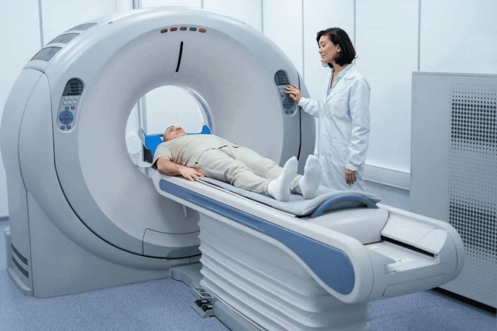
CT scans help find many health issues, like injuries, cancers, and heart problems. The tech behind it is advanced. It involves moving X-ray detectors around the patient to get detailed images from all sides.
Technology Behind CT Scanning
CT scanning works by using X-rays differently in various tissues. Detectors around the patient capture these differences from many angles. Then, computers turn this data into detailed images.
Key parts of CT scanning tech are:
- X-ray Tube: Sends X-rays to the patient.
- Detectors: Catch the X-rays that go through the patient.
- Computer System: Makes the images from the data.
How CT Scans Generate Cross-Sectional Images
CT scans make images by taking X-ray data from many angles. This data is then turned into detailed pictures of inside structures. This helps doctors diagnose and plan treatments accurately.
Typical CT Scan Procedure
A CT scan usually takes about 5 minutes. The patient lies on a table that moves into a big, ring-shaped machine. It’s important for the patient to stay very quiet and not move.
| Procedure Step | Description | Duration |
| Preparation | Patient is positioned on the CT table. | 2-3 minutes |
| Scanning | CT scanner captures images. | 5 minutes |
| Image Reconstruction | Computer reconstructs images. | 5-10 minutes |
Knowing how a CT scan works helps us see its value in medicine. It gives doctors detailed images to diagnose and treat many health problems well.
Abdominal Ultrasound Explained
Abdominal ultrasound technology is key in medical care. It lets us see inside the body without surgery. We use it to check on organs like the liver, gallbladder, and kidneys. This helps us understand how well they’re working.
Principles of Ultrasound Technology
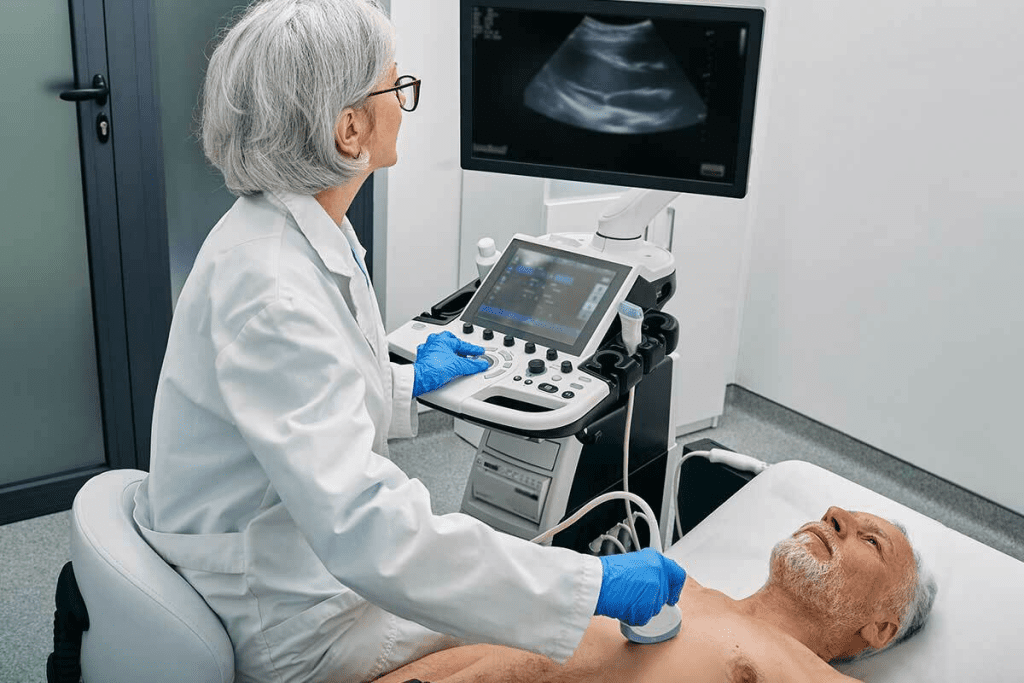
Ultrasound uses sound waves to create images inside the body. A transducer sends out these sound waves. They bounce back and give us clear images in real-time.
Key Components of Ultrasound Technology:
- Transducer: Emits and receives sound waves.
- Sound Waves: High-frequency waves that penetrate tissues.
- Image Processing: Converts received sound waves into visual images.
The Process of Conducting an Abdominal Ultrasound
To do an abdominal ultrasound, we first apply gel to the abdomen. This helps sound waves move better. Then, we use the transducer to take pictures of the organs inside.
The whole process takes about 15-30 minutes. Our skilled team does the ultrasound. They make sure the images are clear and accurate.
Types of Abdominal Ultrasounds
There are many types of abdominal ultrasounds. Each one has its own purpose. Here are a few common ones:
| Type | Description | Clinical Use |
| Complete Abdominal Ultrasound | Comprehensive examination of all abdominal organs. | General assessment of abdominal health. |
| Limited Abdominal Ultrasound | Focused examination of specific organs or areas. | Targeted assessment for specific conditions. |
| Doppler Ultrasound | Assesses blood flow through vessels. | Diagnoses vascular conditions and monitors blood flow. |
A medical expert notes, “Ultrasound is vital because it’s safe and doesn’t use harmful radiation. It’s great for pregnant women and for checking on people often.”
“Ultrasound technology has changed how we diagnose and treat diseases. It gives us images right away that help us make decisions.”
Difference #1: Image Resolution and Detail
CT scans and abdominal ultrasounds differ mainly in their image quality. This affects how well they can diagnose and treat diseases.
CT Scan’s Superior Cross-Sectional Imaging
CT scans are known for their detailed, cross-sectional images. They are great for spotting small issues and understanding complex body parts.
Key benefits of CT scan imaging include:
- High-resolution images of internal organs and structures
- Ability to detect small lesions and abnormalities
- Detailed visualization of complex anatomy
For example, CT scans can show the liver, pancreas, and kidneys clearly. This helps doctors see any problems with these organs.
Ultrasound’s Real-Time Imaging Advantages
Ultrasounds, on the other hand, offer live images. This is great for watching how organs move and for guiding treatments.
Real-time ultrasound imaging is beneficial for:
- Guiding needle biopsies and other interventional procedures
- Assessing blood flow and vascular function
- Evaluating organ movement and function in real-time
Clinical Impact of Resolution Differences
The quality of images from CT scans and ultrasounds matters a lot in medicine. CT scans are better for finding small or complex problems. Ultrasounds are better for watching how things move and for guiding treatments.
| Imaging Modality | Image Resolution | Clinical Applications |
| CT Scan | High-resolution, cross-sectional images | Detecting small abnormalities, complex anatomy, and assessing disease extent |
| Ultrasound | Real-time images, lower resolution than CT | Guiding interventions, assessing dynamic processes, evaluating organ movement |
Knowing these differences helps doctors pick the best imaging method for each patient. This improves diagnosis and care.
Difference #2: Radiation Exposure and Safety Concerns
When we talk about diagnostic imaging, we must think about radiation exposure. This is a big difference between CT scans and ultrasounds. Medical professionals need to consider the risks and benefits of each.
Ionizing Radiation in CT Scans
CT scans use X-rays to make detailed images of the body. This means they expose patients to ionizing radiation. This kind of radiation can damage DNA and raise cancer risks, mainly in kids and young adults.
The amount of radiation from a CT scan depends on the type and the body part being scanned. For example, an abdominal CT scan might give a patient about 10 millisieverts (mSv) of radiation. This is like the background radiation a person gets in a year from nature. While one CT scan is usually safe, many scans can add up and increase lifetime exposure.
Radiation-Free Nature of Ultrasound
Ultrasound imaging doesn’t use ionizing radiation. It uses sound waves to see inside the body. This makes ultrasound safer for people who need many scans or are in sensitive groups like pregnant women and kids.
Ultrasound is often chosen for safe imaging, like in pregnancy or checking soft tissues. But, it might not show as much detail as CT scans for some issues, like complex injuries or detailed blood vessels.
Risk-Benefit Analysis for Different Patient Groups
Choosing between CT scans and ultrasounds depends on the patient’s needs and risks. Pregnant women and kids usually get ultrasounds because they’re radiation-free.
For others, the choice is based on weighing the benefits of the scan against the risks. Here’s a table that shows the main differences in radiation and safety between CT scans and ultrasounds:
| Characteristics | CT Scan | Ultrasound |
| Radiation Exposure | Ionizing radiation (varies by dose) | No ionizing radiation |
| Safety for Pregnant Women | Generally avoided unless necessary | Safe |
| Safety for Children | Potential long-term cancer risk | Safe |
Knowing these differences helps us make choices that protect patients while meeting their medical needs. This way, we can give the best care possible.
Difference #3: Diagnostic Accuracy for Various Conditions
CT scans and ultrasounds have different levels of accuracy for different health issues. This affects how doctors make decisions. Knowing these differences helps pick the best imaging test for each patient.
CT Scan Superiority for Tumors, Bleeding, and Trauma
CT scans are better at finding problems like tumors, bleeding, and trauma. They give detailed views of inside injuries. This is very helpful in emergency situations where quick diagnosis is key.
Key advantages of CT scans include:
- High-resolution imaging of internal organs and structures
- Rapid scanning time, critical in emergencies
- Can spot many acute conditions, like tumors and bleeding
Ultrasound Advantages for Soft Tissue and Gallstones
Ultrasounds are great for checking soft tissues and finding gallstones. They show real-time images, which helps with biopsies and checking organ work. They’re safe because they don’t use harmful radiation, making them good for pregnant women and for long-term monitoring.
The benefits of ultrasound include:
- Real-time imaging for dynamic assessment of organs
- No harmful radiation, safe for repeated use
- Good for finding gallstones and soft tissue issues
Comparative Sensitivity and Specificity in Common Diagnoses
CT scans and ultrasounds have different strengths for common health issues. The right choice depends on the patient’s situation and what the doctor suspects.
| Condition | CT Scan Sensitivity | Ultrasound Sensitivity | CT Scan Specificity | Ultrasound Specificity |
| Tumors | High | Moderate | High | Moderate |
| Bleeding | High | Low | High | Moderate |
| Gallstones | Moderate | High | High | High |
| Trauma | High | Low | High | Moderate |
In summary, CT scans are better for finding acute problems like tumors and bleeding. Ultrasounds are better for soft tissues and gallstones. Knowing these differences helps doctors give the best care to patients.
Difference #4: Procedure Duration and Patient Experience
It’s important to know how CT scans and ultrasounds differ in terms of time and patient experience. These factors greatly affect how happy patients are and how well they follow treatment plans. This, in turn, impacts the success of the diagnosis.
Quick CT Scans vs. More Interactive Ultrasounds
CT scans are fast, taking about 5 minutes. This makes them great for emergencies when every second counts. Ultrasounds, on the other hand, take around 15 minutes. They involve more back and forth between the patient and the doctor.
Patient Preparation and Comfort
How long a procedure takes also affects how ready patients need to be and how comfortable they feel. For CT scans, patients must stay very quiet for a short time. Sometimes, a special dye is used to make images clearer. Ultrasounds need patients to be in a certain position for the best pictures. This might mean the doctor needs to touch the skin with a probe, which might need gel.
To understand these differences better, let’s look at a comparison of key points related to time and patient experience:
| Aspect | CT Scan | Ultrasound |
| Procedure Duration | Approximately 5 minutes | Approximately 15 minutes |
| Patient Interaction | Limited, requires remaining very quiet | More interactive, involves positioning and sometimes feedback |
| Preparation Requirements | May require contrast agent, fasting, or specific attire | Generally requires less preparation, may need to drink water |
Healthcare providers can make better choices by thinking about these points. Whether it’s a CT scan or an ultrasound, it’s key to match the procedure to the patient’s needs. This way, the best results are achieved for everyone involved.
Difference #5: Cost Considerations and Insurance Coverage
It’s important to know the costs of CT scans and ultrasounds. This knowledge helps patients and doctors make better choices. The cost of tests affects how easy it is to get care and how much it costs.
Procedure Costs in the United States
Prices for CT scans and ultrasounds change a lot in the U.S. CT scans usually cost more because of the advanced tech used.
| Procedure | Average Cost Range |
| CT Scan | $1,000 – $3,000 |
| Ultrasound | $200 – $800 |
Many things can affect these prices. These include where the test is done, who does it, and the tech used.
Insurance Coverage Patterns
Insurance for CT scans and ultrasounds varies. Most plans cover both, but how much they cover can differ.
- CT Scans: Usually covered for certain health issues, but might need approval first.
- Ultrasounds: Often covered for regular checks and some tests.
Knowing what your insurance covers is key to avoid surprise bills.
Long-term Economic Impact
Choosing between CT scans and ultrasounds affects money in the long run. CT scans give detailed images but are pricier and use radiation. Ultrasounds are cheaper and don’t use radiation, but might not be as detailed.
Thinking about these points helps decide the best test. It’s about finding a balance between getting the right diagnosis and keeping costs down.
When picking a test, we must weigh short-term needs against long-term effects on health and money.
Difference #6: Accessibility and Availability
When it comes to medical diagnostics, CT scans and ultrasounds have big differences. These differences are key in choosing the right imaging modality for different clinical settings.
Hospital vs. Outpatient Settings
CT scans usually happen in imaging centers within hospitals or outpatient facilities. These places have the right setup for CT scanning’s complex tech. Ultrasounds, though, can happen in many places like emergency rooms, clinics, and even at the bedside.
Ultrasound’s flexibility makes it great for quick and easy diagnoses, which is super helpful in emergencies or for patients who can’t move to imaging centers.
Equipment Requirements and Availability
CT scans need special, big machines that require skilled techs to run. Ultrasound machines, on the other hand, are smaller, easier to use, and can go in many healthcare places.
| Imaging Modality | Equipment Requirements | Typical Settings |
| CT Scan | Complex, large machinery | Dedicated imaging centers |
| Ultrasound | Portable, less complex | Various settings, including bedside |
Emergency vs. Scheduled Imaging Considerations
In emergencies, how fast and where you can get imaging matters a lot. Ultrasounds are often the go-to because they’re easy to move and quick to set up. For planned imaging, both CT scans and ultrasounds can be used, depending on what’s needed and the patient’s situation.
“The portability and ease of use of ultrasound make it an invaluable tool in emergency medicine, allowing for quick assessments and decisions.”
”A renowned emergency medicine specialist.
Looking at accessibility and availability, it’s clear that choosing between CT scans and ultrasounds depends on many things. These include the situation, what the patient needs, and what each imaging modality can do.
Difference #7: Clinical Applications and Limitations
CT scans and abdominal ultrasounds serve different purposes in healthcare. Knowing their uses and limits helps doctors make the right choice for each patient. This choice depends on the patient’s condition and what the doctor needs to know for treatment.
When CT Scans Are the Preferred Choice
CT scans are best in emergencies, like trauma cases, where fast and detailed images are key. They’re also top for complex issues like tumors, internal injuries, and blood vessel diseases. Their clear images help doctors see how severe an injury or disease is.
Emergency Situations: For severe injuries or sudden abdominal pain, CT scans offer quick, detailed images. These images help doctors make fast treatment decisions.
When Abdominal Ultrasounds Are More Appropriate
Ultrasounds are better for checking soft tissues, monitoring pregnancies, and looking at liver or gallbladder issues. They’re also safer for long-term checks because they don’t use harmful radiation.
Soft Tissue Evaluation: Ultrasounds are great for soft tissues and organs like the liver, gallbladder, and kidneys. They give real-time images that help spot problems like gallstones or liver disease.
Complementary Use of Both Technologies
Often, doctors use CT scans and ultrasounds together for better results. A CT scan might first spot a problem, then an ultrasound follows for more detailed checks. This team effort helps doctors get a clearer picture of what’s going on.
Understanding the strengths and weaknesses of CT scans and ultrasounds helps doctors choose the best imaging for each patient. Using these technologies wisely, either alone or together, is key to accurate diagnoses and effective treatments.
How Doctors Choose Between CT Ultrasound Scan and Abdominal Ultrasound
Doctors have to decide between CT scans and ultrasounds based on many factors. They consider what the patient needs and the best way to see inside the body. This choice helps them find the right way to diagnose and treat patients.
Decision-Making Factors for Physicians
Doctors look at several things when choosing between CT scans and ultrasounds. They think about the patient’s situation and what they need to find out. For example, CT scans are better for seeing detailed images of the body, like tumors or injuries.
A study on NCBI shows that the right choice can make a big difference in finding the right diagnosis.
Diagnostic accuracy is key. CT scans are better for finding things like bleeding or broken bones because they show more detail. Ultrasounds, on the other hand, are great for looking at soft tissues and the gallbladder because they show things in real time.
Patient-Specific Considerations
What’s best for the patient also matters a lot. For example, people with kidney disease might do better with an ultrasound because CT scans use contrast agents. Pregnant women and kids usually get ultrasounds to avoid radiation.
Patient comfort and safety are also important. Ultrasounds are safer because they don’t use radiation and are less invasive.
The Future of Integrated Diagnostic Approaches
The future of medical imaging is combining different methods, like CT scans and ultrasounds. This mix can make diagnoses more accurate and help patients get better faster. It lets doctors see more clearly what’s going on inside the body.
As technology gets better, we’ll see more of these combined approaches. This will help doctors tailor treatments to each patient’s needs.
Conclusion
We’ve looked at how CT ultrasound scans and abdominal ultrasounds differ. Each has its own strengths and weaknesses. The right choice depends on things like image quality, radiation, accuracy, and the situation.
Healthcare experts can pick the best imaging method for each case. CT scans are great for detailed views and accuracy in some cases. On the other hand, abdominal ultrasounds are safe, affordable, and work well for others.
As technology gets better, using CT scans and ultrasounds together will keep being key to good care. It’s important to tailor imaging to each patient’s needs. This way, patients get the best care possible.
In short, knowing the differences between CT scans and ultrasounds helps doctors make better choices. This leads to better care and outcomes for patients.
FAQ
What’s the main difference between a CT scan and an ultrasound of the abdomen?
CT scans use X-rays to create detailed images. Ultrasounds use sound waves to show internal organs. This makes them different in how they work.
Which is safer, a CT scan or an ultrasound for pregnant women?
Ultrasounds are safer for pregnant women. They don’t use harmful radiation. This makes them better for checking on the baby and the mother.
How do CT scans and ultrasounds compare in terms of diagnostic accuracy for detecting tumors?
CT scans are better at finding tumors, thanks to their detailed images. But, ultrasounds are good for guiding biopsies and checking some tumors.
Are CT scans or ultrasounds more cost-effective for routine abdominal assessments?
Ultrasounds are cheaper for regular checks. They’re good for soft tissues and finding gallstones. They also don’t use radiation.
Can both CT scans and ultrasounds be used together for diagnostic purposes?
Yes, they can work together. For example, a CT scan might find a tumor. Then, an ultrasound could help take a biopsy from it.
How does radiation exposure from CT scans compare to that from ultrasounds?
CT scans use harmful radiation. Ultrasounds don’t. This makes ultrasounds safer for people who need many scans or are sensitive, like pregnant women and kids.
What are the typical procedure times for CT scans and ultrasounds?
CT scans are quick, done in about 5 minutes. Ultrasounds take longer, around 15 minutes. This lets ultrasounds be more interactive.
How do the costs of CT scans and ultrasounds compare in the United States?
CT scans cost more because of the technology and the need for special equipment and people. Ultrasounds are cheaper.
Are CT scans or ultrasounds more accessible in emergency settings?
CT scans are common in hospitals and used in emergencies for quick, detailed images. Ultrasounds are also used, often for bedside checks.
Can ultrasounds detect conditions that CT scans might miss?
Yes, ultrasounds can find things like gallstones or soft tissue issues that CT scans might not show. Their real-time images are very helpful.
How do doctors decide between ordering a CT scan or an ultrasound?
Doctors choose based on the situation and the patient. CT scans are often used for injuries or complex cases. Ultrasounds are better for soft tissues or checking on a pregnancy.
What’s the future of using CT scans and ultrasounds in diagnostic medicine?
The future is combining both for better care. New technology will make them even more useful. This will help doctors make more accurate diagnoses.
References
“A comparison of the Accuracy of Ultrasound and Computed Tomography for common abdominal diagnoses. (n.d.). https://pmc.ncbi.nlm.nih.gov/articles/PMC3101356/


