
Last Updated on October 30, 2025 by Bilal Hasdemir
Learning about the large intestine is key for medical students and experts. It’s a big part of our digestive system. Its many parts need to be labeled right for us to learn well.Learn about the diagram of large bowel with labeled parts and functions explained in simple terms.
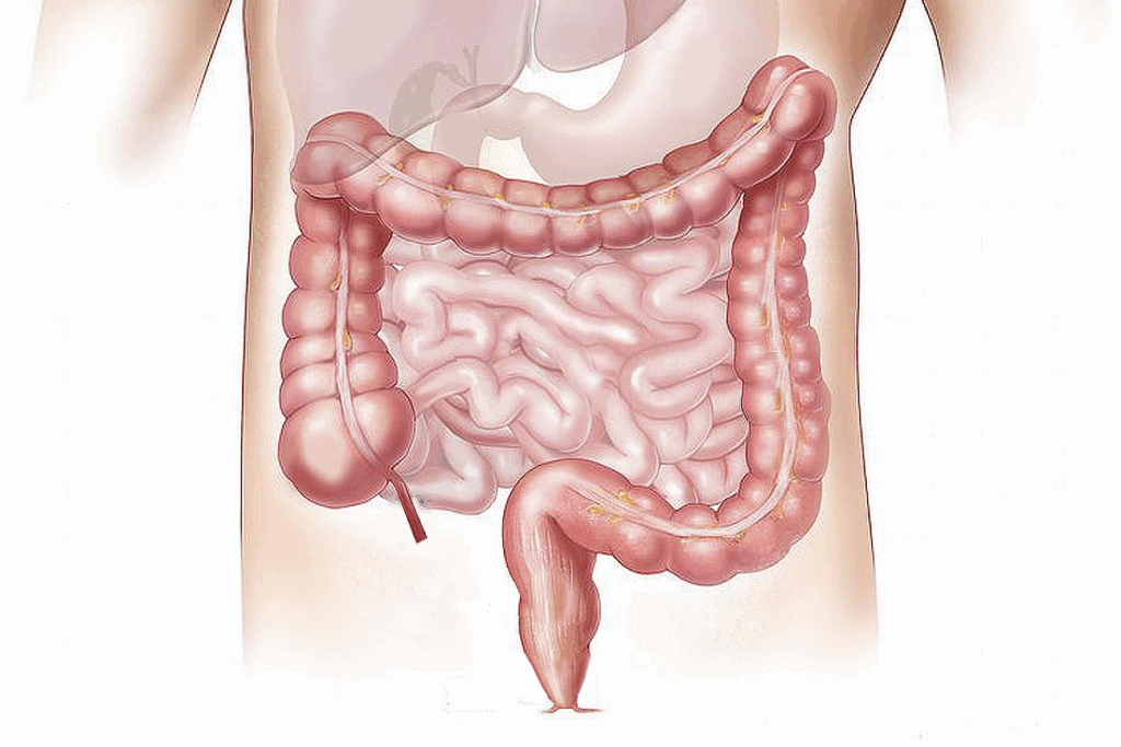
A detailed labeled large intestine diagram helps us spot important parts. These include the cecum, appendix, and the ascending, transverse, descending, and sigmoid colon. Also, the rectum and anal canal. Liv Hospital says clear learning tools are vital for good care.

Knowing the large intestine’s structure is key for future doctors. The large intestine, or colon, is vital in digestion. It’s a must-know for medical students.
Labeling the large intestine’s parts accurately is vital. Correct identification of areas like the cecum and anus is essential. The large intestine has four main sections, each with unique features.
In medical school, precise labeling is critical. It helps students grasp the complex relationships between different parts. This skill is vital for exams and real-world practice, where mistakes can cause serious issues.
The large intestine is about 150cm long, from the ileocecal junction to the anus. Its size and where it sits are key to its anatomy. It’s split into four parts: ascending, transverse, descending, and sigmoid colon.
Knowing the large intestine’s size and position is important. It helps students understand its path in the abdomen. This knowledge is key for diagnosing and treating issues like colon cancer.
By learning the large intestine’s anatomy, students can better understand the digestive system. This knowledge boosts their clinical abilities.

The large intestine is key in the digestive system. It helps process food and absorbs nutrients. It also makes sure waste is removed correctly.
Food moves smoothly from the small intestine to the large intestine. The ileocecal valve controls this move. It lets chyme flow into the large intestine.
This step is important. It starts the large intestine’s work. This includes absorbing water, storing feces, and fermentation by gut bacteria.
The large intestine has several main jobs:
Knowing these roles helps us see why the large intestine is vital. It keeps us healthy and prevents digestive problems.
The large bowel diagram is key for medical education. It shows the large intestine’s parts clearly. Knowing the large intestine’s anatomy is vital for students, and a detailed diagram helps a lot.
To use large intestine diagrams well, start by knowing the main parts. Look for the cecum, colon, and rectum. Labeling key structures makes them stick in your memory.
A labeled large intestine diagram helps you see how parts fit together. This is great for exams or surgery prep.
Looking at the large intestine from different angles helps you get a better picture. 3D models and cross-sectional diagrams give more insight into its structure and function.
| Perspective | Description | Benefits |
| Anterior View | Front-facing view of the large intestine | Provides an overview of the major structures |
| Lateral View | Side view of the large intestine | Helps understand the spatial relationships between structures |
| 3D Model | Three-dimensional representation of the large intestine | Enhances understanding of complex anatomical relationships |
Using both 2D diagrams and 3D models helps students grasp the large intestine’s anatomy better. This is key for success in school and in the clinic.
The cecum is a sac-like structure at the start of the large intestine. It’s key in digestion. It gets chyme from the ileum, the last part of the small intestine.
The cecum has a pouch-like shape. It’s found in the right lower quadrant of the abdomen. It’s a key spot in large intestine anatomy, showing where the small intestine ends.
Its location is important for intestine labeling. It connects to the ileum through the ileocecal valve. This valve controls what goes into the large intestine.
The cecum’s main job is to absorb water and salt from the chyme it gets from the ileum. This helps keep the body’s fluids and electrolytes balanced.
It also has many microbes. These microbes help break down undigested carbs. They make short-chain fatty acids, which give energy to the colon’s lining.
| Characteristics | Description |
| Location | Right lower quadrant of the abdomen |
| Shape | Pouch-like or sac-like |
| Primary Function | Receives chyme from the ileum; continues water and salt absorption |
The appendix is a small, winding tube attached to the cecum. It plays a key role in the large intestine’s function. Despite being seen as vestigial, it has caught the eye of medical studies for its possible immunological roles.
The appendix is usually found near where the small and large intestines meet, attached to the cecum. Its exact spot can differ from person to person. It might even drop down into the pelvis or hide behind the cecum.
Key Anatomical Features:
Recent research has revealed the appendix’s role in digestion. Once thought to be useless, it’s now seen as a place for good bacteria. This helps the gut microbiome bounce back after sickness or other gut problems.
| Function | Description |
| Reservoir for beneficial bacteria | Aids in recovery of gut microbiome |
| Immunological role | Contains lymphoid tissue, potentially supporting immune function |
The appendix’s importance in large intestine anatomy is now clear. It plays a part in keeping the intestine healthy. It’s vital to accurately label the large intestine in studies to grasp the appendix’s role and its health benefits.
The colon is a key part of the large intestine. It has three main segments: the ascending colon, transverse colon, and descending colon. These parts are vital for absorbing water, storing waste, and fermentation.
The ascending colon starts at the cecum and goes up on the right side of the abdomen. It absorbs water and electrolytes from undigested material.
Key Features of the Ascending Colon:
The transverse colon is the second segment. It crosses the abdomen from right to left. It’s the longest part of the colon and is very mobile.
The transverse colon’s position and mobility make it a critical area for the mixing and movement of intestinal contents.
The descending colon is the third segment. It goes down the left side of the abdomen. It continues to absorb water and stores waste until it reaches the sigmoid colon.
| Colon Segment | Location | Primary Function |
| Ascending Colon | Right side of abdomen | Water and electrolyte absorption |
| Transverse Colon | Across the abdomen | Mixing and movement of contents |
| Descending Colon | Left side of abdomen | Water absorption and waste storage |
Knowing about the anatomy and functions of these segments is key for diagnosing and treating large intestine issues. The roles of the ascending, transverse, and descending colon show how complex and important the colon is in digestion.
The sigmoid colon is a key part of the large intestine. It connects the descending colon to the rectum with its unique S-shape. This shape helps move and store waste before it’s eliminated.
The sigmoid colon has an S-shape that fits in the pelvic cavity. This shape is not just for looks; it helps store fecal matter temporarily. It’s located between the descending colon and the rectum, linking the large intestine.
This location is important. It helps waste move from the descending colon to the rectum efficiently. The sigmoid colon’s position in the pelvis also makes it somewhat mobile, which is important in some medical situations.
The sigmoid colon has several key roles before waste leaves the body. Its main job is to hold fecal matter temporarily. This allows for the absorption of water and electrolytes, which is essential for staying hydrated and balanced.
Key Functions of the Sigmoid Colon:
| Function | Description |
| Storage | Temporary holding of fecal matter |
| Absorption | Water and electrolyte absorption |
| Movement | Facilitating waste towards the rectum |
In conclusion, the sigmoid colon is a vital part of the large intestine. Its S-shape is key to the digestive process. Knowing about its anatomy and functions helps us understand the human digestive system better.
The rectum and anal canal are the last parts of the large intestine. They are key for storing and getting rid of waste. Together, they help finish the digestive process.
The rectum is the last part of the large intestine. It holds feces until it’s time to get rid of them. Its dilated structure lets it hold waste until it’s ready to go.
The rectal wall has mucous membranes. These help move stool along.
The anal canal is the last part of the digestive system. It connects the rectum to the anus. It plays a big role in keeping waste in and letting it out when needed.
The anal canal has sphincter muscles around it. These muscles control when stool comes out. This ensures waste is released at the right time.
The anal canal meets the outside world at the anal verge. Knowing how the rectum and anal canal work is important. It helps doctors diagnose and treat problems in these areas.
The large intestine has unique features that help us understand it better. These features are key for accurate labeling in diagrams. They also play a big role in how the large intestine works.
The haustra are pouches in the large intestine’s wall. They form due to the taenia coli, three narrow muscle bands. These bands run along the large intestine’s length.
Haustra make the large intestine look unique. They are vital for identifying it in drawings.
The taenia coli are three bands of smooth muscle. They are shorter than the large intestine. This causes the intestine to pucker and form haustra.
These muscle bands are key for moving contents through the large intestine.
Epiploic appendages are small pouches filled with fat. They are attached to the large intestine’s outer wall.
They are a normal part of the large intestine’s anatomy. You can see them in anatomical diagrams.
| Feature | Description |
| Haustra | Sacculations formed by the contraction of taenia coli |
| Taenia Coli | Three longitudinal bands of smooth muscle |
| Epiploic Appendages | Fat-filled pouches attached to the outer wall |
Large intestine diagrams have many uses in medicine. They help doctors diagnose and plan surgeries. These diagrams show the large intestine’s layout, which is key for understanding many gut problems.
Diagnosing gut issues is easier with large intestine diagrams. They help spot problems like colon cancer, diverticulitis, and inflammatory bowel disease. These diagrams make it easier to see where and how big the issue is.
For example, if someone thinks they have colon cancer, a detailed diagram helps surgeons know where to cut. This ensures they remove the tumor safely.
Large intestine diagrams are key for planning surgeries. Surgeons use them to understand the patient’s gut and plan the best surgery. This is very important for complex surgeries like colectomies.
These diagrams also help explain the surgery to patients. This makes patients more informed and ready to follow their treatment plan.
Modern research has greatly improved our understanding of the large intestine. Advanced imaging techniques have helped us see its complex structures and functions more clearly.
New imaging methods have changed how we study the large intestine. 3D modeling and virtual colonoscopy allow us to see its detailed structures.
These methods have also made diagnosis more accurate. They help with preoperative planning and educational training for doctors.
New studies have revealed more about the large intestine. They focus on its role in gut microbiota and health.
Important findings include new subtle anatomical features. We now understand its role in many gastrointestinal disorders better.
These breakthroughs are important for both doctors and researchers.
Learning the large intestine’s anatomy is key for medical pros to shine. It’s vital to know its complex parts and how they work. This knowledge helps in diagnosing and treating gut problems accurately.
Studying the large intestine’s anatomy is a big part of medical education. It’s important to label and see these structures clearly. This helps doctors understand the digestive system better.
Knowing the details of large intestine anatomy boosts doctors’ skills. It leads to better care for patients. As medical education grows, studying the anatomy will stay essential for top-notch care.
The main parts of the large intestine include the cecum, appendix, and ascending colon. Also, the transverse, descending, and sigmoid colon, rectum, and anal canal are labeled.
Medical students need to know the large intestine’s parts well. This knowledge helps them diagnose and treat digestive problems effectively.
The large intestine’s main job is to take in chyme from the small intestine. It absorbs water and electrolytes and turns them into feces.
Students can study large intestine diagrams by learning about each part and how they work together. Using 3D models helps visualize these structures better.
The cecum is the first part of the large intestine. It receives chyme from the small intestine. Knowing its anatomy and location is key to understanding its role in digestion.
Today, we think the appendix might help keep enteric bacteria in the gut. Even though it’s seen as vestigial, it could have a useful role.
The colon has three parts: the ascending, transverse, and descending colon. These segments are important for absorbing water and electrolytes.
The sigmoid colon’s S-shape and where it is located are key to its function. It connects the descending colon to the rectum and helps with waste elimination.
The large intestine can be identified by its haustra, taenia coli, and epiploic appendages. These features are important for understanding its anatomy.
Large intestine diagrams are useful in diagnosing digestive issues and planning surgeries. They help doctors understand the anatomy of the large intestine.
New research and imaging techniques, like 3D modeling, have greatly improved our view of the large intestine’s anatomy. This has deepened our understanding of its structures.
Khalil, H. M., et al. (2021). Biliary leakage following cholecystectomy: A prospective population study. Journal of Research in Medical and Dental Science, 9(5), 289-296. Retrieved from https://www.jrmds.in/articles/biliary-leakage-following-cholecystectomy-a-prospective-population-study-84919.html