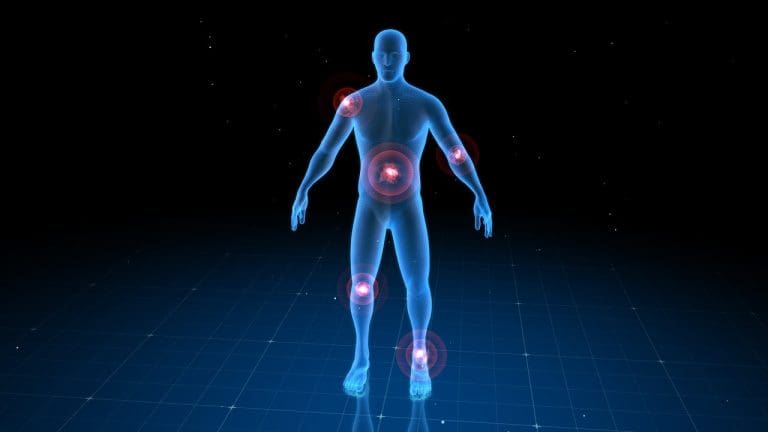
Medical imaging has changed how we diagnose diseases. PET scans and MRI are at the forefront of this change. Every year, over 2 million PET scans are done in the U.S. They help doctors diagnose and treat complex diseases.
PET scans are great at showing how active cells are. On the other hand, MRI gives detailed pictures of the body’s structure. Knowing the difference between PET scan and MRI helps both patients and doctors make better choices.
Key Takeaways
- PET scans and MRI serve different diagnostic purposes.
- PET scans assess metabolic activity, while MRI provides anatomical details.
- Understanding the differences is key to informed decision-making.
- Each imaging technique has its unique applications.
- The choice between PET scan and MRI depends on the medical condition.
Understanding Medical Imaging Technologies
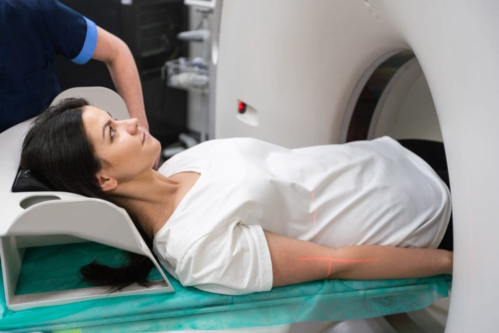
Medical imaging technologies have made big strides, helping doctors make better decisions. These advancements have changed how we diagnose diseases. They’ve brought us from simple X-rays to complex scans like PET and MRI.
The Evolution of Diagnostic Imaging
Diagnostic imaging has grown a lot, from simple X-rays to advanced scans. Now, we can see different tissues and even how they work. This is thanks to new technologies.
“New imaging technologies have changed medicine a lot,” says a top radiology expert. “They help doctors find and treat diseases better.”
How Physicians Choose Imaging Methods
Doctors pick imaging based on the patient’s condition and what they need to see. For example, MRI is great for soft tissues. PET scans are used to see how tissues work.
- Clinical presentation and patient history
- The suspected underlying condition
- The need for functional versus structural imaging
The Importance of Appropriate Imaging Selection
Picking the right imaging is key for accurate diagnosis and care. Wrong choices can cause delays, harm from too much radiation, and higher costs.
Choosing the right imaging means knowing what each method can do. For example, MRI and PET scans show different things. The right choice depends on the situation.
As imaging tech gets better, knowing how to use it is more important. By picking the best imaging, doctors can give better care and improve results.
What is a PET Scan?
Positron Emission Tomography, or PET scan, is a cutting-edge medical imaging method. It shows how active different parts of the body are. This tool is key for spotting problems and understanding how the body works.
The Science Behind Positron Emission Tomography
PET scans use a small amount of radioactive tracer. This tracer goes to areas where cells are very active, like in cancer. It then releases positrons, which collide with electrons to make gamma rays.
These gamma rays are caught by the PET scanner. It uses this info to make detailed pictures of the body’s inner workings.
Radioactive Tracers and Metabolic Activity
The tracers in PET scans target specific body processes. For example, Fluorodeoxyglucose (FDG) goes to cells based on how much glucose they use. This helps spot areas with high activity, like tumors.
Choosing the right tracer depends on what you want to find out. Different tracers show different things, making PET scans very useful.
PET Scanner Equipment and Technology
PET scanner machines are advanced devices that catch gamma rays. They use new tech to make clear images of the body’s activity. Some scanners work with CT or MRI to show both function and structure at once.
As tech gets better, so do PET scanners. They’re getting more accurate and detailed, helping doctors more than ever.
What is an MRI?
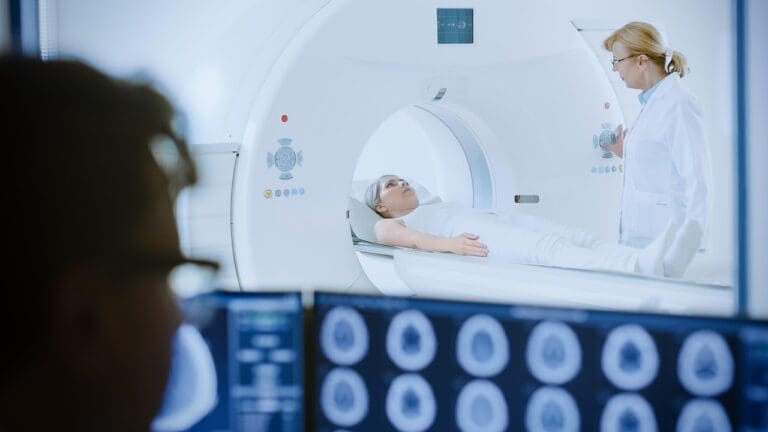
Magnetic Resonance Imaging (MRI) is a cutting-edge medical imaging method. It uses strong magnetic fields and radio waves to show detailed pictures of the body’s inside parts.
Magnetic Resonance Imaging Fundamentals
MRI works on the idea of nuclear magnetic resonance. When you get an MRI, you sit in a strong magnetic field. This field lines up the hydrogen nuclei in your body.
Then, radio waves are sent to mess with these nuclei. This makes them send out signals. These signals help make the images you see.
The strength of the magnetic field is very important. Modern MRI machines have magnets that are 1.5 or 3 Tesla strong. This helps make clear images.
How Magnetic Fields Create Images
Creating MRI images involves a few steps. First, the magnetic field lines up the hydrogen nuclei. Next, radio waves are used to upset these nuclei.
This makes them send out signals. The MRI machine catches these signals. It then uses them to make detailed images.
The secret to MRI’s power is its ability to see the differences in how nuclei relax. This lets it show clear images of different tissues. For example, it can show tumors and healthy tissue apart.
Design and Components
An MRI machine has a few main parts. These include a big cylindrical magnet, a patient table, and a computer system.
| Component | Description |
| Main Magnet | Creates the strong magnetic field needed for MRI |
| Gradient Coils | Help encode the magnetic field, adding spatial information to the signals |
| Radiofrequency Coils | Send and receive the radio signals to disturb and detect the nuclei |
| Patient Table | A table that moves, letting patients get into the right spot in the magnet |
MRI machine design keeps getting better. The goal is to make images clearer, scans faster, and make patients more comfortable.
PET Scan vs MR: Key Differences
PET scans and MRI are both used to see inside the body. But they do different things and have their own strengths. Knowing these differences helps doctors pick the best tool for each patient.
Functional vs. Structural Imaging
PET scans show how active tissues are. They’re great for finding and tracking cancer because they spot metabolic changes. This is key for seeing if a tumor is growing.
MRI, on the other hand, gives detailed pictures of body parts. It’s best for looking at soft tissues like joints and the brain. This makes MRI perfect for diagnosing many conditions.
Resolution and Detail Comparison
MRI gives clearer pictures of soft tissues than PET scans. This is why MRI is better for detailed body scans. But, PET scans are better at catching early signs of disease. They’re also good for seeing how well treatments are working.
Radiation vs. Non-Radiation Technology
PET scans use a tiny bit of radiation. This is because they use radioactive tracers. MRI, on the other hand, doesn’t use radiation. This makes MRI safer for people who need many scans or are worried about radiation.
Time Requirements for Each Procedure
The time needed for PET scans and MRI can differ. MRI scans often take longer, sometimes over an hour. PET scans usually take about 30 minutes to an hour, including getting ready.
It’s important for doctors to know the differences between PET scans and MRI. This helps them choose the best imaging method. This way, patients get the right diagnosis and treatment.
When is a PET Scan Recommended?
PET scans are key in modern medicine. They help see how different parts of the body work. This is very useful for diagnosing and tracking diseases like cancer, brain disorders, and heart issues.
Cancer Detection, Staging, and Monitoring
PET scans are a big help in fighting cancer. They show how active tumors are. This info is key for knowing how serious the cancer is and if treatments are working.
Key applications in cancer:
- Tumor detection and localization
- Cancer staging
- Monitoring treatment response
- Detecting recurrence
| Cancer Type | PET Scan Application | Benefits |
| Lymphoma | Staging and monitoring | Accurate assessment of disease spread and treatment response |
| Lung Cancer | Diagnosis and staging | Helps in identifying tumor extent and planning treatment |
| Breast Cancer | Monitoring treatment response | Assesses the effectiveness of chemotherapy and other treatments |
Neurological Applications (Alzheimer’s, Epilepsy)
PET scans help in neurology too. They help find and track diseases like Alzheimer’s and epilepsy. They show how the brain works and its metabolism.
Key neurological applications:
- Diagnosing Alzheimer’s disease
- Assessing epilepsy
- Evaluating brain function in various neurological conditions
Cardiac Function Assessment
In cardiology, PET scans check how well the heart works. They help find out if the heart is getting enough blood. This is important for diagnosing heart disease and checking the heart’s health.
Cardiac applications:
- Assessing myocardial viability
- Evaluating coronary artery disease
- Monitoring cardiac function
Infection and Inflammation Identification
PET scans can spot infections and inflammation. They do this by showing where the body is working harder. This is useful for finding conditions like bone infections and some infections.
Key benefits:
- Early detection of infection
- Monitoring response to treatment
- Guiding clinical management
When is an MRI Recommended?
Magnetic Resonance Imaging (MRI) is a key tool in medical diagnostics. It gives detailed views of the body’s inside without harmful radiation. This makes it a top choice for many health checks.
Soft Tissue Evaluation and Joint Problems
MRI is great for checking soft tissue injuries and joint issues. It shows clear images of ligaments, tendons, and cartilage. This helps doctors spot problems like torn ACLs and rotator cuff injuries.
Its high detail helps orthopedic doctors plan the best treatments. This is key for fixing joint problems.
Brain and Spinal Cord Abnormalities
In neurology, MRI is key for brain and spinal cord issues. It finds tumors, cysts, and multiple sclerosis lesions. MRI is also great for spinal injuries and herniated disks.
It shows how these problems affect the spinal cord. This is vital for treatment plans.
Abdominal and Pelvic Organ Assessment
MRI also checks the abdominal and pelvic organs. It spots liver diseases and tumors in different organs. It looks at reproductive organs too.
The detailed images help plan surgeries and check treatment success. This is very important.
Vascular Imaging Applications
Lastly, MRI helps with blood flow and vascular diseases. Magnetic Resonance Angiography (MRA) shows blood vessels clearly. This helps find aneurysms, stenosis, and malformations.
This info is key for vascular surgeries and treatments. It’s very important.
In summary, MRI is used for many health checks. It shows soft tissues, joints, and organs clearly. Its use in neurology, orthopedics, and vascular medicine shows its value.
PET Scan for Different Body Regions
PET scanning technology is used in many medical fields. It helps image various body regions. This gives valuable information for diagnosing different conditions.
Brain PET Scan Applications and Findings
PET scans are great for checking brain function and finding problems. Brain PET scans help diagnose and track diseases like Alzheimer’s and Parkinson’s. They use special tracers to show how active different brain parts are.
In Alzheimer’s, FDG-PET scans show how glucose is used in the brain. This helps doctors diagnose early and see if treatments are working.
Whole Body PET Scan Procedures
A whole body PET scan looks at the whole body to find disease, like cancer. It’s good for spotting where cancer has spread and how much is present. The scan uses a radioactive tracer that’s injected into the body.
After waiting, the patient is scanned while moving through the PET scanner. This shows where the tracer is in the body.
Whole body PET scans are key in oncology. They help stage cancer, see how treatments are working, and find if cancer has come back. They give a full picture of the body’s activity, helping doctors decide the best care.
Abdomen and Chest PET Imaging
PET scans of the abdomen and chest check organs and tissues for diseases like cancer and infections. In the abdomen, they spot tumors in organs like the liver and pancreas. In the chest, they look at lung nodules and lymph nodes.
PET scans are good at finding active disease because they show metabolic changes. They give detailed images of both structure and function. This helps doctors make accurate diagnoses and plans for treatment.
MRI Applications by Body Region
Magnetic Resonance Imaging (MRI) is key in medical diagnostics. It’s used in many body areas. Its detailed images help doctors a lot.
Brain and Neurological MRI Procedures
MRI is great for the brain and nervous system. It spots Alzheimer’s disease, multiple sclerosis, and brain tumors. MRI’s clear pictures help doctors see how bad the damage is and plan treatment.
Musculoskeletal and Joint MRI
MRI checks joints, tendons, and ligaments. It’s good for the knee, shoulder, and hip. MRI finds the exact injury, helping doctors treat it right.
Abdominal and Pelvic MRI Examinations
MRI looks at the belly and pelvic areas. It finds problems with the liver, pancreas, and reproductive organs. MRI’s images show tumors, cysts, and inflammation.
MRI is vital in medicine. It gives clear and detailed images. This helps doctors diagnose and treat better.
Is a PET Scan Better Than an MRI?
To figure out if a PET scan is better than an MRI, we need to look closely at what they can do. Both have changed medicine a lot, giving us new ways to see inside the body.
Diagnostic Accuracy Comparison
PET scans and MRI work differently for different health issues. PET scans are great at showing how active cells are. This makes them very useful for finding and tracking cancer.
| Condition | PET Scan Accuracy | MRI Accuracy |
| Cancer Detection | High | Variable |
| Neurological Disorders | Moderate | High |
| Cardiac Function | High | High |
The table shows PET scans are top-notch for finding cancer because they spot active cells. MRI, though, is better at showing soft tissues. This makes it great for brain and muscle problems.
Scenarios Where PET Excels Over MRI
PET scans shine when we need to see how active cells are. For example, they’re better than MRI for finding cancer spread.
“PET scans are very useful in cancer care. They help see how active tumors are. This is key for knowing how far cancer has spread and how well treatment is working.” – Dr. Jane Smith, Oncologist
Conditions Where MRI is Superior to PET
MRI is better for looking at soft tissues. It’s the best choice for diagnosing things like multiple sclerosis, spinal injuries, and some muscle problems.
The Complementary Nature of Both Technologies
PET scans and MRI are not just alternatives; they work well together. Often, using both gives a clearer picture of what’s going on with a patient.
Using PET and MRI together is a big step forward in medicine. It lets doctors see more clearly what’s happening inside the body.
In short, whether a PET scan is better than an MRI depends on the situation. Knowing what each can do helps doctors choose the best test for each patient.
PET Scan vs MRI for Cancer Detection
PET scans and MRI are key tools in fighting cancer. They help doctors find and track cancer in different ways. Each tool gives unique insights that are vital for patient care.
Early Detection Capabilities
Finding cancer early is key to beating it. PET scans are great at spotting cancer early. They show where cancer cells are active. MRI, on the other hand, gives detailed pictures of inside the body. It helps find tumors by their size and where they are.
PET Scan Advantages: It’s very good at catching cancer early by showing where cells are most active.
MRI Advantages: It gives clear pictures of soft tissues. This helps find tumors in tricky spots.
Tumor Characterization
Knowing what a tumor is like is important for treatment. MRI is better at showing the tumor’s size and where it is. PET scans, though, show how active the tumor is. This tells doctors how aggressive it might be.
Treatment Response Monitoring
It’s important to see how well a tumor responds to treatment. PET scans are great for this. They show changes in how active the tumor is early on. This tells doctors if the treatment is working.
Recurrence Detection
Finding cancer again early is critical for quick action. Both PET scans and MRI can spot cancer coming back. But PET scans are better at catching it early because they can see metabolic changes.
| Aspect | PET Scan | MRI |
| Early Detection | Highly sensitive to metabolic changes | Provides detailed anatomical images |
| Tumor Characterization | Indicates metabolic activity | Detailed size and location information |
| Treatment Response | Shows early changes in metabolic activity | Monitors anatomical changes |
| Recurrence Detection | Sensitive to early metabolic changes | Detects anatomical signs of recurrence |
Hybrid Technologies: PET/CT and PET/MRI
Hybrid imaging technologies like PET/CT and PET/MRI combine different imaging methods. They use the strengths of various technologies to give detailed diagnostic insights.
How Combined Imaging Systems Work
PET/CT mixes PET’s functional info with CT’s anatomical details. This combo boosts diagnosis accuracy by linking metabolic activity with exact body locations. PET/MRI, on the other hand, pairs PET with MRI for better soft tissue contrast and functional data without CT’s radiation.
Hybrid imaging systems take images from both modalities, either one after the other or at the same time. Advanced software then aligns and merges these images. This creates a detailed view of the body’s structure and function.
Clinical Advantages of Multimodal Imaging
Hybrid imaging brings several benefits, like better diagnosis accuracy and patient convenience. It also aids in better treatment planning. By combining functional and anatomical info in one session, it reduces the need for multiple tests.
Key Benefits:
- Enhanced diagnostic accuracy through the fusion of functional and anatomical imaging
- Improved patient convenience by reducing the number of separate imaging procedures
- Better treatment planning due to detailed insights into disease extent and characteristics
Cost-Benefit Analysis of Hybrid Approaches
Hybrid imaging systems are a big investment, but they can save costs and improve patient outcomes in the long run. A thorough cost-benefit analysis is key to understanding their value.
| Aspect | PET/CT | PET/MRI |
| Diagnostic Accuracy | High | Very High |
| Cost | High | Very High |
| Patient Convenience | High | High |
| Radiation Exposure | Moderate | Low |
The table shows PET/CT and PET/MRI’s similarities and differences. Both are costly but offer high accuracy and convenience. PET/MRI is best for its low radiation, making it great for some patients.
Patient Experience and Preparation
Understanding what to expect is key when it comes to PET scans and MRI. These tools are vital in medical imaging but need different preparations. Each offers a unique experience for patients.
Preparing for a PET Scan
Getting ready for a PET scan involves several steps. You’ll likely need to fast before the scan, but water is okay. Also, tell your doctor about any meds or health issues.
On the day of the scan, wear comfy clothes and no metal. The radioactive tracer is injected, and the scan takes hours.
Preparing for an MRI
MRI preparation is about safety and comfort. Wear loose, comfy clothes without metal. Remove all metal items, like jewelry and glasses, as they can harm the MRI.
Tell your doctor about any metal implants or pacemakers. If you have claustrophobia, open MRI machines might be better.
Managing Claustrophobia and Anxiety
If you have claustrophobia or anxiety during MRI, there are ways to cope. Open MRI machines are more spacious. Sedation might be an option. Relaxation techniques, like deep breathing, can also help.
Recovery and Post-Procedure Considerations
After a PET scan, you can usually go back to normal activities right away. Some might feel drowsy or have other side effects. Drinking lots of water helps get rid of the tracer.
For MRI, there are no restrictions after the procedure. You can go back to your usual activities immediately. Always follow your healthcare provider’s post-procedure care instructions.
Safety Considerations and Risks
PET scans and MRI are important tools for doctors. But, they also have safety risks. It’s key for patients and doctors to know these risks to make smart choices.
Radiation Exposure in PET Scanning
PET scans use a small amount of radiation. This comes from the radioactive tracer used. The dose is usually measured in millisieverts (mSv).
Even though the dose is low, it’s important to think about the risks. This is true for young patients or those needing many scans.
To lower radiation, doctors use the least amount of tracer needed. They also try to make the scan as short as possible. Patients should talk to their doctor about their own risks.
MRI Contraindications
MRI is usually safe, but there are some things to watch out for. Metal implants and certain devices can be a problem. If you have a pacemaker or other metal implants, you might not be able to have an MRI.
| Contraindication | Description | Safety Consideration |
| Pacemakers | Some pacemakers are not compatible with MRI | Check for MRI compatibility before the scan |
| Metal Implants | Certain metal implants can heat up or move during MRI | Inform the MRI technician about any metal implants |
| Claustrophobia | Fear of enclosed spaces can cause anxiety during MRI | Consider open MRI machines or sedation |
Contrast Agents and Allergic Reactions
Both PET scans and MRI might use contrast agents. These agents are usually safe but can cause allergic reactions in some. If you have allergies, tell your doctor.
Managing allergic reactions is important. This includes watching the patient closely during the scan. If you have a severe allergy, you might need a different test.
Pregnancy and Pediatric Considerations
Imaging tests during pregnancy and in kids need extra care. PET scans can be risky for the baby, so doctors often choose other tests. MRI is safer but might not be right for everyone.
Choosing between PET scans and MRI for kids depends on many things. It’s about the type of test needed, the child’s ability to stay calm, and the risks involved.
Cost and Accessibility Factors
Knowing the cost and how easy it is to get PET scans and MRI is key for smart health choices. The cost of these tests is a big worry for both patients and doctors. It affects which test they choose.
Average Costs in the United States
PET scans and MRI prices vary a lot in the U.S. A PET scan can cost between $1,000 and $5,000. An MRI might cost between $400 and $3,500. Prices depend on where the test is done, the technology used, and if contrast agents are needed.
Insurance Coverage for Different Scan Types
Insurance is very important for getting PET scans and MRI. Most plans cover these tests, but how much they cover can differ. Medicare and Medicaid have their own rules and costs. It’s important for patients to check their insurance to know what they’ll pay.
Availability and Wait Times
How easy it is to get a PET scan or MRI also matters. Cities usually have more places and shorter waits than rural areas. Big cities might offer quick appointments, while small towns might have longer waits.
Geographical Access to Advanced Imaging
Where you live affects your access to these tests. People in remote areas might have to travel far to get them. This is hard for those who need many tests or have trouble moving. New ideas like telemedicine and mobile units could help reach more people.
In summary, the cost and ease of getting PET scans and MRI depend on many things. These include the average price, insurance, how easy it is to get them, wait times, and where you live. Understanding these helps patients and doctors make better choices.
Future Developments in Medical Imaging
Medical imaging is on the verge of a big change. This is thanks to new PET and MRI technologies. We can look forward to better diagnosis and care for patients.
Technological Advancements
New PET and MRI technologies are very promising. Advancements in detector technology and image reconstruction algorithms are making images clearer and scans faster. For example, new PET scanners use time-of-flight to improve image quality.
MRI is also getting better with high-field strength magnets and advanced coil designs. These changes help spot and track medical conditions more accurately.
Artificial Intelligence in Image Interpretation
Artificial intelligence (AI) is becoming key in medical imaging. AI algorithms can analyze images, find problems, and suggest diagnoses. In PET and MRI, AI helps with improving image segmentation, quantifying metabolic activity, and predicting patient outcomes.
AI in image interpretation boosts accuracy and reduces the workload for radiologists. It automates simple tasks, letting doctors focus on complex cases and patient care.
Emerging Applications and Research
Research is finding new uses for PET and MRI in medicine. For instance, functional MRI tracks brain activity, and PET imaging is being studied for assessing cardiac viability and detecting inflammation.
New tech like hybrid imaging (combining PET and MRI) is being developed. It could lead to more detailed diagnoses in one session. These advancements promise to improve patient care and results.
Conclusion: Making Informed Decisions About Imaging
It’s important to know the differences between PET scans and MRI. This knowledge helps patients make better choices about their health. By understanding what each technology offers, patients can find the right option for them.
When deciding between a PET scan and an MRI, talking to doctors is key. They can help figure out which one is best for your condition. Things like the type of condition and what you need from the scan matter a lot.
Knowing how PET scans and MRI work helps patients be more involved in their care. With the right information and doctor’s advice, patients can choose the best option. This leads to better diagnosis and treatment.
FAQ
What are the emerging applications of PET and MRI technologies?
New uses include better image quality, AI for reading images, and new tracers or agents for more info.
Can MRI be used for all parts of the body?
Yes, MRI can image many areas like the brain, spine, and joints. It shows both soft tissues and bones well.
How reliable are PET scans for diagnosing conditions?
PET scans are very good at finding areas of high activity. They’re great for spotting and tracking cancer.
What is the difference between PET/CT and PET/MRI?
PET/CT uses PET and CT for better location info. PET/MRI uses PET and MRI for detailed soft tissue and metabolic info.
Can PET scans or MRI detect all types of medical conditions?
Both are powerful tools, but they’re not perfect. The right choice depends on the condition and what your doctor needs to know.
How do I prepare for a PET scan or MRI?
You might need to fast or avoid certain meds. Wear comfy clothes. Your doctor will give you specific instructions based on your scan.
What are the advantages of hybrid imaging technologies like PET/MRI?
Hybrid imaging like PET/MRI combines PET’s metabolic info with MRI’s detailed images. This gives a full view of the body’s structures and activity.
Can I have a PET scan if I’m claustrophobic?
PET scans are less tight than MRI, but some might feel uncomfortable. Talk to your doctor about your claustrophobia to figure out the best option.
How long does a PET scan typically take?
A PET scan usually takes about 30 minutes to an hour. You also need time for getting ready and for a CT scan if you’re having a PET/CT.
Are there any risks associated with MRI?
MRI is usually safe. But, it’s not good for people with metal implants or pacemakers. Some might feel claustrophobic or have allergic reactions to contrast agents.
Do PET scans use radiation?
Yes, PET scans do use a small amount of radiation because of the tracers they use.
Which is better for cancer detection, PET scan or MRI?
Both are good for different things. PET scans spot cancer by showing where cells are most active. MRI gives clear pictures of tumors and the tissue around them.
What is the main difference between a PET scan and an MRI?
PET scans use radioactive tracers to see how active cells are. MRI uses magnetic fields to show detailed images of inside the body.


