Knowing how to check for bone cancer is key to early treatment. At Liv Hospital, we use the latest medical methods and care to diagnose bone cancer correctly. Does PET scan detect bone cancer? Yes, a PET scan is highly effective in detecting bone cancer and metastases. Studies show that PET/CT provides higher accuracy and sensitivity compared to traditional bone scans, making it a crucial tool in diagnosing and staging bone cancer. Using PET scans enables timely diagnosis and treatment planning, improving patient outcomes.
Many tests and scans help find bone cancer. These include X-rays, MRI, CT scans, and PET scans. Studies show that PET/CT scans are very accurate in spotting bone diseases.
To see if a bone growth is cancer, we might take a tissue sample. We’ll look at these bone cancer detection ways closely. We’ll see why they’re so important and accurate.
Key Takeaways
- Early detection of bone cancer is key to good treatment.
- Many imaging tests, like X-rays and PET scans, help find bone cancer.
- PET/CT scans are very good at finding bone diseases.
- A biopsy might be needed to confirm cancer cells.
- Liv Hospital uses the latest medical methods for accurate bone cancer diagnosis.
Understanding Bone Cancer: Types and Warning Signs

Bone cancer is not just one disease; it’s a group of conditions that affect the bones in different ways. Knowing the types, symptoms, and risk factors is key to early detection and treatment.
Common Types of Primary and Secondary Bone Cancer
Bone cancer is divided into primary and secondary types. Primary bone cancer starts in the bones. Secondary bone cancer comes from cancer spreading to the bones from another part of the body.
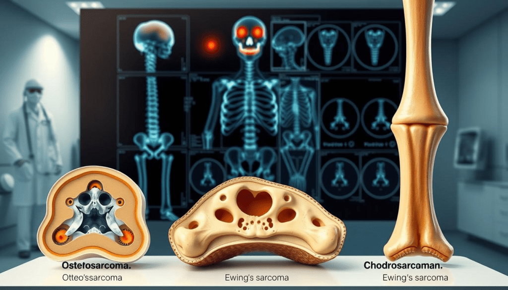
The most common primary bone cancers are osteosarcoma, chondrosarcoma, and Ewing’s sarcoma. Osteosarcoma is the most common, often found in long bones like the femur and tibia.
Secondary bone cancer, or metastatic bone cancer, happens when cancer cells from other parts of the body, like the breast, prostate, or lung, spread to the bones. This type is more common and can cause a lot of pain and complications.
Recognizing Potential Symptoms and Warning Signs
It’s important to recognize the symptoms of bone cancer early. Common signs include persistent bone pain, swelling, and tenderness near the affected bone. Some may notice a lump or mass, while others may have unexplained fractures.
Seek medical attention if these symptoms last or get worse. Bone cancer can also cause systemic symptoms like fatigue, weight loss, and fever. These symptoms can be hard to diagnose because they can look like other conditions.
Risk Factors and High-Risk Groups
Some risk factors increase the chance of getting bone cancer. These include genetic predispositions, like Li-Fraumeni syndrome and hereditary multiple osteochondromas. People who have had radiation therapy or have Paget’s disease of bone are also at higher risk.
Knowing these risk factors helps find high-risk groups. For example, people with a family history of bone cancer or those who have had radiation therapy should watch their bone health closely.
The Initial Diagnostic Pathway for Suspected Bone Cancer

When bone cancer is suspected, knowing the first steps is key to quick and effective treatment. At Liv Hospital, we follow the latest diagnostic and treatment methods. This ensures our patients get the best care possible.
When to Seek Medical Attention
Diagnosing bone cancer starts with a visit to your family doctor. They might do a physical exam and order tests like X-rays and blood tests. If bone cancer is thought of, you’ll see a specialist.
It’s important to see a doctor if you have ongoing bone pain, swelling, or other signs of bone cancer.
- Unexplained bone pain that persists
- Swelling or a lump in the affected area
- Weakened bones leading to fractures
What to Expect During Your First Oncology Consultation
Your first oncology visit will be a detailed check-up. You’ll talk about your medical history, symptoms, and get a physical exam. The oncologist will explain the next steps, which might include more tests or biopsies.
A team of experts is key in treating bone cancer. This team includes orthopedic surgeons, medical oncologists, and radiation oncologists. They work together to give you the best care.
The Importance of a Multidisciplinary Approach
A team effort is vital in diagnosing and treating bone cancer. This teamwork leads to accurate diagnoses and treatment plans made just for you.
Understanding the first steps in diagnosing bone cancer helps patients on their journey. At Liv Hospital, we aim to provide top-notch healthcare. We support and guide our patients every step of the way.
Does Bone Cancer Show Up in Blood Work?
Bone cancer isn’t always found through blood tests. But some tests can show it’s there. Blood tests are a first step in finding many health issues. Knowing how they help find bone cancer is key.
Common Blood Tests for Bone Cancer Screening
Several blood tests can hint at bone cancer. These include:
- Complete Blood Count (CBC)
- Blood chemistry tests
- Tests for specific biomarkers
A CBC can spot odd blood cell counts that might mean bone marrow issues. Blood chemistry tests can show changes in calcium or liver function linked to bone cancer.
Alkaline Phosphatase and Other Bone-Specific Biomarkers
Alkaline phosphatase (ALP) is found in bones and other tissues. High ALP levels in the blood might mean bone activity or damage, which could be from bone cancer.
| Bone Marker | Significance |
| Alkaline Phosphatase (ALP) | Elevated levels may indicate bone growth or damage |
| N-telopeptide (NTx) | High levels can indicate bone resorption |
| C-terminal telopeptide (CTX) | Elevated levels may suggest bone metastasis |
A medical expert says, “Bone markers like ALP aren’t alone in diagnosing but offer useful info with other tests.”
Limitations and False Negatives in Blood Testing
Blood tests have limits in finding bone cancer. Some with bone cancer might have normal blood tests, leading to false negatives.
Limitations include:
- Not all bone cancers release detectable biomarkers
- Blood test results can be influenced by other conditions
- Early-stage bone cancer might not cause significant changes in blood markers
We must remember these limits when looking at blood test results. We need to use more tests to confirm or rule out bone cancer.
Bone Cancer Lab Tests Beyond Blood Work
Blood tests are key in the early stages of diagnosing bone cancer. But other lab tests are also vital. They help doctors understand the disease better.
Urine Tests and Bone Turnover Markers
Urine tests measure bone turnover markers. These markers show how fast bones are being broken down and built up. N-telopeptide (NTx) and C-telopeptide (CTX) are examples of these markers.
High levels of these markers might mean bone cancer is active. But it’s important to look at all the test results together.
Genetic Testing for Hereditary Bone Cancer Syndromes
Some bone cancers run in families. Genetic testing can find people at risk. For example, Li-Fraumeni syndrome and hereditary multiple osteochondromas raise the risk of certain bone cancers.
This test looks at a person’s DNA for specific mutations. Knowing this can help catch bone cancer early and prevent it.
Interpreting Laboratory Results Accurately
It’s important to understand lab results correctly. Doctors need to look at all the tests together. This includes urine tests, genetic screenings, blood work, and imaging studies.
With a full picture of the results, doctors can create a treatment plan that fits each patient’s needs.
| Lab Test | Purpose | Significance in Bone Cancer Diagnosis |
| Urine NTx/CTX | Measures bone resorption markers | Indicates bone turnover rate, potentially suggesting bone cancer activity |
| Genetic Testing | Identifies hereditary syndromes associated with bone cancer | Helps in early detection and preventive measures for high-risk individuals |
| Blood Tests (e.g., Alkaline Phosphatase) | Measures bone-specific biomarkers | Provides insights into bone metabolism and possible cancer activity |
X-Ray Imaging in Bone Cancer Detection
X-ray imaging is often the first step in finding bone cancer. It’s a common and effective way to spot bone problems that might mean cancer.
Identifying Lytic and Blastic Bone Changes
X-rays are key for spotting lytic and blastic bone changes. Lytic lesions are areas where bone is broken down. Blastic lesions show where bone is growing in the wrong way. Both can hint at bone cancer.
| Lesion Type | Description | Possible Indication |
| Lytic | Bone destruction | Potential bone cancer |
| Blastic | Abnormal bone formation | Possible bone cancer or metastasis |
Can a Knee X-Ray Show Cancer?
A knee X-ray might show signs of bone cancer, like lytic or blastic lesions. But an X-ray alone can’t confirm cancer for sure.
“Imaging studies like X-rays are key for spotting bone issues, but a full diagnosis needs more tests.”
When X-Rays Are Insufficient for Diagnosis
X-rays are great for starting, but they have limits. They might miss early bone cancer or some lesions. More tests like CT scans, MRI, or PET scans are needed to be sure.
In short, X-rays are a big help in finding bone cancer. They give important clues about bone problems. But, they’re just the start of figuring out what’s wrong. More tests and checks are usually needed.
CT Scans: Detailed Bone Structure Assessment
In the fight against bone cancer, CT scans are key. They give us detailed images of bones. This helps us spot problems and see how far the disease has spread.
Revealing Bone Destruction and Masses
CT scans are great at showing bone destruction and tumors. They give us clear pictures of bone lesions. This helps doctors know how to treat the cancer.
CT scans can spot small changes in bones. They help us find lytic and blastic lesions. These are signs of bone cancer.
Accuracy of CT Scans in Diagnosing Bone Cancer
CT scans are good at finding bone problems. But how well they diagnose bone cancer depends on a few things. The scan quality, the radiologist’s skill, and how well it matches with other tests all matter.
We use CT scans a lot because they show us the whole bone and the nearby tissues. But we also use other tests like biopsies and MRIs to make sure of the diagnosis.
The Role of CT in Staging and Treatment Planning
CT scans are very important for staging bone cancer. They give us detailed info on how far the disease has spread. This helps us choose the best treatment.
In planning treatments, CT scans help us guide procedures. They let us target the right areas. This reduces risks and improves patient results.
Can MRI Detect Bone Cancer? Sensitivity and Applications
MRI is top-notch for spotting bone cancer. It’s super sensitive in finding both main and spread-out bone cancers. This makes MRI a key player in fighting bone cancer.
MRI Sensitivity for Primary and Metastatic Bone Lesions
MRI is great at showing bone and soft tissue details. It clearly sees primary bone cancers like osteosarcoma and Ewing’s sarcoma. It also finds metastatic bone cancers well, which often start in breast, prostate, or lung cancers.
MRI is good at spotting bone marrow changes. Bone marrow involvement is key in diagnosing bone cancer. MRI shows these changes clearly, helping doctors tell different types apart.
Visualizing Bone Marrow and Soft Tissue Involvement
MRI is excellent at showing bone marrow and soft tissue involvement. This is vital for seeing how far cancer has spread and planning treatment. It shows how tumors relate to nearby soft tissues, nerves, and blood vessels, which is important for surgery.
Also, MRI is safe for kids and pregnant women because it doesn’t use harmful radiation. Its ability to see soft tissue issues makes MRI a key tool in checking for bone cancer.
When MRI Is the Preferred Imaging Method
Many imaging options are available for bone cancer, but MRI is often the best choice. It’s good for checking soft tissue and bone marrow because of its clear images. It’s also great for tracking how well treatments work and spotting cancer coming back, all without using radiation.
In short, MRI is a strong tool for finding bone cancer. It’s great at spotting main and spread-out cancers, and it shows bone marrow and soft tissue well. We use MRI a lot in diagnosing bone cancer, helping patients get the right treatment.
Does a PET Scan Detect Bone Cancer? Effectiveness and Accuracy
PET scans are key in finding bone cancer. They give us both functional and anatomical details. This new imaging method has changed how we find and understand bone cancer, helping us make better treatment plans.
How PET Scans Work for Bone Cancer Detection
PET scans use radioactive tracers to spot cancer cells. Cancer cells use more energy than normal cells. So, PET scans can find bone cancer by showing where energy is high.
To do this, a tiny amount of radioactive tracer is given to the patient. It goes to areas with lots of activity, like cancer cells. Then, a PET scanner picks up the radiation, making detailed images of the body’s energy use.
PET/CT Combination: Achieving 98% Diagnostic Accuracy
Adding CT scans to PET scans makes them even better. The PET scan shows how the body works, and the CT scan shows the body’s structure. Together, they can spot bone cancer up to 98% of the time, making diagnosis more accurate.
This combo helps pinpoint cancer spots and see how far it has spread. This info is key for planning treatment and making it effective.
Comparing PET Scans to Traditional Bone Scintigraphy
Bone scintigraphy has been used for years to find bone cancer. But, PET scans are better. They give clearer images and can spot some cancers that bone scintigraphy misses.
Also, PET scans can see changes in cancer sooner than bone scintigraphy. This means we can catch cancer early and see how treatments are working. So, many doctors now prefer PET scans for their better accuracy and sensitivity.
Conclusion: Navigating the Bone Cancer Diagnostic Journey
Getting a bone cancer diagnosis can be tough. We’ve looked at the tests and procedures used to find this condition. These include X-rays, blood tests, and advanced scans like MRI and PET/CT.
It’s key for patients and doctors to know about these tools. This way, they can work together to find the right diagnosis and treatment. At Liv Hospital, we use the latest imaging and lab tests to help diagnose and treat early.
Diagnosing bone cancer takes imaging tests, lab tests, and a doctor’s evaluation. Knowing about these tools helps us give patients the best care. Finding the right diagnosis is the first step to effective treatment and better health.
FAQ
Does bone cancer show up in blood work?
Blood tests can hint at bone cancer through certain markers like alkaline phosphatase. But, they’re not always clear. They’re used first to check, but more tests are needed for sure.
Can an MRI detect cancer in the bone?
Yes, MRI is great at finding bone cancer. It shows bone marrow and soft tissue well. This makes it key to seeing how far the cancer has spread.
Will bone cancer show up in blood work?
Blood tests might suggest bone cancer with markers like alkaline phosphatase. But, they’re not always sure. More tests, like imaging and biopsies, are needed for a real diagnosis.
Can MRI detect bone cancer?
Yes, MRI is very good at spotting bone cancer. It’s great for seeing how much soft tissue is involved. This makes it a top choice for some cases.
How do you check for bone cancer?
To find bone cancer, many tests are used. These include X-rays, MRI, CT scans, PET scans, and biopsies. The right test depends on the cancer type and stage.
Can a knee X-ray show cancer?
Yes, a knee X-ray might show bone cancer signs, like changes in the bone. But X-rays might not always confirm it, mainly in early stages or some types.
Does a CT scan show bone cancer?
CT scans can show bone cancer well by showing bone damage and soft tissue. They help in planning treatment and understanding the cancer’s spread.
What is the role of PET scans in detecting bone cancer?
PET scans, with CT scans, are very good at finding bone cancer. They have a high accuracy rate. This makes them a strong tool in diagnosing bone cancer.
Are there specific blood tests for bone cancer?
There’s no single blood test for bone cancer. But some markers, like alkaline phosphatase, can hint at bone issues. These tests are used with others for a full picture.
How accurate are CT scans in showing bone cancer?
CT scans are very accurate in showing bone cancer. They’re great for seeing bone damage and soft tissue. They’re key in planning treatment and understanding the cancer’s spread.
Can genetic testing identify hereditary bone cancer syndromes?
Yes, genetic testing can find hereditary syndromes linked to bone cancer risk. It’s important to understand genetic test results for diagnosis and treatment.
References
- Lessa, R., de Souza, M., Pereira, M., Silva, T., & Santos, K. (2024). Clinical decision-making in bone cancer care management using computed tomography. Lung Cancer Management, 13(3), 221-233. https://www.ncbi.nlm.nih.gov/pmc/articles/PMC11570866/
- Aarthy, R., & Vali, M. S. (2024). Detection of bone cancer based on a four-phase gated convolutional neural network model. Biocybernetics and Biomedical Engineering. https://www.sciencedirect.com/science/article/pii/S111001682400992X


