
Knowing the facial nerve‘s detailed anatomy is key in today’s medicine. It’s important for planning surgeries and making diagnoses. The facial nerve helps us make facial expressions, sends taste signals, and controls some glands facial nerve genu.
We’ll look at the five major branches of the facial nerve. We’ll dive into their anatomy and how they work. This guide will help you understand the facial nerve‘s complex roles in our body.
Key Takeaways
- The facial nerve has multiple functions, including motor, sensory, and parasympathetic innervation.
- Understanding the anatomy of the facial nerve is key for surgeries and diagnoses.
- The facial nerve controls facial expressions and sends taste signals.
- The nerve also controls some glands.
- Knowing the five major branches of the facial nerve is vital for patient care.
The Facial Nerve: Overview and Significance

The facial nerve, the seventh cranial nerve, plays a key role in our body. It controls movement, senses, and helps our body relax. Let’s dive into its structure and importance.
Cranial Nerve VII: Structure and Function
The facial nerve is complex and unique. It has motor fibers that help us show emotions through facial expressions. It also has sensory fibers that send taste signals from the tongue’s front part.
Its structure includes several important parts. These are:
- The motor root, which supplies the muscles of facial expression
- The sensory root, also known as the nervus intermedius, which carries taste fibers
- Parasympathetic fibers that innervate various glands, including the submandibular and sublingual salivary glands
Motor, Sensory, and Parasympathetic Components
The motor part of the facial nerve controls our facial muscles. The sensory part sends taste signals. The parasympathetic part helps glands work right.
Together, these parts help us in many ways. They control our facial expressions, send taste signals, and help glands work.
- Controlling facial expressions
- Transmitting taste sensations
- Regulating glandular secretions
Knowing about the facial nerve’s structure and function is key. It helps us understand and treat related health issues. By looking at its parts and what they do, we see its big role in our health.
Anatomical Course of the Facial Nerve

The facial nerve’s journey from the brainstem to the face is complex. It goes through both the intracranial and extracranial pathways. Knowing this path helps us understand its functions and clinical implications.
Origin and Intracranial Pathway
The facial nerve starts in the brainstem, from the facial nerve nucleus in the pons. It then goes through the internal acoustic meatus. This is the start of its journey inside the skull.
Intratemporal Course and Segments
Next, the facial nerve enters the temporal bone. It follows a winding path through it. This path is split into three segments: the labyrinthine, tympanic, and mastoid segments. Each segment is near other important structures in the temporal bone.
Segment | Description | Notable Structures |
Labyrinthine | First segment within the temporal bone | Internal auditory canal |
Tympanic | Passes through the middle ear | Tympanic cavity |
Mastoid | Located within the mastoid part of the temporal bone | Mastoid air cells |
Stylomastoid Foramen: Exit Point from the Skull
The facial nerve leaves the skull through the stylomastoid foramen. This is the end of its journey inside the skull and the start of its path outside. The foramen is between the styloid and mastoid processes of the temporal bone.
Extracranial Course Through the Parotid Gland
After leaving the skull, the facial nerve goes into the parotid gland. There, it splits into its final branches. These branches control the muscles of facial expression, which are key for facial movements.
Understanding the facial nerve’s path is vital in medical practice. It helps in diagnosing and treating nerve-related conditions. Healthcare professionals need to know this pathway well.
The Facial Nerve Genu and Geniculate Ganglion
Exploring the facial nerve, we find the facial nerve genu. It’s a key spot linked to the geniculate ganglion. This part of the nerve is more than just a bend; it’s a complex structure with big roles.
Anatomical Significance of the Facial Nerve Genu
The facial nerve genu is crucial for the nerve’s overall function. It’s a turning point in the temporal bone. This bend is not just about shape; it’s where the geniculate ganglion is, key for taste from the tongue’s front parts.
The geniculate ganglion’s spot in the facial nerve genu shows how the nerve’s shape and function are linked. It’s a key spot for doctors, helping them during surgeries in the temporal bone and around the facial nerve.
Geniculate Ganglion: Structure and Function
The geniculate ganglion is a sensory ganglion. It has the cell bodies of the facial nerve’s sensory fibers. It sends taste info from the tongue’s front parts to the brain. It also helps with parasympathetic nerves in the head and neck.
- The geniculate ganglion is involved in taste sensation.
- It contains the cell bodies of the facial nerve’s sensory fibers.
- It plays a role in parasympathetic innervation.
Knowing about the geniculate ganglion’s structure and function is key for diagnosing and treating facial nerve issues. Conditions like Bell’s palsy or geniculate neuralgia need this knowledge. The connection between the facial nerve genu and the geniculate ganglion shows why detailed anatomy is vital in medicine.
The Temporal Branch: Anatomy and Innervation
The temporal branch is key in controlling muscles of the forehead and around the eye. It’s a vital part of the facial nerve, helping us show our emotions.
Course and Distribution
The temporal branch starts at the parotid plexus and goes up over the zygomatic arch. It reaches muscles in the forehead and around the eye.
As it moves across the face, it can get hurt. This can cause facial asymmetry. Knowing its path is important for doctors and treatments.
Muscles Innervated
The temporal branch controls important muscles for facial expressions. These include:
- The frontalis muscle, which lifts the eyebrows.
- The orbicularis oculi, for closing the eyes.
- The corrugator supercilii and procerus muscles, for frowning.
These muscles help us show a wide range of emotions. If they don’t work right, it’s hard to express feelings.
Clinical Significance and Common Pathologies
Problems with the temporal branch can come from injuries, infections, or surgery.
“Damage to the temporal branch can lead to significant morbidity, including facial asymmetry and difficulty with eye closure.”
Issues like Bell’s palsy and Ramsay Hunt syndrome can affect it. Knowing how important the temporal branch is helps doctors treat these problems well.
We stress the need for detailed knowledge of the temporal branch. This is key for diagnosing and treating related disorders, ensuring the best results for patients.
The Zygomatic Branch: Anatomy and Innervation
The zygomatic branch is a key part of the facial nerve. It controls muscles around the eyes and cheeks. It helps us make different facial expressions.
Course and Distribution
The zygomatic branch starts in the parotid gland and goes along the zygomatic arch. It sends fibers to muscles around the eye. This helps with eye movements and facial expressions.
Muscles Innervated
The zygomatic branch mainly controls the orbicularis oculi and other eye muscles. These muscles are key for eye movements and expressions.
Muscle | Function |
Orbicularis oculi | Controls eye closure and facial expressions around the eye |
Zygomaticus major | Facilitates smiling and other facial expressions |
Clinical Significance and Common Pathologies
Problems with the zygomatic branch can cause muscle weakness or paralysis. This can make eye closure hard and lead to facial asymmetry.
“Damage to the zygomatic branch can cause noticeable facial asymmetry and affect a patient’s ability to express emotions through facial movements.”
Common issues with the zygomatic branch include Bell’s palsy and nerve injuries. Knowing its anatomy and function is key for treating these problems.
The Buccal Branch: Anatomy and Innervation
We look at the buccal branch to understand its role in facial nerve function. It’s one of the facial nerve’s five branches. It controls the muscles of facial expression, mainly around the cheek and mouth.
Course and Distribution
The buccal branch starts from the facial nerve’s anterior division in the parotid gland. It moves forward, often between the masseter muscle’s superficial and deep heads. It then reaches the cheek muscles.
Key aspects of its course include:
- Emerging from the parotid gland
- Passing over or through the masseter muscle
- Innervating muscles around the cheek and mouth
Muscles Innervated
The buccal branch controls several muscles important for facial expressions and oral functions. These include:
Muscle | Function |
Buccinator | Compresses the cheek against the teeth |
Orbicularis oris | Controls the movement of the lips |
Muscles of the upper lip | Elevate and control the upper lip |
Clinical Significance and Common Pathologies
Problems with the buccal branch can cause big issues, like trouble with facial expressions and oral functions. Common issues include:
- Trauma to the facial nerve
- Infections like Lyme disease or Ramsay Hunt syndrome
- Neurological disorders like Bell’s palsy
Knowing the anatomy and innervation of the buccal branch is key for diagnosing and treating these problems.
The Marginal Mandibular Branch: Anatomy and Innervation
Understanding the marginal mandibular branch is key to grasping the facial nerve’s complex anatomy. It affects lower facial movements. This branch controls the muscles of the lower lip and chin.
Course and Distribution
The marginal mandibular branch starts in the parotid gland. It goes forward, usually below the jaw’s angle. Then, it runs above the jaw’s bottom to reach the lower lip and chin muscles.
Its path varies among people. It often links with the facial artery and vein. This is important for surgeons and doctors to know during neck and face surgeries.
Muscles Innervated
The main muscles this branch controls are the depressor anguli oris, depressor labii inferioris, and mentalis. These muscles help move the lower lip and chin. They are key for facial expressions.
- The depressor anguli oris muscle pulls the mouth’s corner down.
- The depressor labii inferioris muscle lowers and turns out the lower lip.
- The mentalis muscle lifts and sticks out the lower lip, showing doubt or contempt.
Clinical Significance and Common Pathologies
This branch can get hurt during neck and face surgeries. Damage can cause muscle weakness or paralysis. This leads to uneven lower lip and chin, affecting how we look.
Conditions like Bell’s palsy can also harm this branch. Knowing its anatomy and function helps doctors diagnose and treat these issues.
The Cervical Branch: Anatomy and Innervation
Learning about the cervical branch’s anatomy and function is important. It helps us understand its role in facial nerve disorders. The cervical branch is a key part of the facial nerve, controlling our facial expressions.
Course and Distribution
The cervical branch starts near the parotid gland and goes down to the neck. It controls the platysma muscle. This muscle helps with facial expressions and neck movements.
The nerve fibers of the cervical branch spread out in the platysma. This ensures the muscle works well.
Muscles Innervated
The main muscle the cervical branch controls is the platysma. This muscle is a wide, flat sheet on the neck. It helps with:
- Facial expressions, like frowning or showing surprise
- Neck movements, like tensing the skin
Clinical Significance and Common Pathologies
Problems with the cervical branch can cause issues. These include:
- Weakness or paralysis of the platysma muscle, affecting facial expressions and neck movements
- Abnormal nerve regeneration, leading to synkinesis or other movement disorders
Knowing about the cervical branch’s clinical significance is key. It helps in diagnosing and treating facial nerve disorders.
Clinical Applications and Disorders
We look into the facial nerve’s role in health and its problems. Knowing how to check it properly is key. The facial nerve is vital for many health issues, and problems with it can cause a lot of suffering.
Bell’s Palsy: Pathophysiology and Presentation
Bell’s palsy is a common facial nerve issue. It makes facial muscles weak or paralyzed. It starts suddenly, usually on one side, and can cause pain, taste issues, and sensitivity to sound.
The exact reason for Bell’s palsy is not known. But, it’s thought to be due to swelling and pressure on the nerve in the temporal bone.
Key features of Bell’s palsy include:
- Sudden onset facial weakness or paralysis
- Unilateral involvement
- Pain around the jaw or behind the ear
- Altered taste sensation
- Hyperacusis
Doctors diagnose Bell’s palsy by listening to the patient’s history and doing a physical check. It’s important to tell Bell’s palsy apart from other facial nerve problems like infections, tumors, or injuries.
Facial Nerve Examination Techniques
Checking the facial nerve well is key for diagnosing and treating its problems. The check should look at facial muscle strength, taste, and nerve functions.
Examination techniques involve:
- Inspecting for facial asymmetry at rest and during voluntary movements
- Testing facial muscle strength against resistance
- Evaluating taste sensation using sweet, sour, salty, and bitter substances
- Assessing for synkinesis or abnormal facial movements
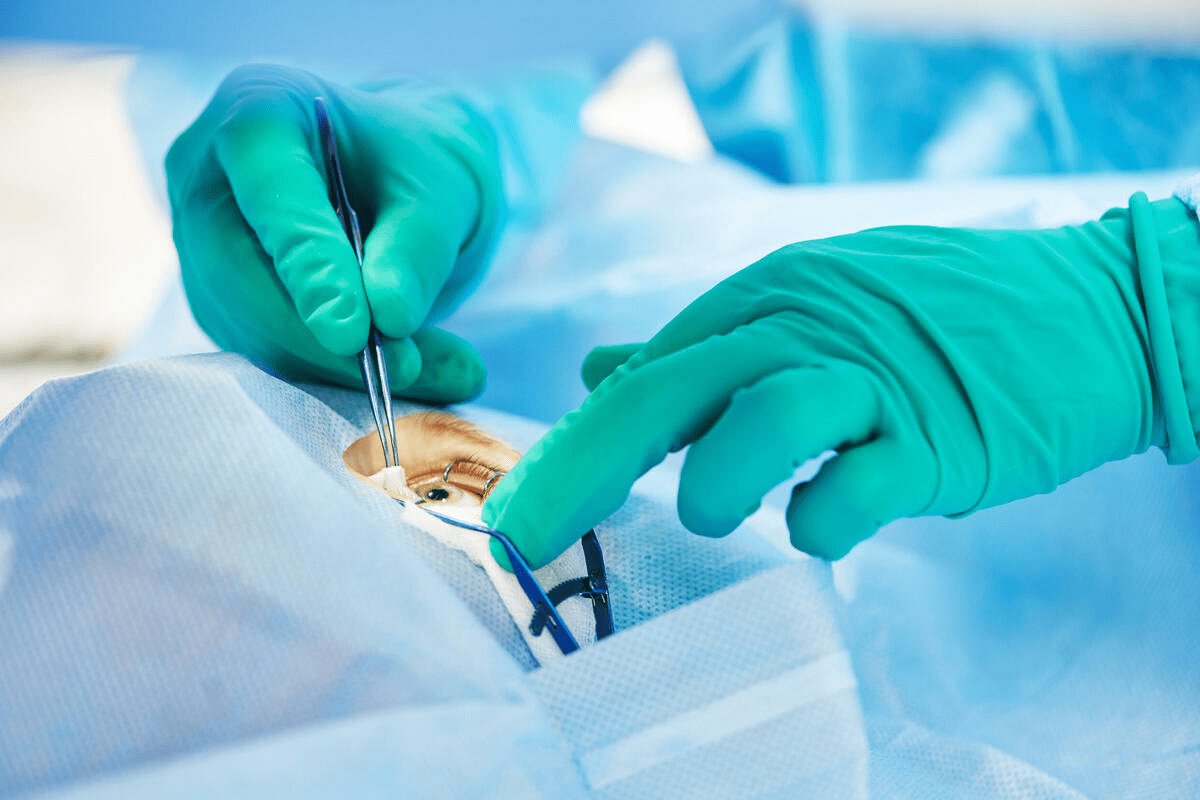
Surgical Considerations and Anatomical Landmarks
When surgery is needed for facial nerve issues, knowing its anatomy is critical. The facial nerve leaves the skull through the stylomastoid foramen, a key landmark.
Surgeons need to know the nerve’s path and any variations to avoid mistakes during surgery. Understanding the facial nerve’s anatomy is essential for planning and doing surgery well.
“Understanding the facial nerve’s detailed anatomy is key for successful surgery in the parotid gland and temporal bone.” –
A renowned neurosurgeon
We’ve talked about the facial nerve’s clinical importance, its disorders, and how to examine and operate on it. Knowing these well is important for better patient care.
Conclusion
The facial nerve, or cranial nerve VII, is very important. It helps control our facial expressions and sends taste information to our brain. It also helps our head and neck by providing parasympathetic innervation.
We’ve looked at how the facial nerve works and its anatomy. It has five main branches: temporal, zygomatic, buccal, marginal mandibular, and cervical. These branches help the nerve do its job.
The facial nerve’s path is key to understanding its role. It starts in the brain and goes out through the stylomastoid foramen. It then spreads out in the parotid gland. Knowing this helps doctors diagnose and treat problems.
Doctors need to know a lot about the facial nerve. Its motor, sensory, and parasympathetic parts are all important. A facial nerve map helps doctors understand its branches and how they work.
In short, the facial nerve is very important. Its complex anatomy and function show why doctors need to know a lot about it. We’ve covered the main points about the facial nerve, showing its significance and complexity.
FAQ
What are the main functions of the facial nerve?
The facial nerve controls our facial expressions. It also transmits taste from the tongue’s front parts. Plus, it helps glands like the salivary glands and the lacrimal gland work.
What are the 5 branches of the facial nerve?
The facial nerve has five main branches. These are the temporal, zygomatic, buccal, marginal mandibular, and cervical. They help us make facial expressions.
Where does the facial nerve exit the skull?
The facial nerve leaves the skull through the stylomastoid foramen.
What is the significance of the geniculate ganglion?
The geniculate ganglion is a key part of the facial nerve. It’s in the temporal bone. It houses the taste fibers of the nerve.
What is Bell’s palsy, and how does it affect the facial nerve?
Bell’s palsy is a condition that weakens or paralyzes facial muscles. It happens when the facial nerve doesn’t work right. This usually affects one side of the face.
How is the facial nerve examined clinically?
Doctors check the facial nerve by looking at muscle strength and taste. They also look for any weakness or paralysis.
What are some common pathologies affecting the branches of the facial nerve?
Trauma, tumors, and inflammation can harm the facial nerve’s branches. This can lead to muscle weakness or paralysis.
What is the course of the facial nerve through the parotid gland?
After leaving the skull, the facial nerve goes through the parotid gland. Inside the gland, it splits into its five branches.
What is the role of the facial nerve in taste sensation?
The facial nerve carries taste from the tongue’s front parts. This is through its chorda tympani branch.
What are the muscles innervated by the temporal branch of the facial nerve?
The temporal branch controls muscles in the forehead. These include the frontalis, procerus, and corrugator supercilii.
References
National Center for Biotechnology Information. Evidence-Based Medical Guidance. Retrieved from https://www.ncbi.nlm.nih.gov/books/NBK554569/

