Last Updated on November 27, 2025 by Bilal Hasdemir

At LivHospital, we’re leaders in advanced diagnostics and care. We see the big role fluorodeoxyglucose positron emission tomography (FDG-PET) plays in medicine today. This method mixes metabolic and anatomical data, giving us key insights into diseases.
FDG-PET, with 18F FDG PET-CT scans, has changed medical diagnostics. It helps us diagnose, stage, and track diseases, like cancers, more accurately. Our use of FDG-PET shows our commitment to top-notch healthcare.
Key Takeaways
- FDG-PET is a key imaging method that combines metabolic and anatomical data.
- 18F FDG PET-CT scans help us diagnose and monitor diseases precisely.
- FDG-PET is very useful in managing different cancers.
- Our hospital is dedicated to using the latest diagnostic technologies.
- FDG-PET scans offer a caring approach to disease management.
The Fundamentals of Fluorodeoxyglucose Positron Emission Tomography

FDG-PET combines nuclear medicine and advanced imaging. It gives us key insights into how our bodies work. This method is vital in fields like oncology, neurology, and cardiology.
How PET-CT Combines Metabolic and Anatomical Imaging
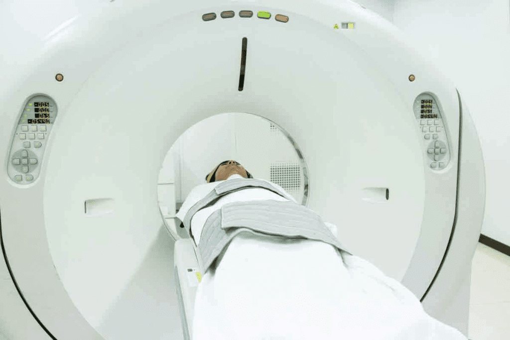
PET-CT scans mix metabolic data from PET with CT’s anatomical details. This gives us a full view of the body’s tissues. It improves diagnostic accuracy by showing both structure and function.
In cancer diagnosis, PET-CT spots tumor activity. It tells us if a tumor is cancerous or not. It also shows where the tumor is. This helps doctors plan treatments and check how well they work.
A study on NCBI shows PET-CT’s power in diagnosis. It has made a big difference in many areas of medicine.
The Historical Development of FDG-PET Technology
The first PET scanners came out in the early 1970s. The use of 18F-fluorodeoxyglucose (FDG) as a tracer was a big step. It let us see glucose metabolism without surgery.
Thanks to better scanners, tracers, and algorithms, FDG-PET is now key in treating many diseases. It gives deep insights into metabolic processes.
Key Fact 1: Understanding 18F Fluorodeoxyglucose as a Radiotracer

To grasp the importance of 18F Fluorodeoxyglucose, we need to look at its chemical makeup and how it works in the body. It acts like glucose, which is why it’s great for finding and tracking diseases.
Chemical Structure and Biological Properties
18F Fluorodeoxyglucose, or Fluorodeoxyglucose F 18 FDG, is made to look like glucose. It has Fluorine-18, a radioactive part that lasts about 110 minutes. This part emits positrons that PET scanners can pick up.
The structure of F-18 Fluorodeoxyglucose is close to glucose’s, but with Fluorine-18 instead of a hydroxyl group. This lets it get into cells like glucose does.
How F-18 Fluorodeoxyglucose Mimics Glucose Metabolism
Inside the cell, F-18 Fluorodeoxyglucose gets changed into F-18 Fluorodeoxyglucose-6-phosphate by hexokinase. But, unlike glucose-6-phosphate, it can’t be broken down further. So, it stays trapped in the cell.
This trapping lets PET scans spot high glucose use areas. These are often seen in cancer, infections, and inflammation. This makes 18F Fluorodeoxyglucose a key tool for doctors.
By showing where glucose use is different, F-18 Fluorodeoxyglucose PET scans help doctors diagnose, plan treatment, and check how well treatments are working.
Key Fact 2: The Mechanism of FDG Fluorodeoxyglucose Uptake
To understand how FDG-PET scans work, we need to know how FDG is taken up by cells. This involves two main steps: getting into the cell and being trapped in tissues that use a lot of sugar.
Cellular Transport and Phosphorylation
Cells take in FDG through glucose transporters, mainly GLUT1, because it looks like glucose. Inside the cell, FDG gets turned into FDG-6-phosphate by hexokinase. This step is key because it keeps FDG inside the cell, as FDG-6-phosphate can’t be used for more sugar processing or sent back out.
The amount of FDG a cell takes in shows how much sugar it uses. Tissues that need a lot of sugar, like some tumors, take in more FDG. This is why FDG-PET scans are so useful in finding and tracking cancer.
Metabolic Trapping in High-Glycolysis Tissues
In tissues that use a lot of sugar, like many cancer cells, FDG gets trapped even more. These cells have more glucose transporters and hexokinase, which helps them take in and process FDG. This leads to a buildup of FDG-6-phosphate, making these tissues show up clearly on PET scans.
The mechanism of FDG uptake and trapping isn’t just for cancer cells. It also happens in other tissues that are very active, like some inflammatory or infectious areas. Knowing how this works is key to using FDG-PET scans correctly and getting the most out of them in different medical situations.
Key Fact 3: Oncological Applications of Fluorodeoxyglucose PET Scans
In oncology, Fluorodeoxyglucose Positron Emission Tomography (FDG-PET) is key for cancer detection and management. It helps us understand cancer diagnosis, staging, and treatment. This is vital for various cancers.
Cancer Detection and Initial Diagnosis
FDG-PET scans are important for catching cancer early. They show where tumors are by looking at glucose metabolism. A top oncologist says, “FDG-PET has changed how we diagnose cancer, making treatment better.” PET scans are great for finding cancers like lymphoma, melanoma, and colorectal cancer.
Staging and Restaging Malignancies
Knowing how far cancer has spread is key for treatment planning. FDG-PET scans help by finding active tumor sites. This info is essential for making treatment plans and predicting outcomes.
FDG-PET also helps after treatment to see if cancer has come back. It’s very useful for cancers like Hodgkin’s and non-Hodgkin’s lymphoma.
Monitoring Treatment Response and Recurrence
FDG-PET scans are also used to check how well treatment is working. They look at glucose changes in tumors. This helps doctors adjust treatments for better results.
Early detection of treatment response or recurrence helps doctors make better choices. This can lead to better survival rates and quality of life. A study found FDG-PET is very useful in cancer management.
In summary, Fluorodeoxyglucose PET scans are vital in cancer care. They give detailed insights into cancer detection, staging, and treatment. Their role in fighting cancer is unmatched.
Key Fact 4: Neurological Uses of 18F FDG PET-CT
The 18F FDG PET-CT has changed how we diagnose and treat brain conditions. It’s a key tool for checking brain function and metabolism. This leads to more accurate diagnoses and better treatments.
Dementia and Neurodegenerative Disease Evaluation
18F FDG PET-CT is very useful for studying dementia and neurodegenerative diseases. It can tell different types of dementia apart. This is because each type shows different glucose metabolism patterns in the brain.
Key applications include:
- Diagnosing Alzheimer’s disease and other dementias
- Assessing the extent of cognitive impairment
- Monitoring disease progression
- Differentiating between neurodegenerative and reversible causes of cognitive decline
Epilepsy Focus Localization
In epilepsy, 18F FDG PET-CT is key for finding seizure foci. It spots areas with abnormal glucose metabolism, which might be where seizures start.
The benefits of using 18F FDG PET-CT in epilepsy include:
- Identifying the seizure focus for possible surgery
- Helping place intracranial electrodes for better localization
- Aiding in understanding the epileptogenic network
Brain Tumor Characterization and Grading
18F FDG PET-CT is also used for brain tumor grading and characterization. Tumors with high glucose metabolism show up as bright spots on scans. This helps figure out how aggressive the tumor is and guides treatment.
The scans are vital for:
- Telling apart tumor recurrence and radiation necrosis
- Figuring out tumor grade and how aggressive it is
- Helping decide on treatments and checking how well they work
Key Fact 5: Cardiovascular Applications of Fluorodeoxyglucose F 18 FDG
We use Fluorodeoxyglucose F 18 FDG for heart health checks with PET-CT imaging. This tool is key for checking heart problems. It helps doctors make better treatment plans.
Myocardial Viability Assessment
FDG-PET is key in cardiology for checking heart muscle health. It finds out if heart muscle is alive or damaged. FDG-PET shows which heart muscle can be saved with treatments like bypass surgery.
It works by seeing how much FDG the heart muscle takes in. High FDG means the muscle is alive. Low FDG means it might be scarred. Doctors use this info to choose the best treatment.
Inflammatory Cardiac Conditions
FDG-PET is also great for finding and managing heart inflammation. Heart inflammation shows up as high FDG uptake. This is very helpful for diseases where inflammation is a big part of the problem.
It helps doctors see how well treatments are working. They can change plans if needed. FDG-PET gives a clearer view of heart inflammation, helping patients get better care.
In short, Fluorodeoxyglucose F 18 FDG PET-CT scans help a lot in heart care. They’re good for checking heart muscle health and finding inflammation. As heart care gets better, FDG-PET will likely play an even bigger role in helping patients.
Key Fact 6: Superior Diagnostic Performance of FDG-PET
FDG-PET is known for its high sensitivity and specificity. It beats traditional imaging methods in detecting diseases. This makes it key for accurate diagnosis and treatment.
Sensitivity and Specificity Compared to Conventional Imaging
FDG-PET outshines other imaging methods in many cases. It’s great at finding tumors and brain disorders. This is thanks to its high sensitivity and specificity.
- Higher Sensitivity: FDG-PET spots metabolic changes early, leading to quicker diagnosis.
- Improved Specificity: It shows where cells are active, helping to tell cancer from non-cancer.
Research shows FDG-PET is better at cancer staging than CT or MRI. This is vital for choosing the right treatment.
Impact on Patient Management and Treatment Decisions
FDG-PET’s top-notch performance changes how doctors manage patients. It gives detailed info on disease spread and activity. This helps doctors tailor treatments to each patient.
- Personalized Treatment Plans: FDG-PET guides treatment plans based on disease metabolism.
- Monitoring Treatment Response: It checks how treatments are working, allowing for changes as needed.
- Detecting Recurrence: Its high sensitivity catches disease return early, leading to quick action.
In summary, FDG-PET’s advanced diagnostic abilities greatly improve patient care. It leads to accurate diagnoses and effective treatments.
Key Fact 7: Limitations and Considerations of FDG PET-CT
FDG PET-CT has many benefits but also some drawbacks. It can give false results and expose patients to radiation. Knowing these limitations is key for making the best decisions for patient care.
False Positive and Negative Results
FDG PET-CT scans can sometimes show false positives and negatives. False positives might happen when inflammation or infection looks like cancer. For example, some diseases or post-surgery inflammation can make the scan think there’s cancer when there isn’t.
False negatives can occur when tumors don’t take up enough F-18 fluorodeoxyglucose. This can be because the tumor is small or has low activity. Some cancers, like mucinous tumors, might not show up on the scan because they don’t take in enough of the tracer.
- False positives can be caused by:
- Inflammatory processes
- Infections
- Post-surgical changes
- False negatives can result from:
- Low metabolic activity in tumors
- Small tumor size
- Specific tumor types with low FDG uptake
Radiation Exposure and Safety Considerations
FDG PET-CT scans also involve radiation exposure. The scan uses a radioactive tracer that emits positrons. While the dose from one scan is usually safe, getting many scans can increase the risk of harm.
To keep patients safe, we follow strict rules. We use the least amount of tracer needed and optimize the scan settings. This helps keep the dose low while getting good images.
Patient Preparation Requirements
Getting ready for an FDG PET-CT scan is important. Patients need to fast and avoid exercise before the scan. This helps get clear results.
Patients with diabetes need special care. Their blood sugar levels can affect how the tracer works. We give them detailed instructions on how to prepare, including any diet or medication changes.
By following these steps, patients help make sure their scan is accurate. This information is vital for their diagnosis and treatment.
The Patient Experience: Undergoing an 18F FDG PET-CT Scan
Knowing what to expect during an 18F FDG PET-CT scan can make you feel less anxious. We aim to walk you through each step of the process. This way, you can feel more prepared and comfortable.
Pre-scan Preparation Protocol
Getting ready for your scan is important. Patients need to fast for 4-6 hours before the scan to keep glucose levels steady. Also, avoid hard exercise for a couple of days beforehand to prevent muscle glucose uptake.
On the day of the scan, wear loose, metal-free clothes and remove any jewelry. Tell your doctor about any medications or health conditions you have. This info helps ensure your safety and the scan’s success.
The Scanning Procedure Step-by-Step
The scanning process starts with a small dose of radioactive glucose (18F FDG) being injected. Then, you wait about 60 minutes for it to spread through your body.
While waiting, you’ll relax in a quiet room to avoid muscle glucose uptake. After the wait, you’ll lie down on a table that moves into the PET-CT scanner.
The actual scan takes about 30 minutes. The scanner takes detailed images of your body’s activity and structure. It’s important to stay very quiet and not move during this time.
Post-scan Instructions and Follow-up
After the scan, you can usually go back to your normal activities unless told not to. Drink lots of water to help get rid of the radioactive tracer.
Follow any post-scan instructions from your healthcare team. This might include avoiding close contact with pregnant women, breastfeeding mothers, or young kids for a bit because of the tracer’s radioactivity.
The scan results will be reviewed by experts. You’ll discuss them with your doctor during a follow-up visit. This meeting is key for understanding your diagnosis and treatment plans.
Emerging Trends in Fluorodeoxyglucose Positron Emission Tomography
New trends in fluorodeoxyglucose positron emission tomography are changing medical imaging. Technology is getting better, leading to big improvements in FDG-PET.
Technological Advancements in PET-CT Equipment
New PET-CT scanners are making images clearer and more accurate. New technologies are being added to make FDG-PET scans better.
Some key improvements include:
- High-resolution PET-CT scanners
- Advanced detector materials
- Improved image reconstruction algorithms
These changes are important for using FDG-PET in more medical areas.
Artificial Intelligence in Image Interpretation
Artificial intelligence (AI) is changing how we look at FDG-PET scans. AI can spot small changes and patterns that humans might miss.
| Benefits of AI in FDG-PET | Description |
|---|---|
| Improved Accuracy | AI makes image interpretation more precise |
| Increased Efficiency | AI does routine tasks, saving expert time |
| Enhanced Detection | AI finds patterns that humans might overlook |
Novel Clinical Applications Under Investigation
Researchers are finding new uses for FDG-PET. Some exciting areas include:
- Studying inflammatory diseases
- Tracking how treatments work in different cancers
- Checking if the heart is working well
As these trends grow, FDG-PET will become even more important in diagnosing and planning treatments.
Conclusion: The Integral Role of FDG-PET in Modern Medicine
Fluorodeoxyglucose positron emission tomography has changed medicine a lot. It gives doctors great info for diagnosing and predicting diseases. 18F FDG PET-CT scans are key in treating many illnesses.
We’ve looked at how FDG-PET works and its uses in cancer, brain, and heart diseases. It’s better than old imaging methods because it’s more accurate.
As medicine keeps getting better, FDG-PET’s role will grow even more. New tech and AI will make it even more useful for doctors.
In short, 18F FDG PET-CT scans are vital in today’s medicine. They help doctors a lot and will keep improving patient care.
FAQ
What is Fluorodeoxyglucose Positron Emission Tomography (FDG-PET) used for?
FDG-PET is a tool for diagnosing diseases. It checks how tissues work, helping find and track cancer. It also helps with some brain and heart issues.
How does 18F Fluorodeoxyglucose work as a radiotracer?
18F Fluorodeoxyglucose acts like glucose, which cancer cells love. It builds up in these cells. This lets doctors see where cancer is through PET scans.
What is the mechanism of FDG uptake in cells?
Cells take in FDG through special transporters. Then, it gets stuck inside, mainly in fast-growing cells like cancer.
What are the oncological applications of FDG-PET scans?
In cancer care, FDG-PET scans help find, stage, and track cancer. They also check how well treatments work and if cancer comes back.
Can FDG-PET be used for neurological conditions?
Yes, it helps with brain diseases like dementia and epilepsy. It also spots brain tumors.
What are the cardiovascular applications of Fluorodeoxyglucose F 18 FDG?
It’s used for heart health. It checks if heart muscle is alive and finds heart inflammation.
How does the diagnostic performance of FDG-PET compare to conventional imaging?
FDG-PET is better at finding and identifying diseases than old methods. This helps doctors make better treatment plans.
What are the limitations and considerations of FDG PET-CT?
It might not always be accurate and can expose you to radiation. You also need to follow certain steps before the scan.
How should I prepare for an 18F FDG PET-CT scan?
You’ll need to fast and avoid exercise. Your doctor will give you specific instructions for the best results.
What can I expect during an 18F FDG PET-CT scan?
You’ll lie on a table that slides into the scanner. Stay very quiet while it takes pictures. You might get contrast agents too.
Are there any emerging trends in Fluorodeoxyglucose Positron Emission Tomography?
Yes, new tech and AI are changing PET-CT. There’s also research into new uses for FDG-PET.
Reference
- Molecular Imaging: PET Technology Overview, Nimmagadda, Molecular Imaging


