
Attention Deficit Hyperactivity Disorder (ADHD) impacts millions globally. Yet, diagnosing it can be tough. Recent studies indicate SPECT scans can reveal important details about ADHD. They might help doctors diagnose it better.
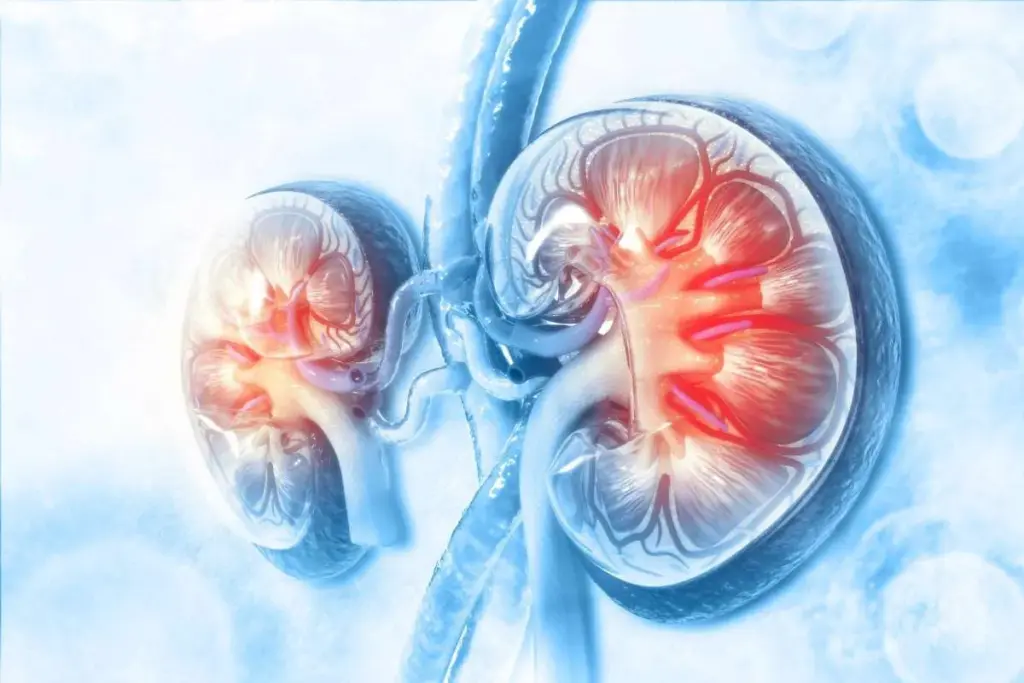
SPECT scans give a peek into how the brain works. They focus on brain perfusion and areas linked to attention issues. By looking at frontal blood flow, doctors can spot signs of ADHD.
Key Takeaways
- SPECT scans can help diagnose ADHD by analyzing brain perfusion patterns.
- ADHD imaging through SPECT scans provides insights into attention disorders.
- Brain perfusion analysis is key to understanding ADHD.
- SPECT scans offer a valuable tool for healthcare professionals.
- Understanding ADHD through imaging can lead to better treatment options.
Understanding SPECT Imaging Technology

SPECT imaging measures blood flow and activity in the brain. It shows how the brain works. This technology is used in clinics and research.
What is a SPECT Scan?
A SPECT scan is a way to see how the brain works. It uses a special kind of imaging called Single Photon Emission Computed Tomography. This method looks at cerebral blood flow in the brain.
To do this, a tiny bit of radioactive tracer is given to the patient. This tracer goes to brain cells based on how active they are.
How SPECT Differs from Other Brain Imaging Methods
SPECT imaging is different from MRI or CT scans. Those scans show the brain’s structure. But SPECT scans show how the brain works.
They are great for checking conditions like ADHD. A researcher said, “SPECT scans show brain activity that other scans can’t. They give a deeper look into brain function.”
The main differences are:
- Focus on functional, not structural information
- Measures cerebral blood flow and activity
- Uses radioactive tracers to see brain function
The Process of Undergoing a SPECT Scan
Getting a SPECT scan involves a few steps. First, a radioactive tracer is given to the patient. Then, the patient rests or does tasks while the tracer is absorbed by the brain.
The scan itself has the patient lying down. A gamma camera moves around their head. It takes pictures of the brain’s activity. The whole thing is safe and gives important information about the brain.
The Neurobiology of ADHD

ADHD’s roots lie in how different brain parts and chemical pathways work together. Knowing this helps us understand the disorder and how to treat it.
Brain Regions Affected in ADHD
Studies have found that ADHD impacts several brain areas. These include parts that help with focus, controlling impulses, and making decisions. The prefrontal cortex, basal ganglia, and anterior cingulate cortex are key areas affected.
Key brain regions affected:
- Prefrontal cortex: involved in decision-making and impulse control
- Basal ganglia: plays a role in control of voluntary motor movements, procedural learning, habit learning, eye movements, cognition, and emotion
- Anterior cingulate cortex: involved in error detection, conflict monitoring, and motivation
Neurotransmitter Imbalances in ADHD
ADHD is linked to imbalances in dopamine and norepinephrine. These imbalances can mess with brain areas that handle focus and impulse control.
“The dopamine hypothesis of ADHD suggests that the disorder is associated with alterations in dopamine neurotransmission.” This idea is backed by how well dopamine-changing meds work for ADHD symptoms.
Structural vs. Functional Brain Differences
ADHD shows up in both the structure and function of the brain. Structural changes might be in the size or thickness of brain areas. Functional changes could be in how the brain acts or connects.
Neuroimaging has given us a peek into these differences. For example, some studies found smaller brain structures in people with ADHD.
“Neuroimaging studies have revealed that ADHD is characterized by abnormalities in brain structure and function, particular in regions related to attention and executive control.”
Source: A leading neuroimaging study on ADHD
Grasping these neurobiological details is key to finding better ways to diagnose and treat ADHD.
Frontal Blood Flow and Its Significance in ADHD
Frontal blood flow in ADHD has caught the eye of many researchers. Studies show a strong link between frontal lobe issues and ADHD symptoms.
The Role of Frontal Lobes in Executive Function
The frontal lobes play a key role in executive function. This includes planning, making decisions, and controlling impulses. Executive function deficits are a hallmark of ADHD, and research points to frontal lobe problems as a cause.
His research shows that reduced frontal lobe activity is linked to ADHD symptoms like inattention and impulsivity.
Patterns of Cerebral Blood Flow in ADHD Patients
Studies using SPECT imaging have looked at cerebral blood flow in ADHD patients. They found distinct patterns of blood flow that set ADHD patients apart from healthy controls.
“SPECT imaging has shown that ADHD patients often exhibit decreased blood flow to the frontal lobes, which is associated with executive function deficits.”
Hypoperfusion vs. Hyperperfusion in Different Brain Regions
In ADHD, hypoperfusion (reduced blood flow) is common in the frontal lobes. This reduced blood flow is believed to contribute to ADHD’s executive function issues.
On the other hand, some studies have found hyperperfusion (increased blood flow) in areas like the basal ganglia and limbic system. This mix of hypoperfusion and hyperperfusion might explain ADHD’s varied symptoms.
Grasping these blood flow patterns is key to finding better ways to diagnose and treat ADHD.
SPECT Findings in ADHD Patients
SPECT scans are key in understanding ADHD. They show how blood flows in the brain.
Research with SPECT imaging has given us insights into ADHD. It looks at how blood flows in the brain. This helps spot patterns linked to ADHD.
Common Blood Flow Patterns Observed
Studies show ADHD patients have different blood flow in their brains. Hypoperfusion, or less blood flow, is seen in the prefrontal cortex. This area is important for making decisions and planning.
On the other hand, some areas show hyperperfusion, or more blood flow. These changes in blood flow relate to ADHD’s various symptoms.
Differences Between ADHD Subtypes
Each type has its own blood flow patterns. For example, ADHD-Combined type and ADHD-Predominantly Inattentive type show different patterns.
Knowing these differences helps doctors create treatments that fit each patient’s needs.
Comparing ADHD to Neurotypical Brain Scans
Studies comparing ADHD to normal brains using SPECT imaging show big differences. These differences help doctors understand ADHD better.
They also give important clues for diagnosing ADHD. This helps doctors understand the brain’s role in ADHD.
Clinical Applications of SPECT in ADHD Diagnosis
SPECT imaging is now used more in diagnosing ADHD. It shows how the brain works and blood flows. This is key in understanding ADHD’s neurobiology.
Right now, ADHD is diagnosed through clinical checks, behavior tests, and patient reports. Adding SPECT scans gives more solid data to back these diagnoses.
Current Diagnostic Protocols
Diagnosing ADHD involves a detailed check-up, history, and neuropsychological tests. These methods are good but can differ between doctors.
Using SPECT scans adds an objective look at brain function. People with ADHD often have unique blood flow patterns in areas linked to focus and control.
| Diagnostic Method | Primary Use | Advantages |
| Clinical Evaluation | Initial Assessment | Comprehensive, includes patient history |
| SPECT Imaging | Objective Brain Function Assessment | Provides cerebral blood flow data, objective measure |
| Neuropsychological Tests | Cognitive Function Assessment | Detailed cognitive performance data |
Integration with Other Diagnostic Methods
SPECT imaging works well with other methods to improve ADHD diagnosis accuracy. It combines with clinical checks and neuropsychological tests for a deeper understanding.
A study talked about SPECT’s role in a court case, sparking debate on its use in ADHD diagnosis. This shows SPECT’s role in a multi-faceted diagnostic approach.
Using SPECT with other tools helps in diagnosing and creating treatment plans. It lets doctors choose the best treatments based on a patient’s brain function.
Limitations of SPECT Imaging for ADHD
SPECT imaging has its limits when diagnosing ADHD. Technical issues and challenges in interpretation are key. It’s important to know these to understand SPECT scan results well.
Technical Limitations of the Technology
SPECT imaging faces technical hurdles. Its resolution is lower than MRI or CT scans. This makes spotting small brain changes hard.
Technical constraints also include the need for special equipment and trained staff. The use of radioactive tracers poses risks, mainly for some patients.
Interpretation Challenges
Reading SPECT scans for ADHD is tricky. It’s hard to tell normal brain activity from ADHD signs. ADHD’s varied nature makes diagnosis tough.
Also, interpreting images can vary. Different doctors might see the same scan differently. This shows the need for clear guidelines and more research.
In summary, SPECT imaging gives insights into ADHD’s brain biology. But, its limits must be acknowledged. Knowing these helps doctors use SPECT scans wisely in treatment.
Controversies Surrounding SPECT for ADHD Diagnosis
There are big debates about using SPECT for ADHD diagnosis. The medical world is trying to figure out if it’s right to use this technology for finding out if someone has ADHD.
Scientific Debates in the Medical Community
Scientists are split on SPECT scans for ADHD diagnosis. Some say they help see how the brain works and blood flows in ADHD. Others think it’s not good enough for making accurate diagnoses.
- Validity Concerns: Some doubt if SPECT scans can really tell if someone has ADHD. They say it’s too complex and brains work differently in everyone.
- Comparative Studies: Some studies compare SPECT scans with other ways to diagnose ADHD. They want to see if SPECT is as good.
Ethical Considerations in Brain Imaging
Using SPECT scans for ADHD diagnosis brings up big ethical questions. People worry about radiation, the chance of wrong diagnoses, and how it might affect patients’ minds.
- Radiation Exposure: SPECT scans use a little bit of radiation. This makes some people worried about health risks, even more so in kids.
- Misdiagnosis Risks: There’s a chance SPECT scan results could be misread. This could lead to the wrong treatment for ADHD.
As the debate goes on, it’s important for doctors and patients to know both the good and bad of SPECT scans for ADHD diagnosis.
Research Evidence: What Studies Show About SPECT and ADHD
Studies on SPECT imaging and ADHD have given us a lot of insight. They look at how blood flows in the brain. This helps us understand ADHD better.
Key Research Findings
Many studies have looked into SPECT imaging and ADHD. It shows how brain imaging is key for diagnosing and treating ADHD. The main points are:
- People with ADHD have different blood flow patterns in their brains.
- There are brain activity differences between ADHD subtypes.
- SPECT imaging might help decide on treatments.
These findings show ADHD is complex. We need different ways to diagnose it.
Meta-analyses and Systematic Reviews
Meta-analyses and systematic reviews have looked at SPECT and ADHD together. They’ve:
| Study | Key Findings | Implications |
| Meta-analysis 1 | Significant differences in cerebral blood flow between ADHD and control groups. | Supports the use of SPECT in ADHD diagnosis. |
| Systematic Review | Variability in SPECT findings across ADHD subtypes. | Highlights the need for subtype-specific diagnostic protocols. |
These studies and reviews help us understand ADHD better. They show how SPECT imaging is important for diagnosing and treating it.
Comparing SPECT to Other Neuroimaging Methods for ADHD
Neuroimaging is key in understanding ADHD. Techniques like SPECT, fMRI, and PET scans give different views. The right choice can change how we diagnose and treat ADHD.
SPECT vs. fMRI
Functional MRI (fMRI) and SPECT look at brain function but in different ways. fMRI checks blood oxygen levels to see brain activity. SPECT, on the other hand, looks at blood flow in the brain.
Key differences between SPECT and fMRI include:
- fMRI has better detail and can show changes as they happen.
- SPECT is better at showing activity related to dopamine and other chemicals.
- fMRI is safer because it doesn’t use radiation.
SPECT vs. PET Scans
PET scans and SPECT are both nuclear medicine methods. But they use different tracers and are used for different things. PET scans are great at showing how active the brain is.
Key differences between SPECT and PET scans:
- PET scans have clearer images than SPECT.
- SPECT is cheaper and easier to find than PET.
- PET scans give more details about brain metabolism.
SPECT vs. Quantitative EEG
Quantitative EEG (QEEG) looks at brain electrical activity. It shows brain function in a way that’s different from SPECT or other methods.
Comparison points between SPECT and QEEG:
- QEEG can show quick changes in brain activity.
- SPECT shows where in the brain blood flow is.
- QEEG is safe and doesn’t use radiation.
In conclusion, each neuroimaging method has its own benefits and drawbacks for ADHD. Knowing these differences helps pick the best method for each situation.
The Role of Neurovascular Coupling in ADHD
The link between brain activity and blood flow is key in ADHD research. Neurovascular coupling, or how brain activity affects blood flow, helps us understand brain function and issues in ADHD.
Understanding Brain-Blood Flow Relationships
Neurovascular coupling is vital for brain health. It makes sure active brain areas get enough oxygen and nutrients. This connection changes based on brain and blood vessel health. In ADHD, knowing this helps us grasp the disorder’s underlying brain issues.
Dysregulation Patterns in ADHD
Studies show ADHD can disrupt neurovascular coupling, leading to odd blood flow patterns. These patterns differ in various brain areas and might relate to ADHD symptoms or cognitive problems. For example, low blood flow in some spots could cause attention issues, while too much blood flow in others might lead to hyperactivity.
Implications for Treatment Approaches
Grasping neurovascular coupling and its issues in ADHD is vital for treatment. By spotting specific blood flow patterns linked to ADHD symptoms, doctors can create more focused treatments. This could include medicines to improve blood flow or therapies to help with brain function problems.
Also, tools like SPECT scans are key in checking blood flow and guiding treatments. They help doctors see blood flow patterns, helping them understand a person’s brain better and tailor treatments.
SPECT Scans and ADHD Medication Monitoring
SPECT scans are now a key tool for tracking ADHD medication’s effects on the brain. They show detailed images of blood flow in the brain. This helps doctors see how different meds change brain activity in ADHD patients.
Visualizing Medication Effects on Brain Function
Seeing how ADHD meds change brain function is a big plus of SPECT scans. Research shows that some meds can make brain activity normal in ADHD patients. This leads to better symptoms and function.
For example, a study might compare brain activity before and after a certain ADHD med. SPECT scans show changes in blood flow that match better focus and less hyperactivity.
Predicting Treatment Response
SPECT scans also help predict how well a patient will respond to a med. By looking at the brain’s starting activity, doctors can choose better treatments.
| Medication Type | Effect on Cerebral Blood Flow | Predicted Treatment Response |
| Stimulants | Increased activity in frontal lobes | Highly responsive |
| Non-stimulants | Moderate increase in prefrontal cortex | Moderately responsive |
Personalizing Medication Approaches
Personalizing ADHD treatment is essential, and SPECT scans help a lot. They show how meds affect the brain, helping doctors tailor treatments for each patient.
Personalized medicine with SPECT scans can manage ADHD better. It means fewer trial-and-error meds and less side effects.
Alternative Diagnostic Approaches for ADHD
Diagnosing ADHD requires a detailed approach. It uses many diagnostic tools. Neuroimaging, like SPECT scans, helps understand brain function. But, doctors also use other methods to make sure they get it right.
Clinical Interviews and Behavioral Assessments
Clinical interviews are key in diagnosing ADHD. They help doctors learn about a patient’s symptoms and history. Behavioral assessments add more to this picture by showing how a patient acts in different situations.
These assessments often include questionnaires filled out by patients, family, or teachers. They help spot ADHD traits like not paying attention, being too active, and acting on impulse.
Neuropsychological Testing
Neuropsychological testing is also vital. It checks how well the brain works, including attention and memory. This helps doctors find out if someone has ADHD by looking at their brain’s performance.
Tests like the Conners Continuous Performance Test (CPT) and the Wisconsin Card Sorting Test (WCST) are used. They show what a person’s brain is good at and what it struggles with.
Emerging Biomarkers and Technologies
There’s a lot of research on emerging biomarkers and new technologies. They could change how we diagnose ADHD. Things like genetic markers and brain scans might give doctors clearer signs of ADHD.
Technologies like artificial intelligence could also help. They can look at lots of data and find patterns that doctors might miss.
The Future of Neuroimaging in ADHD Diagnosis
The future of ADHD diagnosis is bright, thanks to new neuroimaging techniques. As research grows, we’ll see better accuracy and more tailored treatments.
Emerging Technologies and Approaches
New tools are being made to improve ADHD diagnosis. Functional near-infrared spectroscopy (fNIRS) is one. It checks brain activity by looking at blood oxygen levels. This method is non-invasive and can show how ADHD affects the brain.
Diffusion tensor imaging (DTI) is another tool. It looks at brain structure. Studies show it can spot differences in ADHD brains, helping with diagnosis.
Machine Learning and AI Applications
Machine learning and AI are changing ADHD diagnosis. They can find patterns in brain scans that humans miss. This could lead to new ways to spot ADHD.
- Machine learning can tell ADHD scans apart from normal ones. It looks for tiny brain differences.
- AI can mix data from various scans. This gives a fuller view of ADHD brain function.
Multimodal Imaging Strategies
Using many imaging types together is a big step forward. SPECT, fMRI, and EEG can all help. Together, they paint a detailed picture of ADHD brains.
This method can find unique brain patterns in ADHD. This could mean treatments that really fit each person’s needs.
Patient Considerations: Benefits and Risks of SPECT Scans
When thinking about a SPECT scan for ADHD, patients need to look at both sides. This method gives deep insights into brain activity. But, it’s key to know what it means for you.
Radiation Exposure Concerns
SPECT scans use a tiny bit of radioactive tracer to see brain activity. The radiation dose is small, but it’s something to think about, mainly for those needing more scans.
The radiation dose from a SPECT scan is about 3-10 mSv. For comparison, a chest X-ray is around 0.1 mSv. Yet, the scan’s benefits often outweigh the radiation risks.
Insurance Coverage and Out-of-Pocket Costs
Costs and insurance coverage for SPECT scans are big concerns. Coverage can change a lot, depending on your insurance and policy.
| Insurance Provider | Average Coverage | Out-of-Pocket Cost |
| Medicare | 80% | $200-$500 |
| Private Insurance | 50-80% | $500-$1,500 |
| Medicaid | Varies by state | $0-$500 |
Questions to Ask Your Healthcare Provider
Before getting a SPECT scan, talk to your doctor about important things. Ask about:
- Why you need a SPECT scan
- The scan’s benefits and risks
- Other ways to diagnose
- How the scan will help your treatment
- Concerns about radiation
- Costs and insurance
Expert Perspectives on SPECT for ADHD
Experts have different views on using SPECT imaging for ADHD diagnosis. This shows how complex ADHD is and how advanced the technology is.
Views from Proponents
Those who support SPECT imaging say it offers deep insights into ADHD. They believe SPECT scans can spot specific brain areas affected by ADHD. This could lead to better treatments.
Experts think SPECT imaging can add to what doctors already know. It helps understand ADHD better.
Critiques from Skeptics
But, some experts have doubts about SPECT for ADHD. They say the technology isn’t just for ADHD. They worry the results might not help each patient much.
They also talk about the risks of radiation, the high cost, and the need for clear rules. These are big concerns for skeptics.
Consensus Statements from Professional Organizations
Groups like the American Academy of Child and Adolescent Psychiatry have spoken out. They see the value of SPECT and other scans in research. But, they urge caution in using them for ADHD diagnosis in everyday practice.
They agree that more study is needed. This will help figure out SPECT’s place in ADHD diagnosis and treatment.
Conclusion: The Current State of SPECT Imaging for ADHD
SPECT imaging is becoming a key tool in understanding ADHD. It shows how the brain works and where blood flows. This helps us see what’s happening in the brain of someone with ADHD.
Today, SPECT scans can spot unique patterns in brain activity in people with ADHD. Studies have found that it can tell ADHD apart from normal brain scans. This is a big step forward in diagnosing ADHD.
Even though SPECT imaging has its limits and debates, it’s a big part of ADHD diagnosis. As technology gets better, combining SPECT with other brain scans will help us understand ADHD better.
The future of SPECT imaging in ADHD looks bright. More research is needed to make it even better for diagnosing ADHD. As we learn more about ADHD, SPECT imaging will keep helping us find the right treatments.
FAQ
What is a SPECT scan, and how is it used in ADHD diagnosis?
A SPECT scan is a way to see how the brain works by looking at blood flow and activity. It helps find patterns in the brain linked to ADHD. This helps doctors diagnose the condition better.
How does SPECT imaging differ from other brain imaging methods like MRI or CT scans?
SPECT scans show how the brain works and blood flows, unlike MRI or CT scans. These scans look at the brain’s structure. SPECT gives insights into brain function that other scans can’t.
What are the benefits of using SPECT scans to diagnose ADHD?
Using SPECT scans for ADHD diagnosis has many benefits. It lets doctors see brain function and blood flow patterns. This helps in diagnosing and planning treatment.
What are the limitations of SPECT imaging for ADHD diagnosis?
SPECT scans have some limits for ADHD diagnosis. There’s the issue of radiation exposure and interpreting scan results. Also, SPECT scans alone can’t diagnose ADHD. They need to be used with other methods.
Can SPECT scans be used to monitor the effects of ADHD medication?
Yes, SPECT scans can track how ADHD medication works. They show changes in brain function and blood flow. This helps tailor treatment plans.
How does neurovascular coupling relate to ADHD, and what role does SPECT play in understanding this relationship?
Neurovascular coupling is the link between brain activity and blood flow. In ADHD, this link is often off. SPECT scans measure blood flow and activity. They offer insights into ADHD’s neurobiology.
What are the risks associated with SPECT scans, and how can they be mitigated?
SPECT scans carry a risk of radiation exposure. To lessen this risk, the amount of radioactive material is controlled. Patients are also given instructions to reduce exposure.
Are SPECT scans covered by insurance, and what are the out-of-pocket costs?
Insurance coverage for SPECT scans varies. It depends on the provider and the situation. Patients should check with their insurance to find out about coverage and costs.
What are the alternative diagnostic approaches for ADHD, and how do they compare to SPECT scans?
Other ways to diagnose ADHD include clinical interviews and neuropsychological tests. These methods can be used alone or with SPECT scans. They help give a full diagnosis.
What is the future of neuroimaging in ADHD diagnosis, and how might SPECT scans evolve?
The future of ADHD diagnosis might include new technologies like AI. SPECT scans could evolve to use these advancements. This could make diagnosis and treatment planning even better.
References
- Amen, D. G., et al. (2021). SPECT functional neuroimaging distinguishes adult ADHD patients without comorbidities from healthy controls. Frontiers in Psychiatry, 12, 71531


