Understanding medical procedures can be tough. But with the right tools, patients can feel more in control. At LivHospital, we help our patients by giving them clear information. We use advanced images and videos to explain complex procedures like heart cath and angioplasty. Explore heart cath and angioplasty images and videos to better understand each procedure.
Heart catheterization and angioplasty are key for treating heart disease. Millions of these procedures happen every year. Thanks to new technology, they’re safer and work better. By looking at these images and videos, you’ll understand these lifesaving steps better.

Imaging coronary arteries is key in diagnosing and treating heart conditions. It gives vital insights into blockages in arteries. Coronary artery disease is a major cause of death worldwide. It’s essential for doctors to understand and manage it well.
Techniques like angiography show the coronary arteries clearly. This lets doctors see how bad the blockages are. This info is vital for making treatment plans that help patients.
Arterial blockages are a big problem in coronary artery disease. Seeing these blockages through imaging, like angiography, is critical for diagnosis. These images of heart catheterization give a clear look at the blockages. This helps doctors choose the best treatment.
Doctors can see where, how big, and how severe the blockages are. This is key for planning treatments like angioplasty or stenting.
Animations showing how blockages restrict blood flow in coronary arteries help us understand the disease better. These animations show how blockages affect blood flow to the heart. This can cause symptoms like angina or even heart attacks.
These visual aids help doctors explain the condition to patients. This improves patient understanding and helps them follow treatment plans. Also, these animations are great for teaching medical students and trainees about coronary artery disease.

In the cath lab, a mix of advanced equipment helps doctors diagnose and treat heart disease. This lab is a special place where technology plays a big role in heart cath and angioplasty procedures.
The cath lab has key parts like fluoroscopy and digital cineangiography systems. These systems give clear images of the heart’s arteries. They help guide tools through blood vessels and place stents or balloons correctly.
The imaging gear is set up on a C-arm or O-arm. This setup lets doctors see the heart and blood vessels well. It’s key for doing detailed and precise procedures.
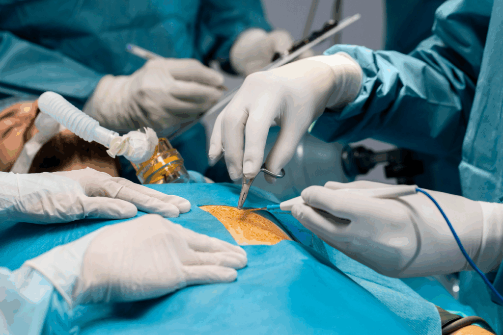
Keeping the lab clean is vital to avoid infections and keep patients safe. Sterile drapes, gowns, and gloves are used to create a clean area. This area stays clean during the whole procedure.
Watching videos on how to keep the area clean and team positions helps a lot. It shows how important these steps are. It helps doctors understand and do their jobs better.
How the team is set up is also very important. It helps everyone work together smoothly and quickly handle any problems. Knowing each person’s role helps improve how well the team works together.
Seeing how heart cath and angioplasty access points work gives us important insights. The right access point is key and depends on the patient’s needs.
We mainly use two access points: the femoral artery and the radial artery. Each has its own benefits and is picked based on the patient and the procedure.
The femoral artery method is a traditional way for these procedures. This method uses a catheter through the femoral artery in the groin. Pictures of this show how the artery is accessed, which is key to understanding the process.
The femoral artery method lets us use bigger catheters for complex tasks. But, it means patients need to rest in bed longer to avoid problems.
The radial approach uses the wrist’s radial artery to access the heart. This method is popular because it’s less invasive and has lower bleeding risks. Videos show how this technique works and its advantages.
| Access Point | Advantages | Considerations |
| Femoral Artery | Larger catheter size possible | Longer bed rest required |
| Radial Artery | Minimally invasive, less bleeding risk | Technical challenges for operators |
By seeing and understanding these access points, patients and doctors can better grasp the heart cath and angioplasty procedures.
The path to the heart is made possible by advanced technology. We use fluoroscopic guidance and anatomical maps to guide the catheter. Real-time imaging is key for precision and safety.
Fluoroscopic guidance gives us a live view of the catheter’s path. It lets us adjust the catheter’s route as needed. Fluoroscopic footage is vital for monitoring and making adjustments.
This technology helps us avoid complications and place the catheter correctly. The real-time feedback is essential for the procedure’s success.
Anatomical roadmapping adds to the precision of catheter navigation. It creates a detailed map of the blood vessels. This map, combined with fluoroscopy, makes navigating easier and more accurate. Anatomical roadmapping videos help us understand the patient’s anatomy better.
These advanced imaging techniques improve cardiac cath procedure outcomes. The mix of real-time fluoroscopy and anatomical roadmapping is a powerful tool. It helps us provide the best care for our patients.
Coronary angiography is key for seeing the coronary arteries clearly. It helps doctors understand heart health. This lets them make the best treatment plans.
This test uses contrast dye to show the arteries. It’s vital for spotting blockages and disease severity.
The dye injection is a critical part of the test. It helps see the arteries and find blockages. The dye is injected in a special order for the best images.
Seeing the arteries from different angles is important. This multi-view approach helps doctors understand how severe the blockages are. It guides them in choosing the right treatment.
| View | Description | Diagnostic Value |
| Left Anterior Oblique (LAO) | Provides a clear view of the left main coronary artery | High |
| Right Anterior Oblique (RAO) | Offers a detailed view of the right coronary artery | High |
| Antero-Posterior (AP) | Gives a complete view of both coronary arteries | Moderate |
Coronary angiography gives us a full picture of heart disease. It helps us create effective treatment plans.
Balloon angioplasty is key in heart care. It needs careful steps and clear views for the best results. We’ll look at each step, using detailed visuals to help you understand.
The first step is to guide a wire across the blockage. High-resolution images show how this is done. They help us see the wire’s path through the heart’s arteries.
The wire must be moved carefully to avoid harming the artery walls. Real-time imaging lets us check the wire’s position and make changes if needed.
After the wire is in place, the balloon is inflated. Videos of balloon inflation show how the blockage is pushed against the artery walls. This helps blood flow again.
These videos help us see how the balloon works. They show the pressure it uses and how it changes the artery. We see the immediate effects of inflation on the blockage and artery.
Getting the stent in the right spot is key to fix blood flow. We use special imaging to guide the stent’s placement. This makes sure it fits perfectly in the artery.
Putting the stent in place is a detailed task. Accurate placement is vital to avoid problems. We watch the stent move through the artery and adjust as needed.
The way we release the stent is also important. Controlled release helps the stent expand evenly. This is good for the patient’s health.
After it’s in, we check how the stent expands and fits against the artery. Optimal wall apposition stops future issues like stent thrombosis. We look at these images to make sure the stent is fully expanded and fits well.
| Aspect | Description | Importance |
| Stent Positioning | Accurate placement within the coronary artery | High |
| Release Sequence | Controlled expansion against arterial walls | High |
| Stent Expansion | Full expansion to restore normal blood flow | Critical |
| Wall Apposition | Stent apposition against the arterial wall | Critical |
In summary, stent deployment is a detailed process. We use advanced imaging and careful techniques. This ensures stents are placed correctly, improving blood flow and patient health.
We see the effects of heart cath and angioplasty through before and after angiogram images. These pictures are key in showing how well cardiac treatments work.
Images of angiograms before and after the procedure show the success of heart cath and angioplasty. They show how blood flow to the heart gets better and how the heart works better.
Videos of restored coronary perfusion after angioplasty show the success of the procedure. They show how blood flow to the heart gets better in real time. This proves the positive effects of cardiac treatments.
Looking at before and after angiograms and videos of restored coronary perfusion shows the big impact of heart cath and angioplasty. These visual aids not only show the success of the procedure. They also highlight the critical role these treatments play in keeping the heart healthy.
It’s key to spot complications during heart cath and angioplasty to keep patients safe. These lifesaving procedures can lead to serious issues like dissection, perforation, and thrombus formation.
Quickly identifying and managing these problems is essential. Visual imagery is vital for doctors to grasp the issue and decide the best action.
Dissection and perforation are major risks during cardiac catheterization. Dissection means the artery wall tears, while perforation causes a hole, risking severe bleeding or tamponade.
Spotting these issues early is critical. Tools like fluoroscopy and angiography offer live views to help doctors see dissections and perforations.
A study in the Journal of the American College of Cardiology stresses imaging’s role. It says, “Quickly spotting and treating coronary artery perforation is key to avoiding severe outcomes.”
“The use of imaging modalities during cardiac catheterization is vital for early spotting of complications like dissection and perforation.”
Journal of the American College of Cardiology
Thrombus formation is another risk during heart cath and angioplasty. Thrombi can block blood flow, causing ischemic events or procedure failure.
Videos showing thrombus formation and resolution offer insights. They help doctors see how these issues work and the best ways to manage them.
| Complication | Imaging Modality | Management Strategy |
| Dissection | Fluoroscopy | Stent placement |
| Perforation | Angiography | Covered stent or coil embolization |
| Thrombus Formation | Intravascular ultrasound | Antithrombotic therapy |
By using these imaging tools and knowing what to look for, doctors can make procedures safer and better for patients.
Cardiac intervention imaging has seen big changes, making heart treatments better. We’ve looked at the imaging methods and tech that make these treatments safer and more precise.
The future looks bright for cardiac cath and angioplasty imaging. New tech aims to make images clearer, use less radiation, and work with other tests. These steps will make cardiac intervention imaging even better.
Looking ahead, cardiac imaging will be more important for patient care. Knowing how it’s changing helps doctors and patients see its value and benefits.
New technologies will keep changing cardiac imaging. This will help doctors give better care and improve how patients do after treatment.
Images and videos help patients understand the procedures better. They also help doctors learn and improve. This reduces anxiety and improves outcomes.
Visual aids show blockages and how blood flows. This helps doctors plan the best treatment. It makes treatment more effective.
Coronary angiography gives clear images of the arteries. It helps doctors see how severe the disease is. This guides their treatment choices.
Seeing where the doctor accesses the artery helps patients and doctors. It shows the techniques used. This improves understanding and training.
These technologies guide the catheter precisely. They show the path to the heart. This is key for the success of the procedures.
Balloon angioplasty is a key treatment. It opens the artery by pushing plaque aside. This improves blood flow and patient health.
Visuals of stent placement and expansion are important. They ensure the stent is correctly placed. This improves patient outcomes.
Comparing angiograms shows the success of the procedure. It clearly shows how the treatment helps patients.
Visuals of complications like dissection and thrombus help doctors. They can plan how to manage these issues. This prevents bad outcomes.
Cardiac imaging has made big strides. New technologies are making procedures safer and more precise. Future advancements will keep improving care.
Images and videos teach doctors and patients about the procedures. They provide a clear view of the equipment and steps involved. This improves training and education.
Sterile protocols keep the area safe and clean. This reduces the risk of complications. It ensures patient safety.
Subscribe to our e-newsletter to stay informed about the latest innovations in the world of health and exclusive offers!
WhatsApp us