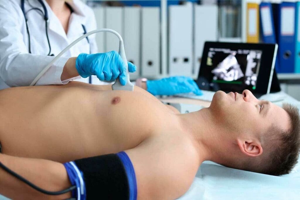
At Liv Hospital, we know how key accurate diagnosis is in treating heart issues. Heart imaging tests are vital. They help doctors see how the heart works and its structure.
These tools are key for spotting heart problems like coronary artery disease and cardiomyopathy. We use many cardiac imaging tests to give our patients the best care.
We’re dedicated to top-notch heart care. We use the latest heart diagnostic imaging methods. This article will dive into the top 5 heart imaging tests. We’ll look at how they work and what they can find.
Key Takeaways
- Heart imaging tests are key to accurate heart condition diagnosis.
- The top 5 heart imaging tests include echocardiograms, cardiac CT scans, cardiac MRI, nuclear cardiology studies, and chest X-rays.
- These tests help diagnose various heart conditions, such as coronary artery disease and cardiomyopathy.
- Advanced cardiac imaging tests enable healthcare professionals to provide complete care.
- Liv Hospital is committed to delivering exceptional cardiovascular care through the use of advanced diagnostic techniques.
The Critical Role of Heart Imaging in Cardiac Diagnosis
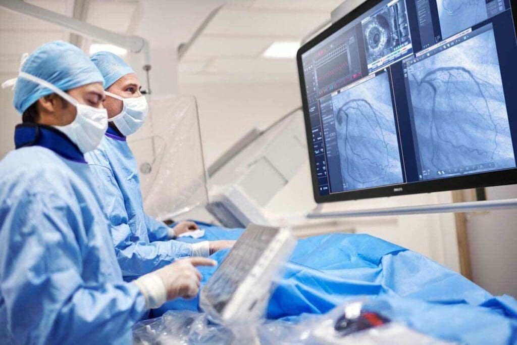
Advanced cardiac imaging techniques are changing cardiology. They give detailed images of the heart, improving diagnosis. These tests help us understand the heart’s structure and function, key to diagnosing and managing heart disease.
Why Cardiac Imaging Is Essential for Heart Disease Detection
Cardiac imaging is key to catching heart disease early. It lets doctors see the heart’s anatomy, spotting issues before symptoms show. This early catch allows for timely treatment, improving patient results.
Key benefits of cardiac imaging include:
- Early detection of heart disease
- Accurate diagnosis of cardiac conditions
- Guidance for treatment planning
- Monitoring of disease progression
How Imaging Guides Treatment Decisions
Cardiac imaging helps not just in diagnosing but also in treatment planning. It gives detailed images of the heart, helping doctors choose the best treatment for each patient.
“The choice of imaging modality can significantly impact patient management, influencing decisions regarding medication, intervention, or surgery.”
For example, imaging tests show how severe coronary artery disease is. This helps decide if angioplasty or bypass surgery is needed. It also checks heart valve function, guiding decisions on repair or replacement.
| Imaging Test | Clinical Application | Guiding Treatment Decisions |
| Echocardiogram | Assesses heart valve function and structure | Informs decisions about valve repair or replacement |
| Cardiac CT Scan | Evaluates coronary artery disease | Guides decisions about angioplasty or bypass surgery |
| Cardiac MRI | Assesses heart function and viability | Influences decisions about revascularization or medical therapy |
Risk Factors That Indicate the Need for Heart Imaging
Certain risk factors mean you might need heart imaging tests. These include a family history of heart disease, high blood pressure, high cholesterol, diabetes, and smoking. People with these risk factors can benefit from cardiac imaging to check their heart health and catch issues early.
Understanding heart imaging’s role in cardiac diagnosis shows its importance in patient care. It’s not just a tool for diagnosis; it’s a key to effective treatment and better health outcomes.
Common Heart Conditions Diagnosed Through Imaging
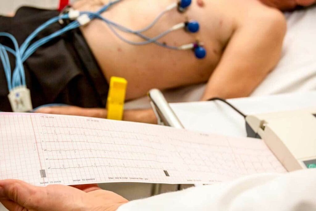
Cardiac imaging is key in finding heart problems that affect many people. We use new imaging methods to see the heart’s shape and how it works. This helps us find and treat heart diseases well.
Coronary Artery Disease
Coronary artery disease (CAD) is a big heart problem we find with imaging. It happens when the heart’s blood supply gets blocked by plaque. Cardiac imaging tests like coronary angiography and cardiac CT scans help us see how bad CAD is.
These tests show us where the blockages are. Then, we can choose the best treatment. This could be medicine, angioplasty, or surgery.
Valvular Heart Disease
Valvular heart disease affects the heart valves. Echocardiography is a main imaging tool for finding this problem. It lets us check how well the valves work.
With cardiovascular tests and procedures, we can spot issues like mitral regurgitation and aortic stenosis. Then, we can plan the best treatment.
Cardiomyopathy and Heart Failure
Cardiomyopathy is a disease of the heart muscle that can cause heart failure. Cardiac MRI and other tests help us find and measure cardiomyopathy. This lets us see why the heart might fail and how to fix it.
Congenital Heart Defects
Congenital heart defects are heart problems that babies are born with. Cardiac imaging is very important in finding these defects. It helps us plan surgeries and care for these babies.
With different heart test procedures, we can find problems like septal defects and tetralogy of Fallot. Then, we can give each child the right care for their heart issue.
Echocardiogram: Ultrasound Visualization of the Heart
Echocardiograms are key in cardiology, giving a non-invasive look at the heart. They use ultrasound waves to show the heart’s structure and function. This helps doctors diagnose heart conditions accurately.
Procedure and Patient Experience
A technician applies gel to the chest and uses a transducer for images. The process is painless and takes 30 to 60 minutes. Patients wear a gown and lie on a table for the imaging.
Types of Echocardiogram Procedures
There are different echocardiogram types, each for specific heart information:
- Transthoracic Echocardiogram (TTE): The most common, with the transducer on the chest.
- Transesophageal Echocardiogram (TEE): A transducer is inserted into the esophagus for closer heart views.
- Stress Echocardiogram: Done before and after stressing the heart, often through exercise or medicine.
- Contrast Echocardiogram: Uses a contrast agent to improve heart structure images.
Clinical Applications and Diagnostic Capabilities
Echocardiograms help diagnose heart conditions like valvular disease and cardiomyopathy. They show the heart’s chambers, valves, and walls, and its function.
| Condition | Echocardiogram Findings | Clinical Implication |
| Valvular Heart Disease | Abnormal valve function or structure | Need for valve repair or replacement |
| Cardiomyopathy | Altered heart chamber size or function | Management of heart failure symptoms |
| Congenital Heart Defects | Structural anomalies in the heart | Surgical planning and intervention |
Advantages and Limitations
Echocardiograms are non-invasive, low-cost, and provide real-time heart images. But image quality can be affected by body type and lung disease. They are very informative but may not offer the detailed tissue information of cardiac MRI.
In conclusion, echocardiograms are a valuable tool in cardiology. They offer a safe way to see the heart and diagnose various conditions. Understanding the different types and their uses helps healthcare professionals make better decisions for patient care.
Cardiac CT Scan: 3D Imaging of Heart Structure
The cardiac CT scan is a non-invasive test that uses X-rays to create detailed 3D images of the heart. It’s a key tool for checking the heart’s structure and finding heart problems.
Procedure and Patient Experience
A cardiac CT scan is done in a special room with a CT scanner. It’s quick, lasting about 10-15 minutes. Patients lie on a table that slides into the scanner. They might get a contrast dye to help see better.
Patient preparation is key to a good scan. This means avoiding some foods or meds before and wearing loose clothes.
Types of Cardiac CT Procedures
There are several types of cardiac CT scans, including:
- Coronary Calcium Scan: This scan checks for calcium plaque in the coronary arteries.
- Coronary CT Angiography: It gives detailed images of the coronary arteries to find blockages.
- Cardiac CT Scan with Contrast: It makes the heart’s structures clearer.
Clinical Applications and Diagnostic Capabilities
Cardiac CT scans are great for finding and managing heart issues like coronary artery disease and cardiomyopathy. They give detailed info about the heart’s anatomy, helping doctors plan treatments.
“Cardiac CT scans have become an essential tool in cardiology, providing a non-invasive way to see the heart’s structure and function.”
Advantages and Limitations
Cardiac CT scans are non-invasive, quick, and give high-quality images. But they involve radiation and might need contrast dye, which isn’t good for everyone.
Choosing between cardiac CT scans and other tests like echocardiograms depends on what’s needed and the patient’s situation. Knowing the good and bad of each test is important for the best care.
Cardiac MRI: Advanced Heart Imaging Test for Complete Check-Up
The Cardiac MRI is a top-notch tool for checking the heart’s health. It’s a non-invasive test that uses a strong magnetic field and radio waves. These tools help create detailed pictures of the heart’s structure and how it works.
Procedure and Patient Experience
During a Cardiac MRI, you lie on a table that moves into a big, cylindrical magnet. The test is usually painless, but some might feel claustrophobic. We make sure you’re comfortable and know what’s happening.
The test can last from 30 to 90 minutes, based on how detailed the scan needs to be. You’ll need to stay very quiet to get clear images.
Types of Cardiac MRI Techniques
There are different Cardiac MRI techniques, each for a specific use:
- Standard Cardiac MRI: Shows detailed pictures of the heart’s structure and how it works.
- Stress Cardiac MRI: Checks how the heart works when it’s stressed, usually with medicine.
- Cardiac MRI Angiography: Looks at the coronary arteries and finds any blockages or problems.
Clinical Applications and Diagnostic Capabilities
Cardiac MRI helps find many heart problems, like cardiomyopathy, congenital heart defects, and coronary artery disease. It gives doctors a lot of information about the heart’s health. This helps them plan the best treatment.
“Cardiac MRI has changed how we diagnose and treat heart disease, giving us amazing insights into the heart’s health.”
Famous medical expert, Cardiologist
Cardiac MRI can diagnose many conditions, including:
| Condition | Diagnostic Capability |
| Cardiomyopathy | Looks at the heart’s structure and function, finding muscle problems. |
| Congenital Heart Defects | Shows detailed images of the heart, spotting structural issues. |
| Coronary Artery Disease | Sees the coronary arteries, finding blockages and checking blood flow. |
Advantages and Limitations
Cardiac MRI is non-invasive, gives high-quality images, and checks the heart’s function and structure. But it might not be right for everyone. Some people might feel claustrophobic, and it’s not safe for those with certain metal implants or pacemakers.
We consider the pros and cons of Cardiac MRI for each patient. We make sure it’s the best choice for their heart condition.
Nuclear Cardiology Studies: Evaluating Heart Function
Nuclear cardiology studies are a detailed way to check the heart’s health. They use a small amount of radioactive material to see the heart and its blood flow. This helps find different heart problems.
Procedure and Patient Experience
The process starts with a small radioactive tracer injected into the blood. This tracer goes to the heart, letting a camera take pictures of its structure and function. Patients might do stress tests, either by exercising or taking medicine, to see how the heart works under stress.
During the test, patients lie on a table while the camera takes pictures from all sides. Most people find it okay, but some might feel uncomfortable or nervous.
Types of Nuclear Imaging Procedures
There are many nuclear imaging tests used in cardiology:
- Myocardial Perfusion Imaging (MPI): Checks blood flow to the heart muscle, helping find coronary artery disease.
- Cardiac Positron Emission Tomography (PET): Gives detailed pictures of the heart’s structure and function, often used to check heart tissue.
- Radionuclide Ventriculography (RNVG or MUGA): Measures the heart’s pumping ability and can spot heart wall motion problems.
Clinical Applications and Diagnostic Capabilities
Nuclear cardiology studies help diagnose and manage heart issues like coronary artery disease, heart failure, and cardiomyopathy. They give important info on the heart’s structure and function, helping doctors make better treatment plans.
These studies can check blood flow to the heart, look at left ventricular function, and spot scar tissue or healthy heart muscle.
Advantages and Limitations
The good points of nuclear cardiology studies include their ability to show how the heart works, being very good at finding coronary artery disease, and checking if treatments work.
But, there are downsides like radiation exposure, possible artifacts, and needing special equipment and skills. Even with these challenges, nuclear cardiology studies are a key tool in heart disease diagnosis and treatment.
Chest X-ray: Basic Heart Imaging for Initial Assessment
The chest X-ray is a key test for checking heart health. It’s a simple, non-invasive way to see the heart’s size, shape, and position. It also looks at the lungs and nearby areas.
Procedure and Patient Experience
Getting a chest X-ray is easy. Patients stand or sit in front of the machine. The technician takes the picture from behind a screen.
“The chest X-ray is a simple, painless test that provides critical information about the heart and lungs,” says a medical expert, a cardiologist at XYZ Hospital. “It’s an essential tool in our diagnostic arsenal.”
What a Heart X-ray Called a Chest X-ray Can Detect
A chest X-ray can show important heart and lung details. It can spot:
- Enlargement of the heart, which may indicate heart failure or other conditions
- Abnormalities in the shape or position of the heart
- Fluid accumulation around the heart or lungs
- Lung conditions that may be related to heart disease, such as pulmonary edema
When Chest X-rays Are Recommended
Chest X-rays are often the first step for heart disease symptoms. These include shortness of breath, chest pain, or swelling in the legs. They also help monitor patients with known heart conditions.
Limitations and Need for Advanced Imaging
Though useful, chest X-rays have limits. They don’t show detailed heart structure or function. For more detailed checks, tests like echocardiograms, CT scans, or MRI scans are needed.
As a Medical consultant, a radiologist notes: “A chest X-ray is just the starting point. Depending on the findings, we may need to proceed with more specialized tests to get a clearer picture of the heart’s condition.”
Choosing the Right Heart Imaging Test: Comparative Analysis
Choosing the right heart imaging test is key to diagnosing and treating heart diseases. There are many options, like echocardiograms, CT scans, MRI, and nuclear cardiology studies. It’s important to pick the best test for accurate diagnosis and effective treatment.
Echocardiogram vs CT Scan: When to Use Each
Echocardiograms and CT scans are two common tests for the heart. An echocardiogram uses ultrasound to show the heart’s structure and function. It’s great for checking heart valves and detecting problems.
A CT scan uses X-rays to create detailed images of the heart and blood vessels. It’s best for finding coronary artery disease and planning treatments. Doctors choose between these tests based on the patient’s condition and what they need to know.
- Echocardiogram: Ideal for assessing heart valve function and detecting cardiac abnormalities.
- CT Scan: Highly effective in detecting coronary artery disease and assessing cardiac anatomy.
Radiation Exposure Considerations
When picking a test, think about radiation exposure. Tests like CT scans and nuclear cardiology studies use radiation, which can raise cancer risk. Echocardiograms and MRI don’t use radiation, making them safer for those needing repeated tests or who are sensitive to radiation.
To reduce radiation, doctors might choose low-dose CT scans. This way, they can lower radiation exposure while keeping image quality good.
Cost and Accessibility Factors
The cost and availability of tests also matter. Echocardiograms are cheaper and more common, making them easier for many to access. But tests like MRI and nuclear cardiology studies are pricier and less common because they need special equipment and skills.
Doctors must weigh the benefits of each test against the patient’s financial situation and access to healthcare. Insurance coverage and out-of-pocket costs are key when choosing a test.
Working with Your Cardiologist to Select the Appropriate Test
Choosing the right test is a team effort between the patient and cardiologist. By knowing the patient’s medical history and symptoms, doctors can suggest the best test. Patients should talk openly with their cardiologist about their needs and concerns.
Together, patients and doctors can make smart choices about heart imaging tests. This ensures accurate diagnosis and effective treatment for heart diseases.
Conclusion: Advances in Cardiac Imaging Technology
Cardiac imaging technology has made big strides, changing how we diagnose and treat heart disease. Echocardiography, CT scans, and MRI have gotten better. This means doctors can now make more accurate diagnoses and plan better treatments.
The fast growth of cardiac imaging is helping patients a lot. New heart imaging tests let us spot heart problems more clearly. This cuts down on wrong diagnoses and makes patients’ outcomes better.
As cardiac imaging tech keeps getting better, we’ll see even more advanced tools. Healthcare pros will keep giving top-notch care by using the newest cardiac imaging. This will help them diagnose and treat patients even better.
The future of heart care is bright because of these tech advances. We’re excited to see how these improvements will help both patients and doctors.
FAQ
What is a heart imaging test?
A heart imaging test uses technology to see the heart’s inside. It helps doctors find and treat heart problems.
What are the top 5 heart imaging tests?
The top 5 heart imaging tests are echocardiogram, cardiac CT scan, cardiac MRI, nuclear cardiology studies, and chest X-ray. Each test has its own way of showing the heart’s details.
What is the difference between an echocardiogram and a CT scan?
An echocardiogram uses sound waves to see the heart. A CT scan uses X-rays to make detailed 3D images. The right choice depends on what the doctor needs to see.
What is a cardiac MRI used for?
Cardiac MRI gives detailed pictures of the heart. It’s great for finding heart problems like cardiomyopathy and congenital heart defects.
Are heart imaging tests safe?
Most heart imaging tests are safe. But some, like CT scans and nuclear cardiology studies, use radiation. Always talk to your doctor about the risks and benefits.
How do I choose the right heart imaging test?
Choosing the right test depends on your condition and medical history. A cardiologist can help pick the best test for you.
What is nuclear cardiology used for?
Nuclear cardiology studies check heart function and blood flow. They help find coronary artery disease and check if treatments work.
Can a chest X-ray detect heart problems?
A chest X-ray can spot some heart issues,ike an enlarged heart. But it might not show all the details of the heart’s structure and function.
How do advances in cardiac imaging technology impact heart disease diagnosis and treatment?
New cardiac imaging tech makes diagnosing heart disease better. It helps doctors plan better treatments. This has greatly improved cardiology and patient care.
What are the risk factors that indicate the need for heart imaging?
Heart imaging is needed if you have a family history of heart disease, high blood pressure, high cholesterol, diabetes, or symptoms like chest pain or shortness of breath.
What is the role of cardiac imaging in diagnosing coronary artery disease?
Cardiac imaging is key in finding coronary artery disease. It shows the heart’s details, helping doctors understand the disease and plan treatments.
References
- Lakshmanan, S., et al. (2023). A comparison of cardiovascular imaging practices in Africa: Highlighting inequalities in cardiac disease diagnosis. Cardiovascular Journal of Africa, 34(7), 350-356. https://pmc.ncbi.nlm.nih.gov/articles/PMC11195774/


