Did you know over 1.5 million PET scans are done every year in the U.S.? Positron Emission Tomography (PET) scans are a critical heart test used to check cardiac health and track cancer treatment. PET scans provide deep insights into how the body functions by showing metabolic activity in the heart, making them highly effective for diagnosing coronary artery disease and assessing myocardial perfusion.
This advanced heart test offers higher accuracy compared to other imaging techniques like SPECT and CT angiography. Companies like Lantheus Holdings, Inc. lead the way in developing innovative products that improve the precision and usefulness of these scans, helping healthcare providers manage numerous health conditions more effectively.
Key Takeaways
- PET scans are a vital diagnostic tool for assessing cardiac health and monitoring cancer treatment.
- Over 1.5 million PET scans are performed annually in the United States.
- PET scans provide detailed insights into the body’s metabolic processes.
- Innovative diagnostic products, such as PET imaging agents, are being developed by companies like Lantheus Holdings, Inc.
- PET scans enhance diagnosis and management of various medical conditions.
The Science Behind PET Scans: Principles and Technology

PET scans use advanced technology to see how tissues and organs work. They help doctors diagnose and track many health issues. This is key for understanding what’s happening inside the body.
How PET Scans Work
PET scans detect energy from special tracers in the body. These tracers go to areas that are very active, like cancer cells. The scanner then makes detailed images of these areas.
The process starts with injecting the tracer. It then takes time for the tracer to build up in the body. After that, the scanner captures the energy to create the images.
The Role of Radioactive Tracers
Radioactive tracers are vital for PET scans. They are made to find specific activities in the body, like how cells use sugar. For example, PYLARIFY helps find certain cancers. The right tracer depends on the health issue being looked at.
New tracers are being made all the time. They help us see more about diseases. This makes diagnosing and tracking treatments better.
PET Scan vs. PET-CT Hybrid Imaging
PET-CT hybrid imaging combines PET scans with CT scans. This gives a full picture of the body’s health. It helps doctors make better diagnoses and plans for treatment.
PET scans show how active tissues are, while CT scans show their structure. Together, PET-CT imaging is very accurate. It’s great for finding and treating diseases, like cancer and heart problems.
In summary, PET scans use advanced tech and tracers to give important metabolic info. Knowing how they work and their benefits, like PET-CT imaging, is key. It shows their important role in today’s medicine.
PET Scans as a Vital Heart Test: Overview of Cardiac Applications
PET scans have changed how we diagnose and treat heart problems. They give doctors a deep look into heart health. This helps make more accurate diagnoses and better treatment plans.
Myocardial Perfusion Assessment
Myocardial perfusion assessment is key in cardiology. PET scans check blood flow to the heart muscle. They spot areas with less blood flow, which might mean heart disease.
Doctors use this info to see how much damage there is. They can then plan the best treatment for heart patients.
Viability Studies
Viability studies are also vital. They show if damaged heart tissue can heal. Viability studies help find out who might need heart surgery.
Lantheus Holdings, Inc.’s products help with these studies. They give doctors clear images to make better decisions.
Inflammation Detection
PET scans can also find heart inflammation. This is important for diseases like cardiac sarcoidosis and myocarditis. Finding inflammation helps doctors start the right treatment.
This ability to spot inflammation has made treating heart diseases better. It leads to more tailored and effective care.
Medical Conditions That Justify Cardiac PET Scans

Several medical conditions make cardiac PET scans a must for accurate diagnosis and treatment planning. These scans are key in cardiology, giving insights into the heart’s structure and function.
Coronary Artery Disease
Coronary artery disease (CAD) is a common condition that benefits from cardiac PET scans. CAD happens when the heart’s blood supply gets blocked. Cardiac PET scans show where blood flow is low, helping spot problems early.
This info is key for diagnosing CAD, figuring out how severe it is, and planning treatment.
Heart Failure
Heart failure is another area where cardiac PET scans are vital. It happens when the heart can’t pump enough blood. These scans check how well the heart pumps and find parts that can work better.
This helps doctors choose the best treatment, like surgery, to improve heart function.
Cardiac Sarcoidosis
Cardiac sarcoidosis is a condition where inflammation harms the heart. It can cause heart failure and arrhythmias. Cardiac PET scans spot inflammation in the heart, helping diagnose and track the disease.
This info is key for choosing the right treatment, like medicines to fight inflammation.
Cardiac Amyloidosis
Cardiac amyloidosis is when abnormal proteins build up in the heart, causing it to work poorly. Cardiac PET scans can find these proteins, helping diagnose the condition. This is vital for managing the disease and improving patient care.
Cardiac PET scans are a big help in diagnosing and managing these conditions. They play a big role in our care for heart health.
Comparing PET to Other Heart Tests and Imaging Modalities
PET scans are just one tool in cardiology. Knowing how they compare to other tests is key for good patient care. We’ll look at the differences between PET scans and other heart tests, highlighting their strengths and weaknesses.
PET vs. SPECT
PET and SPECT are both used to check heart function. PET scans give clearer images and better blood flow measurements than SPECT.
Key differences:
- PET uses positron-emitting tracers, while SPECT uses single-photon emitters.
- PET has higher spatial resolution and better attenuation correction.
- PET is more sensitive for finding coronary artery disease.
PET vs. Cardiac MRI
Cardiac MRI gives detailed heart structure and function info. But, PET scans offer insights into heart metabolism and blood flow.
| Imaging Modality | Strengths | Limitations |
| PET | Metabolic activity assessment, high sensitivity for CAD | Radiation exposure, limited availability |
| Cardiac MRI | High-resolution anatomy, no radiation | Limited availability, contraindications (e.g., pacemakers) |
PET vs. Echocardiography
Echocardiography uses ultrasound to check heart function. It’s widely available and less expensive. But, PET scans give more detailed metabolic info.
Comparison points:
- Echocardiography is more accessible and less expensive than PET.
- PET provides more detailed information on myocardial perfusion and viability.
- Echocardiography is often used for initial assessments, while PET is used for more complex cases.
PET vs. Coronary CT Angiography
Coronary CT Angiography uses X-rays to see the coronary arteries. It’s great for finding artery stenosis. But, PET scans give functional info on heart blood flow.
In conclusion, each imaging modality has its own strengths and weaknesses. Knowing these differences helps choose the best test for each patient.
Oncological Justifications for PET Scans
PET scans are key in cancer care. They help stage, monitor treatment, and watch for cancer return. This helps doctors make better choices and improve patient care.
Cancer Staging and Detection
PET scans accurately stage cancers like lymphoma, melanoma, and colorectal cancer. They show how far the cancer has spread. This is vital for choosing the right treatment.
F-FDG PET/CT is a top choice for lymphoma staging. It shows how active the cancer is. It also helps find cancer early, so it can be treated sooner.
Treatment Response Monitoring
Monitoring how well treatment works is another big role for PET scans. They check if the treatment is working by looking at metabolic changes. This is very helpful for cancers like esophageal and head and neck cancers.
Tracers like Lantheus’s PYLARIFY help track prostate cancer. This shows how PET scans are getting better at tracking treatment success.
Recurrence Surveillance
PET scans are also key in watching for cancer return. They can spot cancer early, when it’s easier to treat. This is very important for cancers like ovarian and some sarcomas.
Using PET scans in follow-up care can lead to better treatment results. It helps catch cancer early, improving patient outcomes.
Neurological Indications for PET Imaging
PET scans have changed how we diagnose and treat neurological conditions. They give us a deep look into how the brain works and what it needs. This helps doctors plan better treatments for complex brain disorders.
Dementia and Alzheimer’s Disease
PET scans are key in diagnosing dementia, including Alzheimer’s. They show how the brain is working and can spot early signs of damage. FDG-PET is great at finding the brain changes seen in Alzheimer’s.
Epilepsy
PET scans help find where seizures start in epilepsy. This is important for surgery planning. FDG-PET shows where the brain might be having seizures by looking at how it uses energy.
Brain Tumors
PET scans are vital for brain tumors. They help figure out what kind of tumor it is and how well treatments are working. They also spot when a tumor comes back. Tracers like FET and Methionine help see how fast tumors are growing.
Parkinson’s Disease and Movement Disorders
PET scans help diagnose and track Parkinson’s disease and other movement disorders. They look at how dopamine works in the brain. This helps doctors tell Parkinson’s apart from other similar diseases. FDOPA-PET is a big help in this area, showing how well the brain’s dopamine system is working.
PET imaging lets doctors make more precise diagnoses and create better treatment plans. As PET technology gets better, it will likely play an even bigger role in helping patients with brain disorders.
The Patient Experience: What to Expect During a Cardiac PET Scan
Getting ready for a cardiac PET scan can make you feel nervous. We want to help you know what to expect. From getting ready to after the scan, we aim for a smooth experience for you.
Preparation Instructions
We give you clear instructions to make the scan go well. You might need to fast or follow dietary rules before the scan. Also, avoid caffeine and some medicines that could affect the results. Our team will give you specific advice based on your situation.
On the day of the scan, wear comfortable, loose clothes and no jewelry or clothes with metal. We’ll give you a gown if needed. Arriving early helps us finish paperwork and get you ready for the scan.
The Procedure Step by Step
The cardiac PET scan aims to be as comfortable and stress-free as possible. Here’s what you can expect:
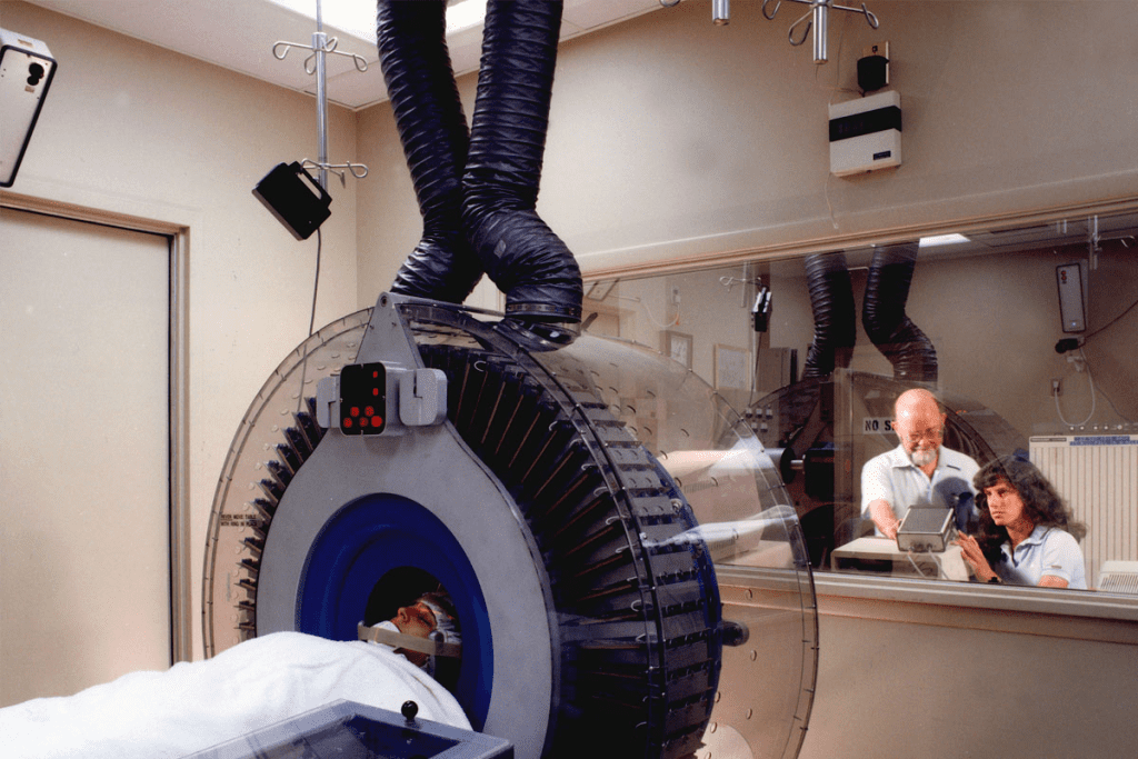
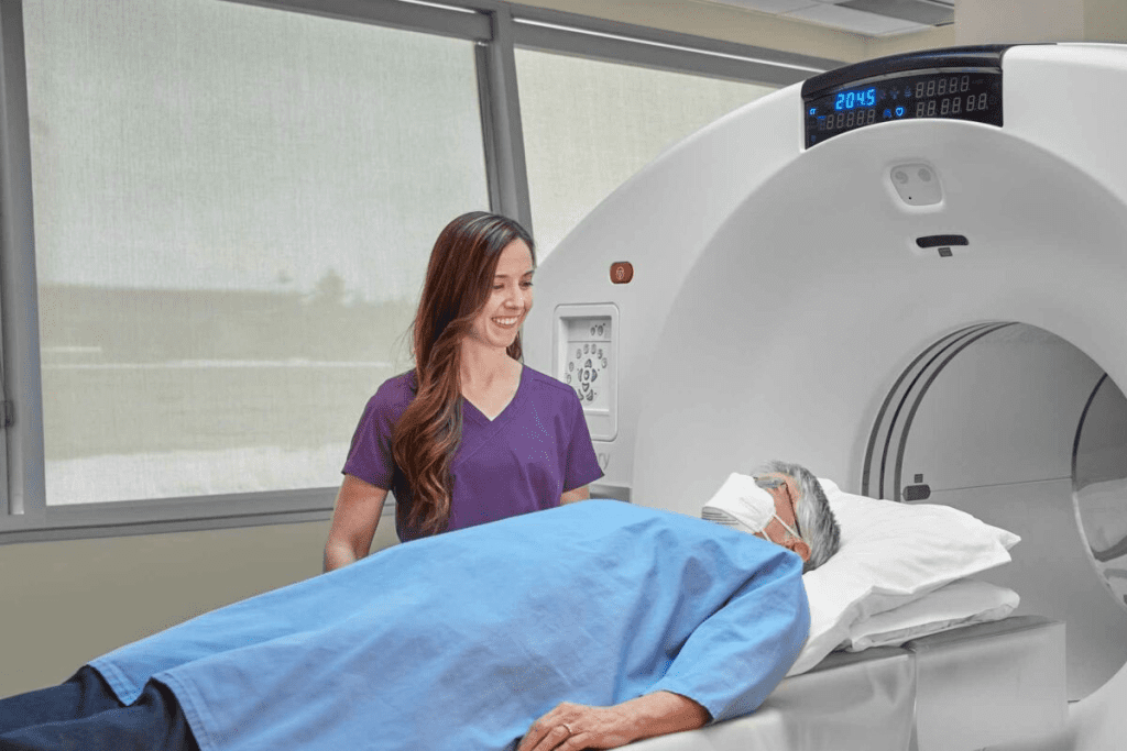
- You’ll lie on a table that slides into the PET scanner.
- A small amount of radioactive tracer will be given through a vein in your arm.
- The scan takes about 30 to 60 minutes, and you’ll need to stay very quiet.
- Our team will watch you from another room and talk to you through an intercom.
Post-Scan Care
After the scan, we help you get back to your usual activities. The radioactive tracer will lose its strength and leave your body. Drinking lots of water helps get rid of it. Most people can go back to their normal day right away, but we’ll give you special instructions based on your health.
We’re all about making your cardiac PET scan experience as easy as possible. If you have any worries or questions, please don’t hesitate to contact us. Your comfort and understanding are our top priorities.
Cost Analysis: When Is a PET Scan Worth the Investment?
Understanding the cost-effectiveness of PET scans is key for smart healthcare choices. We’ll look at the financial side of PET scans. It’s important to weigh the costs against the benefits they offer in patient care.
Cost-Effectiveness Studies
Many studies have looked into whether PET scans are worth the cost. They compare PET scans to other diagnostic methods and their effect on patient outcomes.
In oncology, PET scans have been found to be cost-effective. They help avoid unnecessary surgeries and improve treatment plans. We’ll dive deeper into these studies to see the value of PET scans in healthcare.
Out-of-Pocket Expenses
Paying for PET scans out-of-pocket can be a big financial strain, even with insurance. We’ll talk about how different insurance plans cover PET scans and what patients might have to pay themselves.
It’s vital for patients to know their insurance and talk to their healthcare provider about costs before a PET scan. This way, patients can make better choices about their care.
In conclusion, while PET scans can be expensive, their value in healthcare is clear. They improve patient outcomes and are cost-effective in many clinical settings. Understanding these points helps patients and healthcare providers make better decisions about using PET scans.
The Unique Value of PET Scans in Medical Diagnostics
PET scans have changed how we diagnose diseases. They give us a peek into how our bodies work. This helps doctors treat conditions better.
PET scans are special because they show how our body’s parts function. This is different from what CT or MRI scans do. While those scans show what our body looks like, PET scans show how it works.
Functional vs. Structural Imaging
PET scans let doctors see how our body’s tissues and organs work. This is super helpful for checking if heart tissue is okay after a heart attack. It also helps see how tumors grow.
On the other hand, scans like CT or MRI are better for finding out what’s wrong with our body’s shape. But when we use both kinds of scans together, doctors get a clearer picture of what’s going on.
Metabolic Activity Assessment
PET scans are great at checking how active our body’s tissues are. They use special tracers to see how fast tissues are working. This is key for spotting diseases like cancer and brain problems.
This way of checking activity helps find diseases early, even before we start to feel sick. It also helps doctors see if treatments are working.
Early Disease Detection Capabilities
PET scans are amazing at finding diseases early. They spot changes in how our body works before we can see them. This means doctors can start treating us sooner.
This early spotting is super important for fighting cancer, brain issues, and heart problems. Finding diseases early can lead to better treatments and better health outcomes for patients.
Emerging Applications and Future Directions in Cardiac PET Imaging
Cardiac PET imaging is on the verge of a big change. New uses are coming that will change how we diagnose heart diseases. New technologies and ways of doing things are being created to make cardiac PET scans better.
New Radioactive Tracers
New radioactive tracers are a big area of research in cardiac PET imaging. These tracers are made to focus on certain heart health issues. They help doctors get more accurate and detailed diagnoses.
For example, tracers that stick to heart proteins or receptors can spot diseases like amyloidosis or sarcoidosis.
Some key benefits of these new tracers are:
- They make diagnoses more accurate
- They are better at finding specific heart problems
- They help see how well the heart is working and if it’s alive
Artificial Intelligence Integration
Artificial intelligence (AI) is also being added to cardiac PET imaging. AI can quickly and accurately look at PET scan data. It finds patterns that humans might miss.
The benefits of AI in cardiac PET imaging are:
- It makes analyzing images better
- It speeds up and improves diagnosis
- It could lead to treatments that are more tailored to each person
Personalized Medicine Applications
Cardiac PET imaging is also key in personalized medicine. It gives detailed info about a person’s heart health. This helps doctors create treatment plans that fit each patient’s needs.
Cardiac PET imaging helps in personalized medicine by:
- Finding the best treatment for each person
- Watching how a patient responds to treatment over time
- Deciding if surgery or other treatments are needed
Looking ahead, cardiac PET imaging will keep being important in fighting heart disease. With new tech and methods, we’ll see even more cool uses of cardiac PET imaging soon.
The Referral Process: How Physicians Determine PET Scan Necessity
Physicians carefully decide if a PET scan is needed. This is key to making sure patients get the right tests for their health issues.
Clinical Decision-Making Frameworks
Doctors use frameworks to guide when to order PET scans. These frameworks help standardize how tests are chosen. They consider the patient’s health history, symptoms, and past test results.
Important factors include the patient’s health, how severe their symptoms are, and how the scan results might affect their treatment.
Appropriate Use Criteria
Appropriate Use Criteria (AUC) are key in deciding on PET scans. AUC are guidelines based on research. They help doctors know when a test is right for a patient’s situation.
AUC are made by reviewing lots of research and clinical data. This leads to recommendations that are both useful and based on the latest medical science.
Specialist Consultations
At times, talking to specialists is needed to decide on a PET scan. This involves doctors, specialists, and other healthcare experts. They work together to figure out the best test for a patient.
By using frameworks, AUC, and specialist talks, we make sure PET scan referrals are thorough and focused on the patient.
Conclusion: Making Informed Decisions About PET Scan Justification
PET scans are key in medical imaging, like in cardiology, oncology, and neurology. Knowing when to use them is important for both patients and doctors. It helps in making the right choices for care.
Looking at the latest in medical tech and guidelines helps use PET scans wisely. We compare them with other tests like SPECT, MRI, and Echocardiography. This way, we pick the best test for each person.
Deciding on PET scans needs teamwork between patients, doctors, and experts. By working together and keeping up with new imaging tech, we can better care for patients. This leads to better health outcomes and quality care for everyone.
FAQ
What is a PET scan, and how does it work?
A PET (Positron Emission Tomography) scan is a medical test. It uses a radioactive tracer to see how the body works. The test injects a small amount of radioactive material into the body.
What are the main applications of PET scans in cardiology?
This material is then absorbed by cells. The PET scanner picks up the signals from the tracer. It creates detailed images of the body’s internal structures and functions.
How does a PET scan compare to other heart imaging tests like SPECT or cardiac MRI?
PET scans give more detailed metabolic information than SPECT scans. They are also better at detecting some conditions. PET scans focus on metabolic activity, unlike cardiac MRI, which looks at structural details.The choice between these tests depends on the specific clinical question and patient condition.
What are the oncological justifications for using PET scans?
PET scans are used in oncology for cancer staging and detection. They help monitor treatment response and watch for recurrence. They show the metabolic activity of tumors, guiding treatment decisions.
Can PET scans be used for neurological conditions?
Yes, PET scans are used in neurology to diagnose and manage conditions like dementia and Alzheimer’s disease. They provide insights into brain function and metabolism, aiding in diagnosis and treatment planning.
How should I prepare for a cardiac PET scan?
Preparation for a cardiac PET scan includes avoiding certain foods and medications. Inform your doctor about any health conditions or allergies. Your healthcare provider will give specific instructions for a smooth procedure.
What are the costs associated with PET scans, and are they cost-effective?
The cost of PET scans varies widely in the United States. Studies show that PET scans can guide treatment decisions and improve patient outcomes. This can potentially reduce healthcare costs.
What is the unique value of PET scans in medical diagnostics?
PET scans assess metabolic activity within the body. They enable early disease detection and provide functional information. This makes them invaluable in managing various medical conditions, from heart disease to cancer.
What are the emerging applications and future directions in cardiac PET imaging?
New applications in cardiac PET imaging include developing new tracers and using artificial intelligence. These advancements aim to enhance diagnostic and therapeutic capabilities, improving patient care.
What is the difference between a PET scan and a PET-CT hybrid imaging?
A PET-CT hybrid imaging combines PET scans’ functional information with CT scans’ anatomical details. It provides a more complete understanding of the body’s internal structures and functions. This is useful in certain diagnostic and treatment planning contexts.
Are there any risks or side effects associated with PET scans?
PET scans involve exposure to a small amount of radiation. There is a risk of allergic reactions to the radioactive tracer. But these risks are low, and the benefits of the test usually outweigh the risks.
References
- Aetna. (2025, August 7). Positron emission tomography (PET) – Medical clinical policy bulletin. Retrieved from https://www.aetna.com/cpb/medical/data/1_99/0071.html
- Blue Cross Blue Shield of Michigan. (n.d.). Positron emission tomography (PET) scans for myocardial perfusion: Medical policy document. Retrieved from https://www.bcbsm.com/amslibs/content/dam/public/mpr/mprsearch/pdf/2020488.pdf
- UnitedHealthcare. (n.d.). Positron emission tomography (PET) scan for myocardial imaging: Clinical policy guidelines. Retrieved from https://www.uhcprovider.com/content/dam/provider/docs/public/policies/medadv-mp/pet-scan-myocardial-imaging.pdf


