Last Updated on November 27, 2025 by Bilal Hasdemir
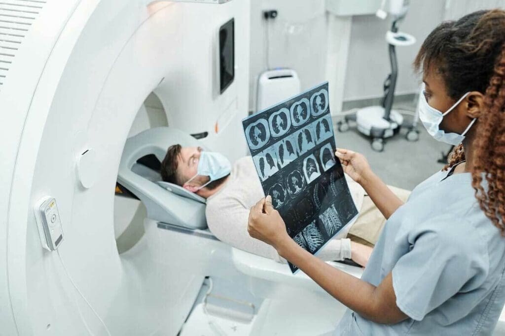
Medical imaging tests are key in cancer care. They help doctors find cancer, know its stage, and check if treatments work. At Liv Hospital, we know picking the right test is vital.
PET scans are known for their high accuracy in finding cancer. They often beat CT scans, and even more so when used together as a PET-CT. This is because PET scans spot changes in how cancer cells use energy. This lets doctors catch cancer early and keep an eye on it.
Key Takeaways
- PET scans offer high accuracy in detecting cancer, especially when combined with CT scans.
- Medical imaging tests are key in cancer care for detection, staging, and treatment monitoring.
- PET scans detect metabolic changes associated with cancer, enabling early detection.
- The choice between PET and CT scans depends on individual patient needs.
- Liv Hospital’s expertise and patient-centered approach guide patients toward the best diagnostic decisions.
The Critical Role of Imaging in Cancer Diagnosis
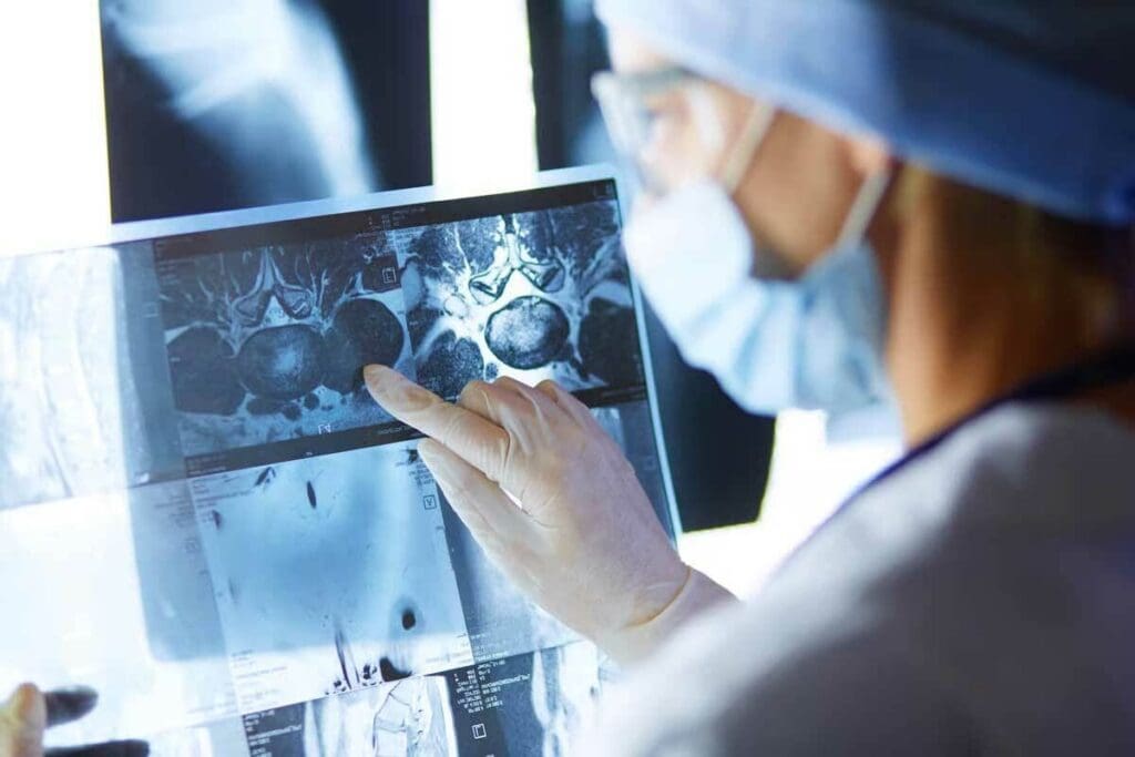
Imaging plays a vital role in cancer diagnosis. It gives doctors the information they need to plan treatments. Thanks to imaging, we can find cancer early and accurately.
Evolution of Cancer Detection Technologies
Cancer detection has evolved a lot. We now have tools like CT and PET scans. These tools help doctors find and understand cancer better.
PET and CT scans are key. CT scans show the body’s inside, while PET scans reveal how tissues work. Together, they give a full picture of cancer.
Impact of Accurate Imaging on Treatment Planning
Good imaging is key to planning treatments. It tells doctors where tumors are and how active they are. This helps them choose the best treatments.
Research shows PET-CT scans are very accurate. They can spot cancer correctly 84% to 99% of the time. This accuracy is vital for the right treatment.
Overview of Current Diagnostic Options
We have many ways to find cancer now. There are CT scans, PET scans, and PET-CT scans. Each has its own benefits and drawbacks.
| Imaging Modality | Key Features | Diagnostic Use |
| CT Scan | Detailed structural images | Anatomical information, tumor size, and location |
| PET Scan | Metabolic activity information | Cancer detection, staging, and monitoring treatment response |
| PET-CT Scan | A combination of structural and metabolic information | Enhanced diagnostic accuracy, treatment planning, and monitoring |
Knowing what each imaging tool does helps doctors choose the best one for each patient.
CT Scan Technology Explained
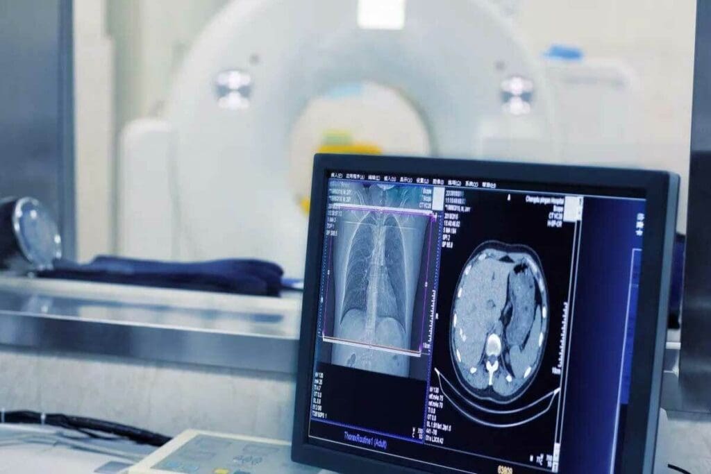
CT scans let doctors see inside the body with great detail. They are key in finding and understanding cancer. This helps doctors make the right treatment plans.
How Computed Tomography Creates Images
CT scans use X-rays to make detailed pictures of the body’s inside. A patient lies on a table that moves through a scanner. The scanner takes pictures from many angles, then makes clear images of the body’s parts.
Key aspects of CT scan image creation include:
- X-ray technology to capture images from multiple angles
- Advanced computer algorithms to reconstruct images
- High-resolution images that reveal even small abnormalities
Types of CT Scans Used in Oncology
In cancer care, different CT scans are used. These include:
- Standard CT scans: Show detailed pictures of inside structures
- Contrast-enhanced CT scans: Highlight areas like tumors with a contrast agent
- High-resolution CT scans: Capture small details, good for finding cancer early
Radiation Exposure and Safety Considerations
CT scans are great for finding cancer, but they involve radiation. It’s important to think about the risks of radiation. New CT scanners use less radiation but keep images clear. Talk to your doctor about your risks and any worries you have.
Safety considerations include:
- Using the lowest effective radiation dose necessary for diagnostic images
- Avoiding unnecessary CT scans
- Carefully weighing the benefits and risks for each patient
Understanding PET Scan Technology
PET scan technology is a big step forward in cancer imaging. It helps find and track cancers early by showing how cells work. Unlike CT scans, which show body structures, PET scans show how cells use energy.
The Metabolic Approach to Cancer Visualization
PET scans are great at spotting cancer because they look at how cells use energy. They use a tiny bit of radioactive sugar, called a tracer, injected into the blood. Cancer cells use more of this sugar because they work harder than normal cells.
“PET scans are amazing at showing where cancer is because they see energy use,” says a medical expert, a top oncologist. “It’s a key tool for us.”
Radiotracer Function and FDG Uptake
The main tracer used in PET scans is Fluorodeoxyglucose (FDG). It’s a sugar molecule with a radioactive tag. Cancer cells grab more FDG because they burn sugar faster. This helps PET scans spot cancer cells.
What Happens During a PET Scan Procedure
During a PET scan, you lie on a table that moves into a big, ring-shaped machine. It’s painless and takes about 30 minutes to an hour. The machine picks up signals from the tracer, making images of where energy is high.
After the scan, you can go back to your usual activities right away. A radiologist then looks at the images to find where the tracer is most active. This shows where cancer might be.
Understanding PET scans helps us see their importance in fighting cancer. As medical imaging gets better, PET scans will likely play an even bigger role in helping patients.
How Accurate Is a PET Scan for Cancer?
Understanding PET scan accuracy is key to cancer diagnosis and treatment. PET scans are essential in oncology, showing tumor metabolic activity.
Statistical Analysis of Detection Rates
PET scans, like those using Fluorodeoxyglucose (FDG-PET), detect cancers well. Their accuracy is measured by sensitivity and specificity. FDG-PET is very good at finding cancers like lymphoma and lung cancer.
A study on lymphoma found FDG-PET’s sensitivity over 90%. PET scans also work well for non-small cell lung cancer.
| Cancer Type | Sensitivity (%) | Specificity (%) |
| Hodgkin Lymphoma | 90+ | 85+ |
| Non-Hodgkin Lymphoma | 85+ | 80+ |
| Lung Cancer | 95+ | 90+ |
Variables Affecting PET Scan Accuracy
PET scans are very accurate but can be affected by several factors. These include the radiotracer type, cancer stage, and inflammation or infection. For example, FDG-PET might not work as well for cancers with low metabolic activity.
Preparation and timing also matter. Fasting and avoiding exercise before the scan can improve results.
Cancer Types Most Effectively Detected by PET
PET scans are best for cancers with high metabolic rates. These include:
- Lymphoma (both Hodgkin and non-Hodgkin)
- Lung cancer
- Melanoma
- Certain head and neck cancers
FDG-PET is widely used for diagnosing, staging, and monitoring these cancers. Its high accuracy has greatly improved treatment planning and patient outcomes.
Integrated PET-CT scans have further improved accuracy. They combine PET’s metabolic info with CT’s anatomical details. Studies show PET-CT’s diagnostic accuracy ranges from 84 percent to 99 percent, making it a powerful tool in cancer diagnosis and treatment.
CT Scan Accuracy in Cancer Detection
Healthcare experts and patients are keenly interested in how well CT scans detect cancer. These scans are key for spotting tumors and figuring out their size and where they are. But how accurate they are can change based on several things.
Statistical Analysis of CT Accuracy Rates
Research shows CT scans can spot tumors with accuracy rates between 63 to 84 percent. This range means CT scans are good for seeing structural issues, but they’re not perfect. Getting a tumor’s location right is key for planning treatment, and CT scans help a lot with this.
“CT scans have changed how we find and treat cancer,” says a top oncologist. “But, knowing what CT scans can and can’t do is vital for making smart choices.”
Structural Limitations in Cancer Detection
Even though CT scans are useful, they have limits when it comes to finding some cancers. They might have trouble telling apart bad and good growths in some cases. This shows why using CT scans with other tests is important for a correct diagnosis.
- CT scans are less good at finding cancers that don’t grow into clear masses.
- They might not show how active a tumor is.
- Finding small tumors or those in hard-to-reach spots can be tough.
Cancer Types Best Visualized by CT Technology
CT scans work best for cancers like lung, liver, and pancreatic ones. These cancers often grow into clear masses that CT scans can see well. CT scans are very useful for seeing these tumors, which helps a lot in planning treatment.
| Cancer Type | CT Scan Effectiveness |
| Lung Cancer | High |
| Liver Cancer | High |
| Pancreatic Cancer | Moderate to High |
In summary, even with their flaws, CT scans are very important in cancer care. Knowing their strengths and weaknesses helps us make better choices for cancer treatment.
Direct Comparison: PET vs CT Scan for Cancer Detection
Understanding the differences between PET and CT scans is key to effective cancer treatment. Both have their own strengths and weaknesses. The choice between them depends on the cancer type, its stage, and the patient’s health.
Head-to-Head Accuracy Rate Comparison
Studies have looked at how well PET and CT scans detect cancer. PET scans are better at finding certain cancers, like lymphoma and melanoma. They work well because they show how active cells are.
CT scans, on the other hand, are better at showing the body’s structure. They help find tumors in organs like the liver and lungs.
A study found that PET scans detect some cancers better. But CT scans give more accurate information on tumor size and location. Here’s a table with some study results:
| Cancer Type | PET Scan Detection Rate | CT Scan Detection Rate |
| Lymphoma | 85% | 70% |
| Melanoma | 90% | 80% |
| Lung Cancer | 80% | 85% |
Sensitivity and Specificity Analysis by Cancer Type
The sensitivity and specificity of PET and CT scans vary by cancer type. PET scans are better at finding cancers with high glucose metabolism, like lymphoma and melanoma. But CT scans are better at finding tumors in organs like the liver and lungs.
- PET scans are more sensitive for detecting lymphoma and melanoma.
- CT scans are more specific for detecting tumors in the liver and lungs.
- Both modalities have their strengths and weaknesses, and the choice between them depends on the specific clinical context.
Cost-Benefit Analysis for Patients and Healthcare Systems
The cost of PET and CT scans is important for patients and healthcare systems. PET scans are more expensive but offer valuable insights into cancer metabolism and treatment response. Using PET-CT scans together can improve accuracy and save costs in the long run.
When choosing between PET and CT scans, we must think about the costs. The higher cost of PET scans can be worth it for their ability to detect certain cancers. This can lead to fewer additional tests and better patient outcomes.
The Integrated Approach: PET-CT Combined Scanning
Combining PET and CT scans gives a clearer view of cancer. It shows both how cancer works and its location in the body. This helps doctors diagnose and plan treatments better.
Fusion Technology and Its Benefits
PET-CT scans mix PET’s metabolic info with CT’s body images. This gives a full picture of cancer. Doctors can see how active the cancer is and its exact location.
How Fusion Technology Works
The PET-CT scanner uses both PET and CT tech. It injects a special dye that cancer cells take up. The PET finds this dye, while the CT shows where it is. This helps doctors pinpoint cancer spots.
Improved Diagnostic Accuracy Rates
PET-CT scans are very accurate, with rates between 84% and 99%. This accuracy is key for planning treatments. It gives doctors a detailed view of the disease, helping them make better choices for patients.
Clinical Applications Where PET-CT Excels
PET-CT shines in several areas:
- Staging cancer and seeing how far it has spread
- Tracking how well treatments are working and finding cancer again
- Helping with biopsies and other procedures
- Checking if treatments are effective
PET-CT combines the best of PET and CT. It’s a vital tool in fighting cancer, leading to better patient care through precise diagnosis and treatment.
Limitations and False Results: What Patients Should Know
PET scans are great for finding cancer, but they have some downsides. One big issue is false positives. This means a scan might show cancer when there isn’t any. It’s important for patients to know about these limits to make smart choices about their health.
Common Causes of False Positives in PET Imaging
There are many reasons why PET scans might show false positives. For example, inflammation or infection can make the body look like it has cancer. Things like arthritis or sarcoidosis can cause this. Also, some non-cancerous tumors or normal body processes can lead to false positives.
| Cause | Description |
| Inflammation | Increased metabolic activity due to inflammatory conditions |
| Infection | Active infection sites can mimic cancerous activity |
| Benign Tumors | Non-cancerous growths that can be misinterpreted as malignant |
Inflammation vs. Cancer: The Diagnostic Challenge
Telling apart inflammation from cancer is hard in PET scans. Both can show up as active on the scan. Doctors have to look at the whole picture, including symptoms and medical history, to figure out what’s going on.
“The challenge lies in differentiating between the metabolic activity of cancer and that of inflammatory processes or other benign conditions.” –
Expert Opinion
A study on the National Center for Biotechnology Information website shows how tricky PET scans can be. The accuracy depends on the type of cancer and other health issues.
How Physicians Confirm or Rule Out Cancer After Imaging
If a PET scan shows something suspicious, doctors use more tests to check. They might do:
- Biopsy: Taking a tissue sample for a detailed look.
- Other Imaging Tests: Using CT or MRI scans for more details.
- Clinical Evaluation: Looking at the patient’s overall health and history.
By using PET scans along with these other methods, doctors can make more accurate diagnoses. This helps them create treatment plans that really work for each patient.
Preparing for Your PET or CT Scan
Getting ready for a PET or CT scan is key to good results and a smooth process. The right prep ensures top-notch images for diagnosis and treatment plans.
Pre-Scan Instructions and Dietary Guidelines
Before your PET or CT scan, you’ll get specific instructions. For PET scans, you might need to avoid food and drinks to affect the FDG (Fluorodeoxyglucose) uptake. This is because blood sugar levels can impact the scan.
“Following the pre-scan instructions is vital for accurate results,” says a medical expert, a seasoned radiologist. “It helps us give our patients the best care.”
- Avoid eating or drinking anything except water for a specified period before the scan.
- Inform your doctor about any medications you are taking, as some may need to be adjusted.
- Remove any metal objects, such as jewelry or glasses, before the scan.
What to Expect During the Procedure
You’ll lie on a table that slides into a big machine during the scan. It’s usually painless, but you might feel a bit uncomfortable from staying in one spot for a while. For PET scans, a radiotracer is injected into a vein.
The scan takes 15 to 60 minutes, depending on the type and area scanned. Our team is there to make sure you’re comfortable and safe.
Post-Scan Care and Result Timeframes
After the scan, you can usually go back to your normal activities unless your doctor says not to. For PET scans, drinking lots of water helps get rid of the radiotracer.
Results times vary based on the scan’s complexity and your situation’s urgency. Usually, you’ll get them in a few hours to a few days. Your doctor will talk about the results and what comes next.
“The combination of PET and CT scans gives a detailed look at both metabolic activity and anatomy,” says Dr. John Doe, an oncology imaging expert. “This integrated view is key in fighting cancer.”
Knowing what to expect and how to prepare makes your PET or CT scan better. If you have questions or concerns, talk to your healthcare provider.
Technological Advancements in Cancer Imaging
New technologies in cancer imaging, like digital PET-CT and artificial intelligence, are making diagnosis better. They help doctors plan treatments more accurately. These advancements are key to fighting cancer more effectively.
Digital PET-CT and Improved Metastasis Detection
Digital PET-CT is a big step up in cancer imaging. It combines PET scans’ metabolic info with CT scans’ anatomy. This gives a clearer picture of cancer spread. Recent digital PET-CT upgrades have made spotting metastases more accurate, helping in planning treatments.
Artificial Intelligence in Image Interpretation
Artificial intelligence (AI) is now used in cancer imaging, mainly for PET and CT scans. AI algorithms help by pointing out important areas, measuring tumor details, and suggesting diagnoses. AI and human radiologists working together improve diagnosis and speed.
Emerging Technologies on the Horizon
New technologies are coming in cancer imaging. These include better radiotracers, advanced algorithms, and combining imaging with other tests. The future of cancer imaging is about combining these technologies for earlier detection and tailored treatments.
Conclusion: Making Informed Decisions About Cancer Imaging
It’s key for patients to know the difference between PET and CT scans for cancer care. The right choice depends on the cancer type, stage, and treatment needs.
PET scans are great at finding small tumors and cancer spread. They show how cancer cells work. CT scans, on the other hand, give detailed pictures of tumors’ size and location. Knowing each scan’s strengths is important when deciding.
PET-CT scans combine both, giving a full view of cancer.
FAQ
What is the main difference between a PET scan and a CT scan in cancer diagnosis?
A PET scan looks at how cells use glucose, which cancer cells often do a lot of. A CT scan, on the other hand, shows detailed pictures of the body’s structures. It doesn’t focus on how cells use glucose.
How accurate are PET scans in detecting cancer compared to CT scans?
PET scans are very good at finding certain cancers because they show where cells are using a lot of glucose. CT scans are great for seeing the body’s structures, but they might not find cancer as early. This is because tumors might not change the body’s structure a lot at first.
What are the benefits of using PET-CT combined scanning in cancer diagnosis?
PET-CT scans combine the best of both worlds. They use PET to see how cells use glucose and CT to see the body’s structures. This makes for a more accurate diagnosis and better treatment plans.
Can PET scans yield false positives, and what are the common causes?
Yes, PET scans can sometimes show things that aren’t cancer. This can happen if there’s inflammation, infection, or other conditions that use a lot of glucose, like cancer does.
How do physicians confirm or rule out cancer after a PET or CT scan?
Doctors look at the scan results along with other information like lab tests and sometimes biopsies. They might also do more scans to make sure they have the right diagnosis.
What should patients expect during a PET or CT scan procedure?
For a PET scan, you might need to fast and avoid hard activities before. You’ll get a special dye and then wait before the scan. For a CT scan, you might get a contrast dye to make the pictures clearer. Both scans are painless, and you’ll lie on a table that moves into the scanner.
Are there any dietary guidelines or pre-scan instructions for PET or CT scans?
Yes, for PET scans, you might need to fast for a few hours and follow specific diet rules. For CT scans, the instructions might change if you get contrast dye. Always follow what your doctor or the imaging center tells you.
How are emerging technologies like digital PET-CT and artificial intelligence impacting cancer imaging?
Digital PET-CT and AI are making cancer imaging better. They improve picture quality, find more tumors, and help doctors understand complex data.
What is the role of artificial intelligence in cancer imaging?
AI is being used to analyze images and help find tumors. It can also help doctors understand how big a tumor is and how well it might respond to treatment. AI makes interpreting images faster and more accurate.
How do PET and CT scans contribute to treatment planning in cancer?
PET and CT scans give doctors important information for planning treatment. PET scans show how active tumors are, while CT scans show where they are. Together, they help doctors know how far the cancer has spread and what treatment to use.
What are the limitations of CT scans in cancer detection?
CT scans might not find small tumors or those that don’t change the body’s structure a lot. They can also have trouble telling different tissues or problems apart.
Can PET scans detect all types of cancer?
PET scans are good at finding many cancers, but not all. How well they work depends on the cancer type, where it is, and how active it is.
References
- Taghipour, M., Vafaei-Najar, A., Salmanian, B., Jahani, M. R., & Ghannadi, A. (2017). Post-treatment FDG-PET/CT versus contrast-enhanced CT in evaluation of oropharyngeal squamous cell carcinoma: A comparative study. BMC Medical Imaging, 17(1), 25. Retrieved from https://pmc.ncbi.nlm.nih.gov/articles/PMC5360462/


