How Many X-Rays Can You Have in a Year?
Medical imaging, such as X-rays, has transformed how doctors diagnose and treat diseases. However, concerns about radiation exposure often lead people to ask — how many X rays can you have in a year?
At Liv Hospital, patient safety is always the top priority. While X-rays are incredibly useful, it’s important to manage radiation exposure carefully. Health experts recommend a maximum of 1 millisievert (mSv) of radiation per year for the general public to stay within safe limits.
The exact number of X-rays you can safely have depends on your health needs and the type of X-ray performed. Our healthcare teams at Liv Hospital ensure every scan is medically necessary and conducted with the lowest possible radiation dose to protect your health.
Key Takeaways
- The safe number of X-rays per year varies based on individual medical needs.
- Regulatory agencies set an annual dose limit of 1 millisievert (mSv) for the general public.
- Radiation workers are subject to different dose limits than the general public.
- Minimizing unnecessary radiation exposure is a priority in medical imaging.
- Liv Hospital is committed to delivering high-quality, patient-centered care.
Understanding Radiation Exposure from Medical Imaging
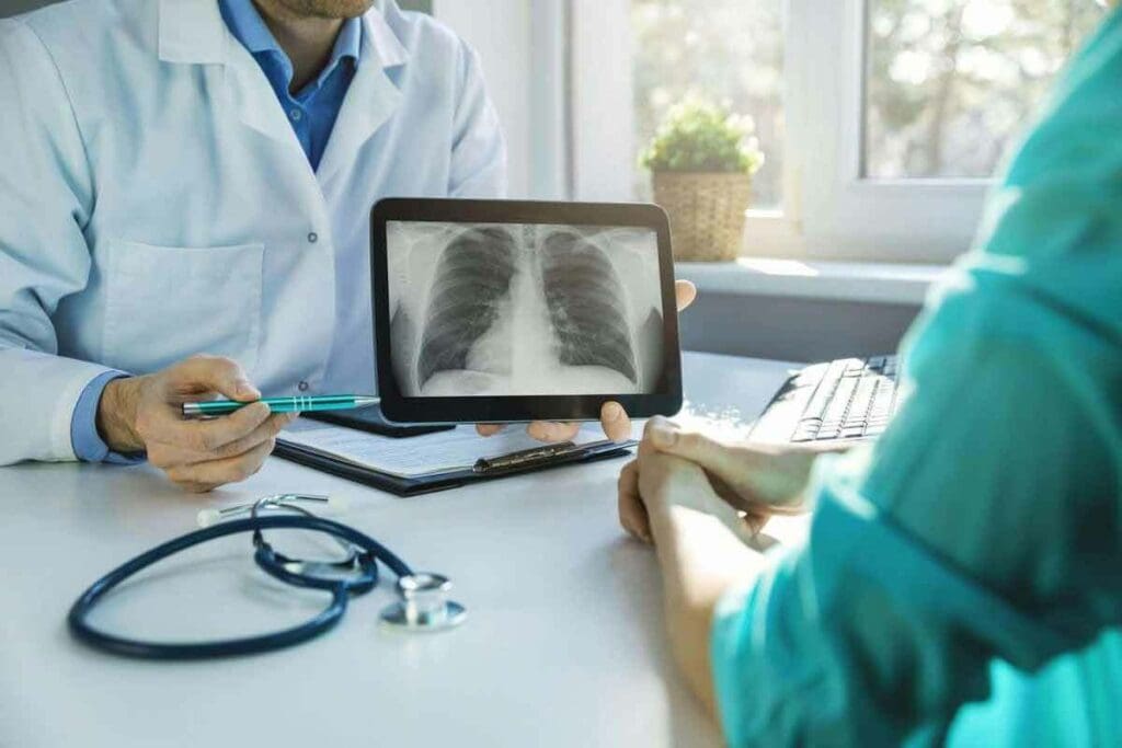
Medical imaging is a big part of how we get exposed to radiation. It’s about 53% of what the average person in Australia gets each year. This shows we need to know the risks and benefits well.
Medical imaging helps us find and treat health problems. But we must think about the risks of radiation. Doctors compare the benefits of imaging to the risk of radiation. They make sure the benefits are worth the risk.
Types of Diagnostic Imaging That Use Radiation
Many imaging methods use radiation to see inside our bodies. These include:
- X-rays: Used for bones, teeth, and some soft tissues.
- Computed Tomography (CT) scans: Give detailed images of the body.
- Fluoroscopy: Shows what’s happening inside us in real time.
- Mammography: Special X-rays for the breasts.
How Radiation Dose Is Measured and Quantified
The dose from imaging is measured in millisieverts (mSv) or milligrays (mGy). The effective dose in mSv considers how different parts of us react to radiation. This gives a full picture of the risk.
| Imaging Modality | Typical Effective Dose (mSv) |
| Chest X-ray | 0.1 |
| CT Scan (Head) | 2.0 |
| Mammography | 0.4 |
Knowing these numbers helps us understand the risks of different scans.
Regulatory Limits: How Many X-Rays Can You Have in a Year
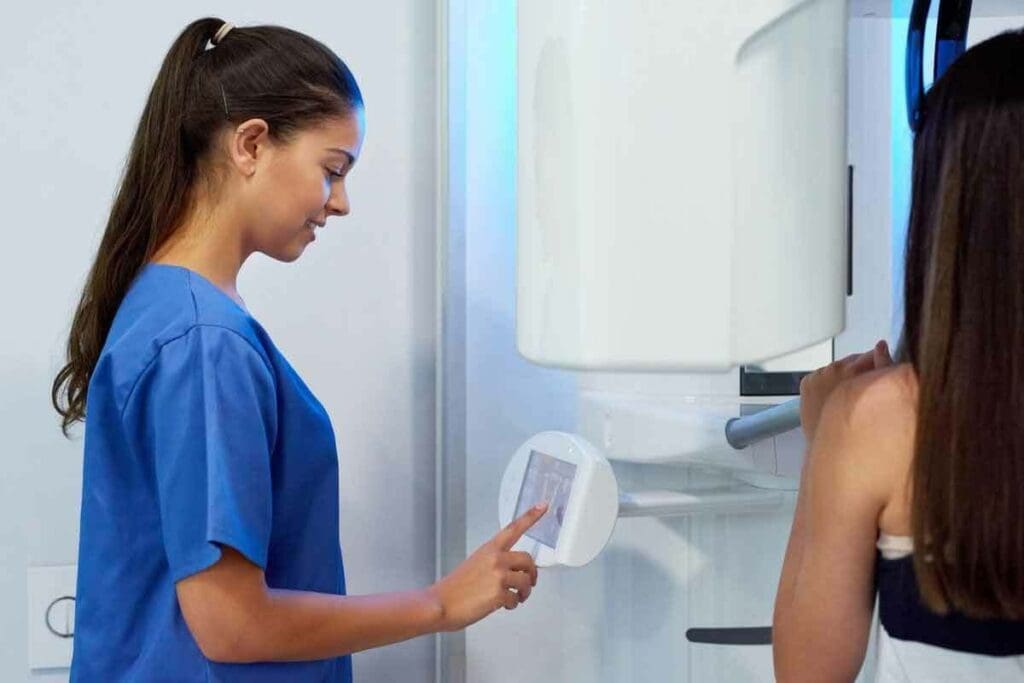
It’s important to know the limits on radiation exposure. This is true for everyone and for those in healthcare. These limits help keep us safe from radiation’s harmful effects. They also let us use medical imaging safely.
Public Dose Limits: The 1 mSv Annual Threshold
The Nuclear Regulatory Commission (NRC) in the U.S. sets a yearly dose limit of 1 millisievert (mSv) for the public. This rule helps keep radiation exposure from medical imaging safe. For example, a chest X-ray has about 0.1 mSv of radiation.
This means you could safely have around 10 chest X-rays in a year. But remember, this limit is for everyone, not just those getting medical tests.
It’s essential to note that these limits are for the general public. They don’t apply to patients getting medical tests. Doctors weigh the benefits of imaging against the risks for each patient.
Occupational Exposure Limits for Healthcare Workers
Workers in healthcare who often get exposed to radiation have different limits. For example, radiologic technologists and interventional radiologists can get up to 20 mSv a year. This is averaged over five years, with no year over 50 mSv. They must wear dosimeters and follow strict safety rules to keep their doses low.
Monitoring and safety protocols are key to keeping workers’ exposure safe. This includes using shields, reducing exposure time, and staying far from radiation sources.
International Variations in Radiation Safety Standards
Though there are global guidelines, like those from the International Commission on Radiological Protection (ICRP), rules can differ by country. For example, some places might have stricter limits for the public or workers.
| Country | Public Dose Limit (mSv/year) | Occupational Dose Limit (mSv/year) |
| United States | 1 | 20 (averaged over 5 years) |
| European Union | 1 | 20 |
| Australia | 1 | 20 (averaged over 5 years) |
The table shows how different countries have different safety standards. Knowing these differences is vital for healthcare providers and patients in today’s global health world.
Typical Radiation Doses from Common X-Ray Procedures
The doses from X-ray exams vary a lot. This depends on the type of procedure and the body part being imaged. Knowing these doses helps us understand the safety and risks of medical imaging.
Dental X-Rays: Exposure and Safety
Dental X-rays are very common. They use low doses of radiation, about 0.005 mSv per exam. This is similar to the background radiation we get in a few hours.
Using digital X-ray technology and proper shielding helps reduce exposure. The American Dental Association says digital X-rays cut radiation by up to 50% compared to film X-rays.
Chest X-Rays: Radiation Levels per Exam
A chest X-ray gives about 0.1–0.2 mSv per exam. This dose is low and often used as a comparison for other imaging.
Chest X-rays help diagnose conditions like pneumonia and lung diseases. Their low radiation dose makes them safe for diagnosis.
Extremity and Bone X-Rays: What to Expect
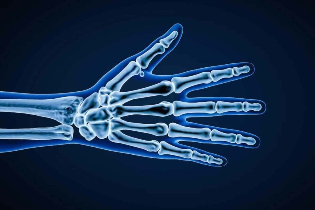
Extremity and bone X-rays have doses similar to or a bit higher than chest X-rays. They range from 0.1 to 1.0 mSv, depending on the procedure and body part.
These X-rays are used to find fractures, bone infections, and other bone issues. The doses are generally safe for diagnosis.
Mammography and Fluoroscopy Procedures
Mammography has a slightly higher dose, about 0.4 mSv per breast. Fluoroscopy, which shows real-time X-ray images, can have higher doses based on the procedure’s length.
Fluoroscopy helps guide procedures and diagnose conditions like gastrointestinal disorders. The dose varies with the procedure’s complexity and length.
| X-Ray Procedure | Typical Radiation Dose (mSv) |
| Dental X-ray | 0.005 |
| Chest X-ray | 0.1-0.2 |
| Extremity/Bone X-ray | 0.1-1.0 |
| Mammography (per breast) | 0.4 |
| Fluoroscopy | Varies with procedure duration |
“The key to safe medical imaging is not to avoid X-rays altogether, but to use them judiciously and with an understanding of their radiation doses.”
— Expert in Radiology
CT Scans: Higher Radiation Concerns
CT scans use more radiation than regular X-rays. This is because they take many X-ray images from different angles. These images help create detailed pictures of the body’s inside.
Why CT Scans Deliver More Radiation Than Conventional X-Rays
CT scans need more radiation because they take many images. This is to get a clear picture of the body’s inside. The images must be of high quality.
The complexity of the scan and the body area being examined affect the radiation dose. For example, scans of the abdomen use more radiation than scans of the arms or legs.
Radiation Doses from Different Types of CT Examinations
CT scans expose patients to different amounts of radiation. Here are some examples:
- Head CT scans: typically around 2 millisieverts (mSv)
- Chest CT scans: usually in the range of 7 mSv
- Abdominal CT scans: often between 8 to 10 mSv
These doses are much higher than regular X-rays. For instance, a chest X-ray gives about 0.1 mSv.
How Many CT Scans Are Considered Safe Annually
Finding a safe number of CT scans per year is hard. It depends on why the scans are needed. But guidelines say the benefits should be greater than the risks.
Most patients benefit from a CT scan. But those needing many scans should talk to their doctor. They should also make sure the scan uses the least amount of radiation needed.
Comparing Medical Radiation to Natural Background Exposure
Every day, we are exposed to some radiation from natural sources. This helps us understand how much radiation we get from medical tests. Natural radiation comes from space and the earth, air, and water around us.
Annual Background Radiation Sources
The amount of natural radiation we get changes based on where we live and how high we are. For example, in Australia, people get about 1.5 mSv of natural radiation each year. In the United States, it’s around 3 mSv, mainly because of radon gas in homes. Radiation safety guidelines use these numbers to explain the risks of medical imaging.
Putting Medical Imaging Radiation in Context
When we talk about the safety of medical imaging, comparing it to natural radiation is useful. A chest X-ray is like a few days of natural radiation. But a CT scan of the abdomen and pelvis is like several years of natural radiation. This helps us understand the risks and benefits of medical imaging.
Looking at natural radiation helps us see the risks and benefits of medical imaging. This comparison is key to understanding the safety and need for medical procedures.
Long-Term Health Effects of Radiation Exposure
Radiation from medical imaging raises health concerns. As imaging grows, knowing its risks is key.
The main worry is cancer risk from imaging. Cancer risk assessment looks at the radiation dose, patient age, and imaging type.
Cancer Risk Assessment from Diagnostic Imaging
Even repeated imaging, like dental X-rays, has a small cancer risk. For example, 10 mSv might cause 1 extra cancer case per 1,000 people. A chest X-ray is about 0.1 mSv, while a CT scan is around 7 mSv.
The risk is based on the linear no-threshold (LNT) model. It says any radiation dose has some cancer risk. But, the LNT model is a safe guess, and real risks might be lower.
Cumulative Exposure Considerations Over a Lifetime
Long-term radiation exposure is a big risk factor. Young patients getting many scans may face higher cancer risks.
Healthcare must balance imaging benefits and risks. Using radiation dose reduction techniques and alternatives can lower exposure.
Understanding radiation’s long-term effects helps us reduce risks. By optimizing imaging, we can keep its benefits while lowering risks.
Special Considerations for Vulnerable Populations
Children and pregnant women need extra care when it comes to radiation from medical imaging. They might be more at risk because of their age, health, and how their bodies are growing.
Pediatric Patients and Radiation Sensitivity
Children are more sensitive to radiation because their bodies are growing and they have a long life ahead. To protect them, pediatric imaging centers use special protocols. These protocols adjust the radiation dose based on the child’s size, not a one-size-fits-all approach.
Key considerations for pediatric patients include:
- Using the lowest possible radiation dose necessary for diagnostic image quality
- Employing size-based protocols for imaging procedures
- Carefully justifying the need for imaging procedures involving radiation
Pregnancy and X-Ray Safety Protocols
Pregnant women need special care when it comes to medical imaging with radiation. The main goal is to protect the fetus from radiation. Healthcare providers follow strict rules, like checking if a woman is pregnant before imaging.
Protocols for pregnant women may include:
- Using alternative imaging modalities that do not involve radiation, such as ultrasound or MRI, when possible
- Minimizing the radiation dose and field size to the minimum required for diagnostic purposes
- Shielding the abdomen with a lead apron when the area of interest is not the abdomen or pelvis
| Imaging Modality | Radiation Exposure | Alternative Options |
| X-ray | Low | Ultrasound, MRI |
| CT Scan | Moderate to High | MRI, Ultrasound |
| Fluoroscopy | Variable | Ultrasound, MRI |
Patients with Chronic Conditions Requiring Frequent Imaging
People with chronic conditions often need many imaging tests over their lives. This can lead to a lot of radiation exposure. It’s important to manage the radiation dose to reduce long-term risks.
Strategies for managing radiation exposure in patients with chronic conditions include:
- Maintaining detailed records of the patient’s imaging history to track cumulative radiation exposure
- Using the lowest dose protocols available for necessary imaging procedures
- Considering alternative imaging modalities that do not involve ionizing radiation
Radiation Dose Reduction Techniques and Technologies
Reducing radiation exposure is a big challenge in medical imaging today. It’s important for keeping patients safe, like children and pregnant women. We need to find ways to lower doses without losing image quality.
ALARA Principle: As Low As Reasonably Achievable
The ALARA principle is key in keeping patients safe from radiation. It means we should use the least amount of radiation needed for good images. This helps us balance quality and safety.
Key strategies for implementing ALARA include:
- Using the lowest necessary dose for diagnostic purposes
- Optimizing imaging protocols for specific patient populations
- Employing dose-reducing technologies when available
Modern Equipment Advancements for Dose Optimization
New technology in medical imaging has helped lower radiation doses. For example, CT scans with new algorithms can cut doses by up to 40%. Modern gear is made to reduce doses while keeping images clear.
Some notable advancements include:
- High-sensitivity detectors that allow for lower doses
- Advanced image processing algorithms that enhance image quality at lower doses
- Automated exposure control systems that adjust dose based on patient size and anatomy
Shielding and Procedural Protocols for Safety
Shielding and following strict protocols are also vital for safety. Using lead aprons and shielding organs can greatly reduce exposure.
Effective procedural protocols include:
- Collimation to limit the X-ray beam to the area of interest
- Proper patient positioning to minimize repeat exposures
- Regular quality control checks on imaging equipment
By using these methods and technologies, we can lower radiation doses a lot. This makes medical imaging safer for everyone. The work on reducing doses is always getting better, helping keep patients safe.
Alternative Imaging Options with Lower or No Radiation
Medical imaging has options that use less or no radiation. We’ve talked about X-rays and CT scans needing radiation. But, not all imaging needs radiation.
For those needing frequent scans or are sensitive to radiation, like kids or pregnant women, there are better choices. Ultrasound and MRI are great alternatives to X-rays and CT scans.
Ultrasound as a Radiation-Free Alternative
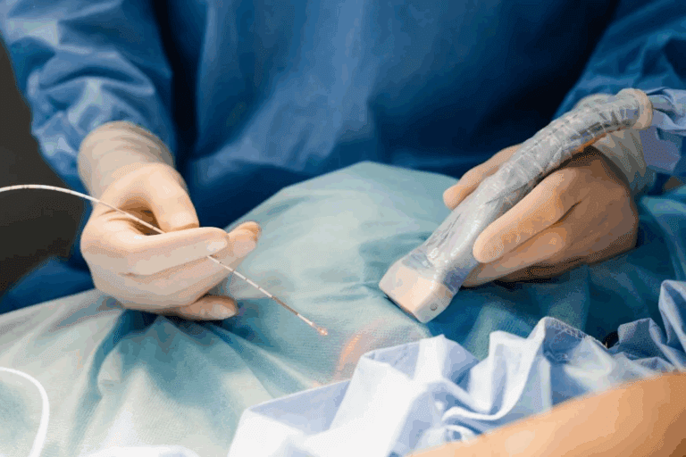
Ultrasound uses sound waves to see inside the body. It’s safe and works well without radiation. Ultrasound is often used for the abdomen, pelvis, and to check on babies during pregnancy. It’s also good for guiding some medical procedures.
Ultrasound is easy to move around and is cheaper than other imaging. But, the quality can depend on the person doing the scan. It might not show as much detail as other methods for some issues.
MRI: When It Can Replace X-Ray or CT Imaging
MRI doesn’t use radiation. It uses a strong magnetic field and radio waves to create detailed images. MRI is great for soft tissue imaging, like the brain, spine, and joints.
MRI can sometimes replace CT scans, which are good for soft tissue. But, MRI takes longer and some people might feel claustrophobic.
When to Ask Your Doctor About Imaging Alternatives
If you’re worried about radiation, talk to your doctor about other options. Ask if there are radiation-free ways to get the needed images. Or if ultrasound or MRI could work instead.
- Know what your condition needs for diagnosis.
- Learn about the pros and cons of different imaging methods.
- Talk about any issues or reasons you can’t have MRI or ultrasound.
Being informed helps you choose the best imaging for you. This might lower your radiation exposure.
Conclusion: Making Informed Decisions About X-Ray Safety
It’s important for patients to know the risks and benefits of medical imaging. This knowledge helps them make smart choices about their health. When thinking about X-rays and other tests, remember the long-term effects of radiation.
Patients should learn about the radiation from X-rays and how to reduce it. Knowing this lets them talk to doctors about their options. This way, they can weigh the good of tests against the risks of X-ray safety and radiation safety.
Choosing wisely about X-rays means looking at safer options like ultrasound or MRI. It’s also key to follow the ALARA principle. This means keeping radiation doses as low as possible.
By focusing on X-ray safety and radiation safety, patients and doctors can improve how tests are done. This way, they can keep the benefits of these important medical tools while reducing risks.
FAQ
How many X-rays can you have in a year?
The safe number of X-rays in a year depends on your health needs and legal limits. For everyone, the yearly limit is 1 mSv. Workers in radiation fields can get up to 20 mSv per year.
How many X-rays are safe?
X-rays are safe based on dose and how often you get them. Most medical X-rays have low doses. This makes the risk very small for most people.
What is the safe limit for X-rays?
Rules for safe radiation exposure are set by agencies. The public limit is 1 mSv a year. Workers can get up to 20 mSv annually.
How many chest X-rays are safe in a year?
Chest X-rays have a low dose of radiation. Getting many chest X-rays in a year is usually safe. But, it depends on your health needs.
How much radiation is in a chest X-ray?
A chest X-ray has about 0.1 mSv of radiation.
Is there a limit to how many CT scans you can have?
There’s no strict limit, but CT scans have more radiation than X-rays. How many CT scans are safe varies by your health needs and risk.
How many CT scans are considered safe?
CT scans are safe when needed for medical reasons. But, it’s important to watch how much radiation you get over time.
Can you have too many X-rays?
Too many X-rays can increase your radiation exposure. But the risk is usually small for most tests. Doctors consider the benefits and risks.
Are there alternatives to X-rays?
Yes, options like ultrasound and MRI don’t use harmful radiation. They’re good choices when needed.
How can radiation exposure be minimized?
To reduce radiation, use the ALARA principle and modern tech. Also, follow proper shielding and procedures.
Are there special considerations for children and pregnant women?
Yes, kids and pregnant women need special care to avoid too much radiation. This is because they’re more sensitive.
How does medical radiation compare to natural background radiation?
Medical radiation doses vary. But, many tests have doses similar to or less than what you get from background radiation each year.


