
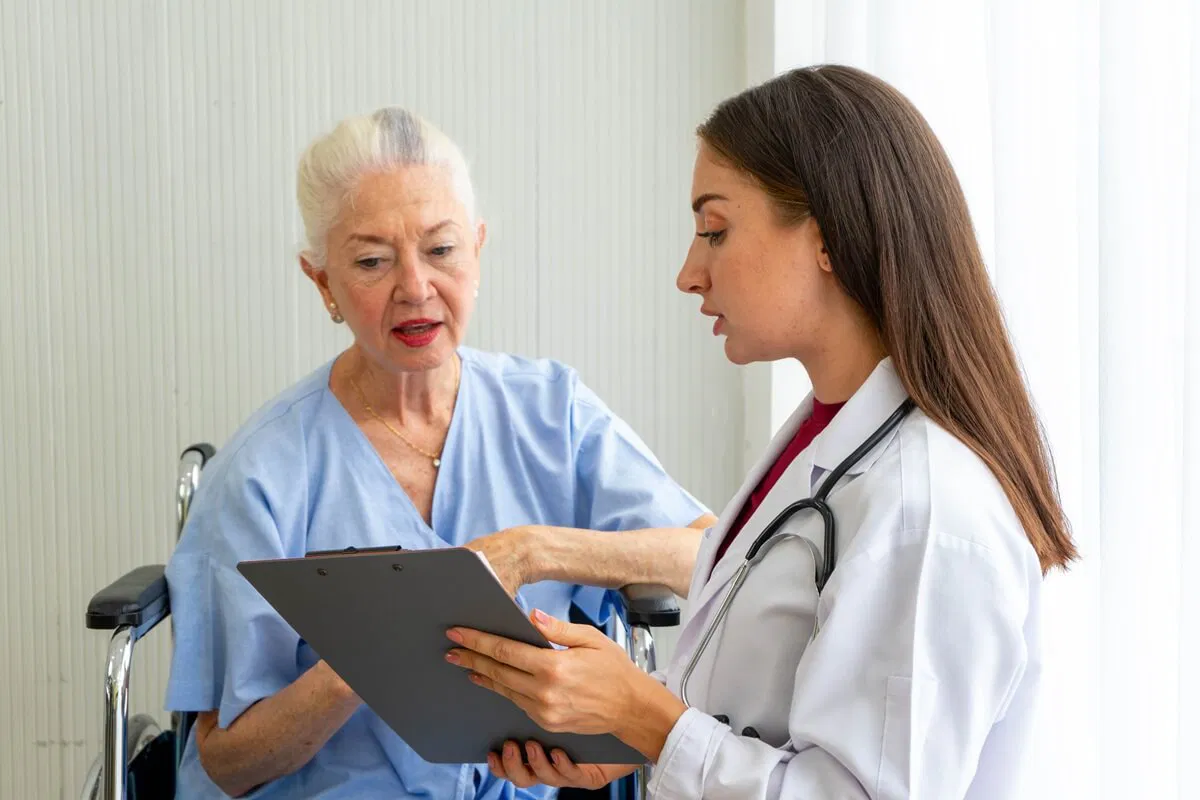
A thorough cranial nerve examination is key in checking the brain’s health. It helps doctors find problems in the brain stem or along the nerves. These nerves start in the brain stem. Learn how to check cranial nerves. Our simple, step-by-step clinical guide makes performing a complete examination easy.
At Liv Hospital, we use special tests that are both precise and gentle. This makes the exam easier for patients. Learning how to do these tests well is important for spotting and treating brain issues.
Key Takeaways
- Cranial nerve examination is essential for neurological assessment.
- Abnormalities in cranial nerve function indicate specific brain stem pathology.
- Systematic testing protocols enhance clinical precision and patient comfort.
- Mastering cranial nerve examination techniques is vital for diagnosing neurological conditions.
- Liv Hospital’s approach prioritizes patient-centered care and clinical expertise.
The Fundamentals of Cranial Nerve Assessment

Checking cranial nerves is key in a neurological exam. It helps doctors find and pinpoint problems. This test is a big part of the general neurologic exam.
Clinical Significance of Cranial Nerve Examination
Assessing cranial nerves gives important info about the nervous system. Issues with these nerves can point to many neurological problems. This includes nerve disorders and brainstem issues.
The importance of cranial nerve exams is huge. They help:
- Find where problems are in the nervous system
- Spot conditions like stroke, multiple sclerosis, and brain injuries
- Watch how diseases get worse or improve with treatment
- Decide on more tests and treatment plans
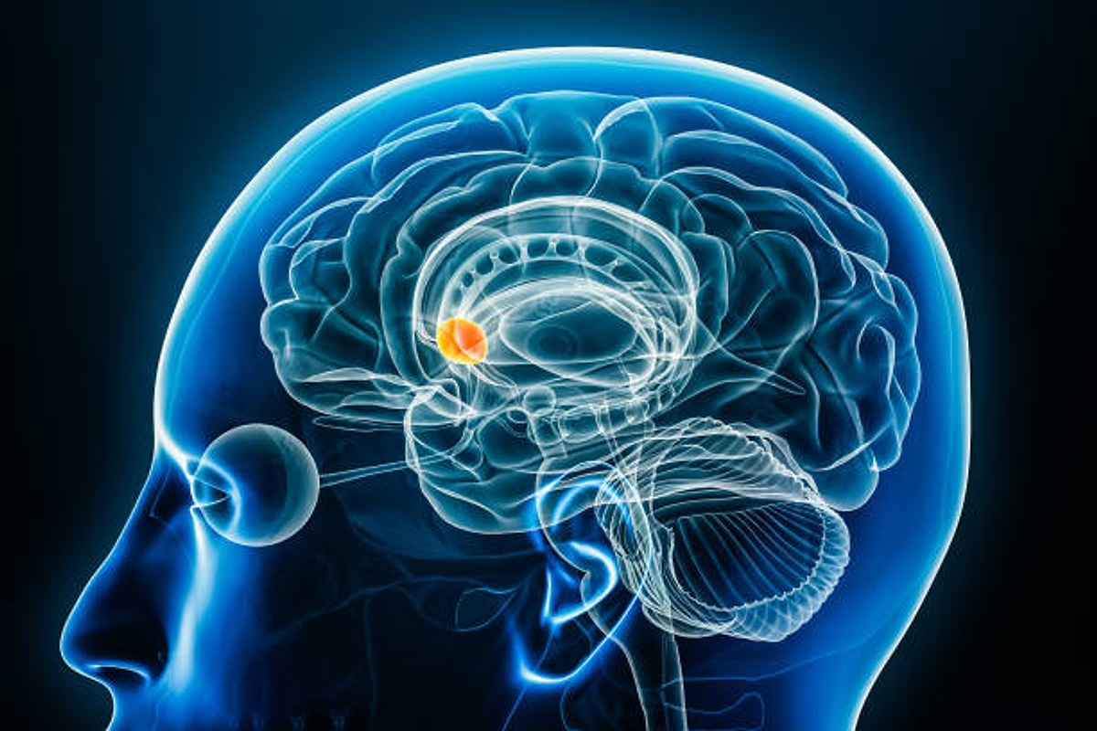
Anatomical Basis of the 12 Cranial Nerves
Knowing the anatomy and role of the 12 cranial nerves is vital. These nerves control many things like feeling, moving, and body functions.
The 12 cranial nerves and their main jobs are:
- Olfactory Nerve (CN I): Smell
- Optic Nerve (CN II): Vision
- Oculomotor Nerve (CN III): Eye movement, pupil size
- Trochlear Nerve (CN IV): Eye movement
- Trigeminal Nerve (CN V): Feeling on the face, chewing
- Abducens Nerve (CN VI): Eye movement
- Facial Nerve (CN VII): Facial expressions, taste
- Vestibulocochlear Nerve (CN VIII): Hearing, balance
- Glossopharyngeal Nerve (CN IX): Swallowing, taste
- Vagus Nerve (CN X): Many body functions, swallowing
- Accessory Nerve (CN XI): Neck and shoulder movement
- Hypoglossal Nerve (CN XII): Tongue movement
When to Perform a Cranial Nerve Examination
Do a cranial nerve exam in many situations. This includes:
- People with neurological symptoms like weakness or numbness
- Trauma patients with head or neck injuries
- Those suspected of having a stroke or transient ischemic attack
- Patients with known neurological conditions
Knowing when and how to do a cranial nerve exam helps doctors give better care. It helps them make smart choices about tests and treatments.
Preparation and Equipment for Cranial Nerve Testing
Before we start checking cranial nerve function, we need to get ready. We must have the right equipment and a good place to work. This makes sure the test goes smoothly and is done right.
Essential Tools for a Complete Examination
The tools needed for cranial nerve testing are simple. Here’s what you need:
- A 256-hertz or 512-hertz tuning fork for checking vibration sense and hearing.
- Ishihara or Hardy-Rand-Ritter plates for color vision tests.
- A Snellen chart to measure how well you can see.
- A penlight or flashlight for testing the pupillary light reflex.
- Cotton swabs or tissue paper for checking facial sensation and corneal reflex.
Having these tools ready makes the test easier. It makes sure we check everything we need to.
Patient Positioning and Environment
How we position the patient and the environment are key for a good test. Here’s what we suggest:
- Put the patient in a comfy sitting or lying down spot, based on their needs.
- Make sure the room is bright and the temperature is just right.
- Keep the room quiet to help the patient focus.
By making the environment better, we can make the patient feel less stressed. This helps us get more accurate results.
Documentation Requirements
It’s important to document the test results well. We need to write down:
Cranial Nerve | Test Performed | Findings |
I (Olfactory) | Smell identification | Normal/Abnormal |
II (Optic) | Visual acuity, color vision | Normal/Abnormal |
III, IV, VI (Oculomotor, Trochlear, Abducens) | Pupillary light reflex, eye movements | Normal/Abnormal |
Good documentation helps us keep track of the patient’s progress. It also helps us share the results with other doctors.
How to Check Cranial Nerves: General Approach
Checking cranial nerves is key in a neurological exam. It needs a detailed method. This way, we make sure all nerves are checked well, giving us a full picture of a patient’s health.
Systematic Examination Sequence
We follow a set order for checking cranial nerves. This method helps us not miss anything and makes sure we cover everything.
- First, we look at the patient’s overall look and any signs of trouble.
- Then, we check each nerve in order, starting with CN I (Olfactory) and ending with CN XII (Hypoglossal).
- For each nerve, we use the right tools and methods, like smell tests for CN I and eye tests for CN II.
Following a set order is key for a complete check. Here’s a simple guide for checking cranial nerves:
Cranial Nerve | Function | Test |
CN I (Olfactory) | Smell | Smell testing with various odors |
CN II (Optic) | Vision | Visual acuity, visual field testing |
CN III, IV, VI (Oculomotor, Trochlear, Abducens) | Eye movements | Pupillary light reflex, extraocular movements |
Patient Communication During Assessment
Talking clearly with the patient is very important during the exam. We need to explain each test well and make sure they understand what to do.
Good instructions are key for getting right results. For example, when checking vision, we tell the patient to cover one eye and read from a chart.
Recognizing Normal vs. Abnormal Findings
Telling normal from abnormal findings is very important. We need to know what’s usual for each test and spot any differences.
A normal pupillary light reflex is quick and even. But, if it’s slow or missing, it could mean there’s a problem.
By using a systematic method, talking well with the patient, and understanding the results, we can do a full check of cranial nerves.
Assessing Olfactory Nerve (CN I): Smell Testing
Our sense of smell, helped by the olfactory nerve (CN I), is often missed in health checks. Yet, it gives us key clues for diagnosing problems. Being able to smell different odors is important for spotting nerve issues early.
Proper Technique for Smell Assessment
To check the olfactory nerve, we use a simple method with common smells. The patient must identify these smells with their eyes shut. It’s key to use a different nostril for each smell test to avoid mixing scents and check each nerve separately.
We often use smells like coffee, vanilla, or lavender. The patient sniffs each one and tries to name it. This test checks how well the olfactory nerve works and how well the patient can smell.
Common Olfactory Abnormalities
Olfactory problems can show up as anosmia (loss of smell), hyposmia (reduced sense of smell), or dysosmia (distorted sense of smell). These can come from things like colds, head injuries, brain diseases, and some medicines.
- Anosmia can really affect someone’s life, making it hard to enjoy food and smell dangers.
- Hyposmia might be a sign of diseases like Parkinson’s or Alzheimer’s early on.
- Dysosmia can make people smell things in a weird or bad way, which can be upsetting.
Clinical Correlations of CN I Dysfunction
Problems with the olfactory nerve can link to many health issues. For example, anosmia after a head injury might mean a broken cribriform plate, where the nerves pass through. In diseases like Parkinson’s or Alzheimer’s, smell problems can show up before other symptoms.
Knowing how CN I problems relate to health helps doctors diagnose and treat better. It shows why checking the olfactory nerve is important in health checks.
Examining Optic Nerve (CN II): Vision Assessment
The optic nerve, or CN II, is key to our vision. It’s examined in many tests to check how well we see. These tests look at different parts of our vision.
Visual Acuity Testing with Snellen Charts
Visual acuity tests how sharp our vision is. We use Snellen charts for this. Patients read letters from far away, like 20 feet.
The results show how well they can see. It’s like a fraction, like 20/20. This means they can read at 20 feet what a normal eye can at 20 feet too.
Color Perception and Ishihara Plates
Color perception is also important. We test it with Ishihara plates. These plates have numbers or shapes in dots.
People with normal color vision can see these. But those with color blindness might not. This test finds color vision problems, which can show optic nerve issues.
Visual Field Assessment by Confrontation
Visual field tests check our side vision. We face the patient and move our fingers into their vision. They tell us when they see it.
This test finds problems like blind spots. These can be signs of optic nerve damage.
Funduscopic Examination Technique
The funduscopic exam looks at the retina and optic disc. We use an ophthalmoscope in a dark room. It shines a light through the pupil to see inside the eye.
This exam checks the optic disc, retina, and blood vessels. It tells us about the optic nerve’s health.
In summary, checking the optic nerve is about more than just vision. It includes tests for sharpness, color, field, and eye health. These tests help diagnose and treat optic nerve problems.
Evaluating Eye Movement Nerves (CN III, IV, VI)
Checking the nerves that control eye movement is key in a full neurological check-up. The oculomotor (CN III), trochlear (CN IV), and abducens (CN VI) nerves work together. If they don’t work right, it can cause big problems with how we see things.
Pupillary Light Reflex Assessment
The pupillary light reflex tells us a lot about CN III. We use a light to test this reflex. Both pupils should get smaller when light hits them. If not, it could mean a problem with CN III or the optic nerve (CN II).
Extraocular Movement Testing
We test eye movement by asking someone to follow a target with their eyes. We check how well they can move their eyes in all directions. This test helps find issues with CN III, IV, or VI.
Ptosis and Lid Position Evaluation
Ptosis, or a droopy eyelid, can show a problem with CN III. We check how the eyelids sit and look for any unevenness or weakness. Ptosis can also happen with other conditions, like myasthenia gravis or Horner’s syndrome.
Identifying Common Ocular Nerve Palsies
Ocular nerve palsies can come from many things, like blood vessel problems, injuries, or tumors. CN III palsy can cause a droopy eyelid, a big pupil, and trouble moving the eye. CN IV palsy affects the superior oblique muscle, leading to vertical double vision. CN VI palsy weakens the lateral rectus muscle, causing horizontal double vision. Spotting these palsies is important for figuring out and treating the real issue.
Testing Trigeminal Nerve (CN V): Sensory and Motor Functions
The trigeminal nerve, or CN V, is key for facial feeling and movement. It’s important in neurological tests. We check its sensory and motor parts to see how well it works.
Three-Division Sensory Testing Technique
We test the trigeminal nerve’s three parts: ophthalmic, maxillary, and mandibular. We use a cotton swab or von Frey hair to touch the face. The patient closes their eyes and tells us where they feel the touch.
Sensory testing helps find problems with the trigeminal nerve. By comparing feelings on both sides, we can find where issues are.
Corneal Reflex Examination
The corneal reflex is a big part of testing the trigeminal nerve. We touch the cornea with a cotton swab to see if the patient blinks. This test checks the trigeminal and facial nerves. If the reflex is weak or missing, it could mean a problem with these nerves.
Jaw Movement and Masseter Strength Assessment
We check the trigeminal nerve’s motor function by looking at jaw movement and masseter strength. The patient clenches their teeth while we feel the muscles. We also check if the jaw moves strangely when they open their mouth. If it does, it could mean a nerve problem.
Recognizing Trigeminal Neuralgia
Trigeminal neuralgia causes sharp, shock-like pain in the face. We diagnose it by the patient’s history of sudden, severe pain. Knowing about trigeminal neuralgia helps us treat it right.
By testing the trigeminal nerve’s sensory and motor parts, we can find and treat problems with this important nerve.
Facial Nerve (CN VII) Examination Techniques
The facial nerve, or CN VII, controls our facial expressions and taste from the tongue’s front part. It’s key for diagnosing many neurological issues.
Facial Expression Testing Protocol
We test the facial nerve by asking patients to make different facial expressions. They smile, frown, show their teeth, and close their eyes hard. We look for any muscle weakness or imbalance.
Key observations include: if the face moves evenly, if there’s drooping, and if they can wrinkle their forehead.
Taste Assessment on Anterior Two-Thirds of Tongue
We check the nerve’s sensory function by testing taste on the tongue’s front part. We use sweet, sour, salty, and bitter tastes on both sides.
It’s essential to compare the taste perception on both sides of the tongue to identify any asymmetry.
Differentiating Upper vs. Lower Motor Neuron Lesions
It’s important to tell apart upper and lower motor neuron lesions. Upper lesions spare the forehead but affect the lower face. Lower lesions cause weakness or paralysis of the whole face side.
- Upper motor neuron lesions: sparing of the forehead, contralateral weakness.
- Lower motor neuron lesions: ipsilateral weakness or paralysis of the entire half of the face.
Bell’s Palsy vs. Central Facial Weakness
We must tell Bell’s palsy, a lower motor neuron issue, from central facial weakness, an upper motor neuron problem. Knowing the difference is key for the right treatment.
Assessing Vestibulocochlear Nerve (CN VIII): Hearing and Balance
Checking the vestibulocochlear nerve is key for hearing and balance. It’s called CN VIII. This nerve is important in neurological exams to spot hearing or balance problems.
Hearing Acuity Testing Methods
We use pure tone audiometry to check hearing. It shows how well a person can hear different sounds. Speech audiometry helps see how well someone understands speech, which is important for talking.
Tuning fork tests are quick and simple. They help doctors check for hearing loss and tell if it’s conductive or sensorineural.
Weber and Rinne Tests for Conductive vs. Sensorineural Hearing Loss
The Weber and Rinne tests use tuning forks. They help figure out if hearing loss is conductive or sensorineural. The Weber test places a tuning fork on the skull. In a normal person, the sound is heard equally in both ears.
The Rinne test compares how well sound is heard through air versus bone. A tuning fork is first held near the ear and then on the mastoid process. Normally, air conduction is better. If bone conduction is better, it means there’s conductive hearing loss.
Basic Vestibular Function Assessment
Vestibular function tests include the Romberg and Unterberger step tests. These tests check balance with eyes closed. If there’s a lot of movement, it might mean vestibular problems.
The caloric test involves water at different temperatures in the ear canal. It causes nystagmus. The type, direction, and length of nystagmus tell us about vestibular function.
Nystagmus Evaluation
Nystagmus is when the eyes move on their own. It can show vestibular system issues. We look at the direction, size, and length of eye movements. Nystagmus in certain positions or during tests like the Dix-Hallpike can help find vestibular problems.
Knowing about nystagmus is key for diagnosing issues like BPPV or central vestibular disorders. By looking at nystagmus and other tests, doctors can find where the problem is and how to treat it.
Examining Lower Cranial Nerves (CN IX, X, XI, XII)
Checking the lower cranial nerves is key in a full neurological check-up. These nerves, like CN IX, CN X, CN XI, and CN XII, help with swallowing, talking, and tongue actions.
Glossopharyngeal and Vagus Nerve Assessment (Gag Reflex, Palate Movement)
The glossopharyngeal and vagus nerves work together. The gag reflex is a key test for these nerves. To check it, gently touch the back of the pharynx with a tongue depressor. A normal gag reflex shows the pharyngeal muscles contracting.
We also check palate movement by asking the patient to say “ah”. This shows if the soft palate moves evenly. Uneven or missing palate elevation might mean a problem with CN IX or CN X.
Spinal Accessory Nerve Testing (Shoulder Shrug, Head Turning)
The spinal accessory nerve is tested by checking the sternocleidomastoid and trapezius muscles. We ask the patient to shrug and turn their head against resistance. Weakness or unevenness in these actions could mean a problem with CN XI.
For example, a CN XI issue might make it hard to turn the head or shrug the shoulder on one side.
Hypoglossal Nerve Examination (Tongue Movement and Strength)
The hypoglossal nerve controls tongue movements. We look for atrophy, fasciculations, and weakness in the tongue. If the tongue deviates to one side when sticking it out, it could mean a CN XII problem.
We also test tongue strength by asking the patient to push their tongue against their cheek. This shows if the tongue is weak.
Bulbar and Pseudobulbar Palsy Recognition
Bulbar and pseudobulbar palsy affect the lower cranial nerves. Bulbar palsy is from a lower motor neuron issue, causing muscle weakness and atrophy. Pseudobulbar palsy is from an upper motor neuron problem, leading to spasticity and weakness without atrophy.
Knowing these conditions helps in diagnosing and treating neurological issues.
Examining the lower cranial nerves needs a detailed and careful method. By checking these nerves, we can spot problems and help our patients.
Conclusion: Mastering the Cranial Nerve Examination
Learning to do a cranial nerve exam is key for healthcare workers. It helps them check the nervous system and find problems. Knowing how to check each of the 12 cranial nerves helps doctors spot issues and plan treatments.
A good cranial nerve exam checks each nerve carefully. It uses different methods and tools. This way, doctors can see how well the nervous system is working.
Using cranial nerve exams well makes doctors better at their jobs. It leads to better care for patients. The cranial nerve exam is a big part of checking the nervous system. It’s very important for diagnosing and treating nerve problems.
FAQ
What is the importance of cranial nerve examination in neurological assessment?
Cranial nerve examination is key to spotting specific problems. It helps in diagnosing and treating neurological issues.
How many cranial nerves are there, and what is their role?
There are 12 cranial nerves. They control many functions like movement, feeling, and automatic body actions.
What is the anatomical basis of the 12 cranial nerves?
Knowing the anatomy and function of the 12 cranial nerves is vital. Each nerve has its own role and path.
When should a cranial nerve examination be performed?
You should get a cranial nerve exam if you notice symptoms like weakness or numbness. Also, if you have vision problems.
What are the essential tools required for a complete cranial nerve examination?
You’ll need a Snellen chart, Ishihara plates, an ophthalmoscope, a tuning fork, and a reflex hammer.
How should a patient be positioned during a cranial nerve examination?
The patient should sit or lie down comfortably. Make sure there’s enough light. The examiner needs to be in the right spot.
What is the systematic examination sequence for checking cranial nerves?
Start with the olfactory nerve (CN I) and go to the hypoglossal nerve (CN XII). This order helps you check each nerve properly.
How do you assess the olfactory nerve (CN I)?
Use smell tests to check the olfactory nerve. Ask the patient to identify different smells.
What is the purpose of visual acuity testing with Snellen charts?
Snellen charts help check the optic nerve (CN II). They also test your vision.
How do you evaluate the eye movement nerves (CN III, IV, VI)?
Check the pupillary light reflex and extraocular movements. Also, look at ptosis and lid position.
What is the significance of testing the trigeminal nerve (CN V)?
Testing the trigeminal nerve is important. It checks facial sensation and jaw movement.
How do you assess the facial nerve (CN VII)?
Use facial expression tests and taste assessments. This helps find upper and lower motor neuron lesions.
What is the role of the vestibulocochlear nerve (CN VIII) in hearing and balance?
The vestibulocochlear nerve transmits sound and balance info. Its assessment includes hearing tests and balance checks.
How do you examine the lower cranial nerves (CN IX, X, XI, XII)?
Examine the glossopharyngeal and vagus nerves, spinal accessory nerve, and hypoglossal nerve. They control swallowing, vocalization, and tongue movement.
References
World Health Organization. Evidence-Based Medical Guidance. Retrieved from https://apps.who.int/iris/handle/10665/44476

