At Liv Hospital, we use a coronary angiogram procedure to see the heart’s blood vessels. This helps us find blockages or narrow spots. It’s a key test for checking heart health.
Studies show that coronary angiography is the best way to find heart disease. Every year, over two million people get this test. We’ll show you how this important test works.
Key Takeaways
- Coronary angiogram is a minimally invasive test to visualize heart blood vessels.
- It detects blockages or narrowing in the heart’s arteries.
- Coronary angiography is the gold standard for diagnosing coronary artery disease.
- Over two million procedures are performed worldwide each year.
- Liv Hospital provides advanced cardiac care with a commitment to safety and innovation.
What Is a Coronary Angiogram and Why It’s Performed

It’s important for both patients and doctors to know about coronary angiograms. This test, also known as a “dye test for heart,” shows the heart’s arteries in detail. It helps find any blockages or problems.
Definition and Diagnostic Purpose
A coronary angiogram uses a thin catheter to reach the heart’s arteries. It injects dye and takes X-ray images. This lets doctors see the arteries clearly and find any issues.
“Coronary angiography is key in diagnosing heart disease,” says a cardiologist. It helps doctors see the heart’s arteries and decide on treatments like angioplasty or stents.
Indications for Coronary Angiography
Doctors suggest coronary angiography for patients with heart disease symptoms. This includes chest pain or trouble breathing. They also recommend it for abnormal stress test results and certain high-risk patients.
Here are the main reasons for a coronary angiography:
- Symptoms of heart disease
- Abnormal stress test results
- Acute coronary syndrome
- High-risk patient profiles
Gold Standard for Coronary Artery Disease Diagnosis
Coronary angiography is the top choice for diagnosing heart disease. It gives clear images of the heart’s arteries. This is vital for deciding on treatments like angioplasty or stents.
A leading cardiologist says, “Coronary angiography is the best way to see heart disease. It helps doctors make the right treatment plans.” This is key for better patient care.
Pre-Procedure Patient Assessment and Preparation

At Liv Hospital, we focus on a detailed pre-procedure check for patients getting a coronary angiogram. This step is key to spotting risks and making sure the procedure is safe and effective.
Medical History Review and Risk Evaluation
We start by looking closely at the patient’s medical history. This includes any heart issues, surgeries, or conditions like diabetes or high blood pressure. This helps us understand the patient’s risk level and plan the best care.
Risk evaluation looks at the patient’s overall health and heart risks. This way, we can make the coronary angiogram safer and more effective for them.
Medication Management Before the Procedure
Managing medications is a big part of getting ready for the procedure. We check the patient’s current meds to see if they need to change or stop them before the angiogram. This reduces the chance of bleeding or other problems.
For example, some patients on blood thinners might need to stop them before the procedure. Our team gives clear instructions on what to do with medications to keep the patient safe.
Fasting Requirements and Laboratory Tests
Patients usually need to fast before the angiogram to avoid complications with anesthesia. We also do blood tests to check the patient’s health and find any issues before the procedure.
Patient Education and Informed Consent
Teaching patients about the procedure is very important. We make sure they know what the coronary angiogram is, its benefits, and possible risks. Our team explains what to expect and what to do after the procedure.
We also get informed consent from the patient. This means they fully understand and agree to the procedure. It helps them feel more at ease and respects their rights.
Equipment and Materials for the Coronary Angiogram Procedure
Coronary angiography needs special tools, like catheters and imaging systems. The test uses a cardiac dye to find blocked arteries. This helps doctors decide if they need to do angioplasty or stent placement. The coronary angiogram relies on advanced imaging and contrast media.
Catheterization Laboratory Setup
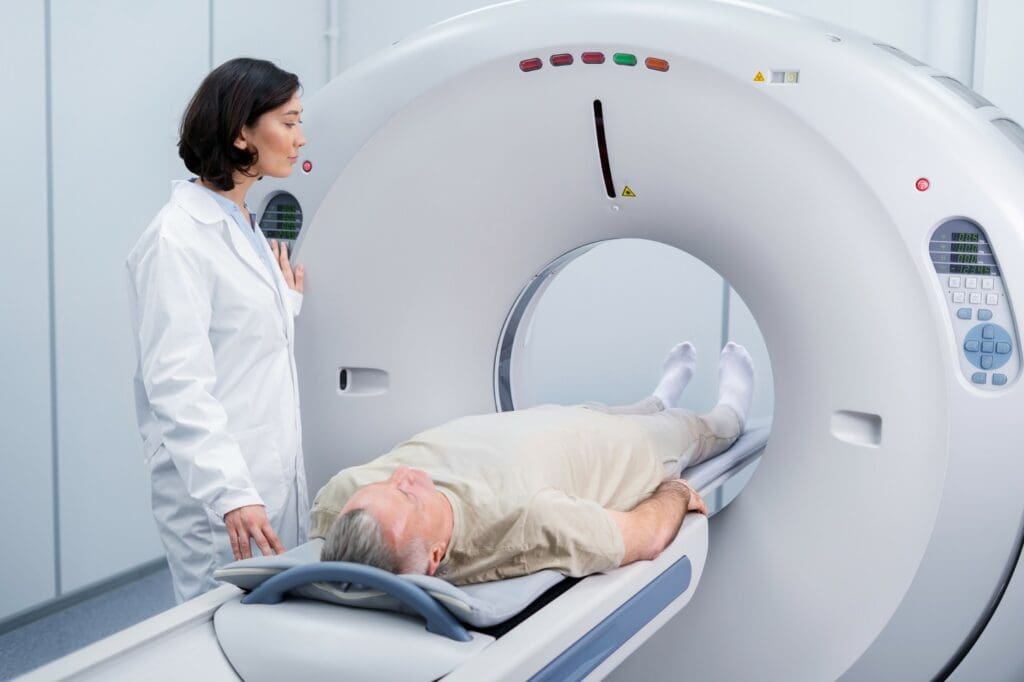
The catheterization lab is where coronary angiograms happen. It has the latest imaging tech, like X-ray machines and monitors. These help doctors see the coronary arteries clearly.
Types of Catheters and Guidewires
Different catheters and guidewires are used in a coronary angiogram. The type chosen depends on the patient’s body and the procedure’s needs.
| Type | Description | Use |
|---|---|---|
| Judkins Catheter | Pre-shaped catheter for coronary angiography | Commonly used for diagnostic purposes |
| Guidewire | Thin, flexible wire to guide catheters | Navigating through blood vessels |
Contrast Media and Imaging Systems
Contrast media make blood vessels show up better on images. Systems like digital subtraction angiography give clear pictures. These are key for making accurate diagnoses.
Step-by-Step Coronary Angiogram Procedure
The coronary angiogram procedure has several key steps. These steps are important for accurate diagnosis and patient safety. We will walk you through each stage, from preparation to the final imaging.
Patient Positioning and Vital Sign Monitoring
Getting the patient in the right position is key for the procedure’s success. We make sure the patient is comfortable and secure on the table. We also keep a close eye on their vital signs to spot any problems quickly.
Research shows that careful positioning and monitoring are vital. These steps help reduce risks and keep the patient safe.
Vascular Access Techniques
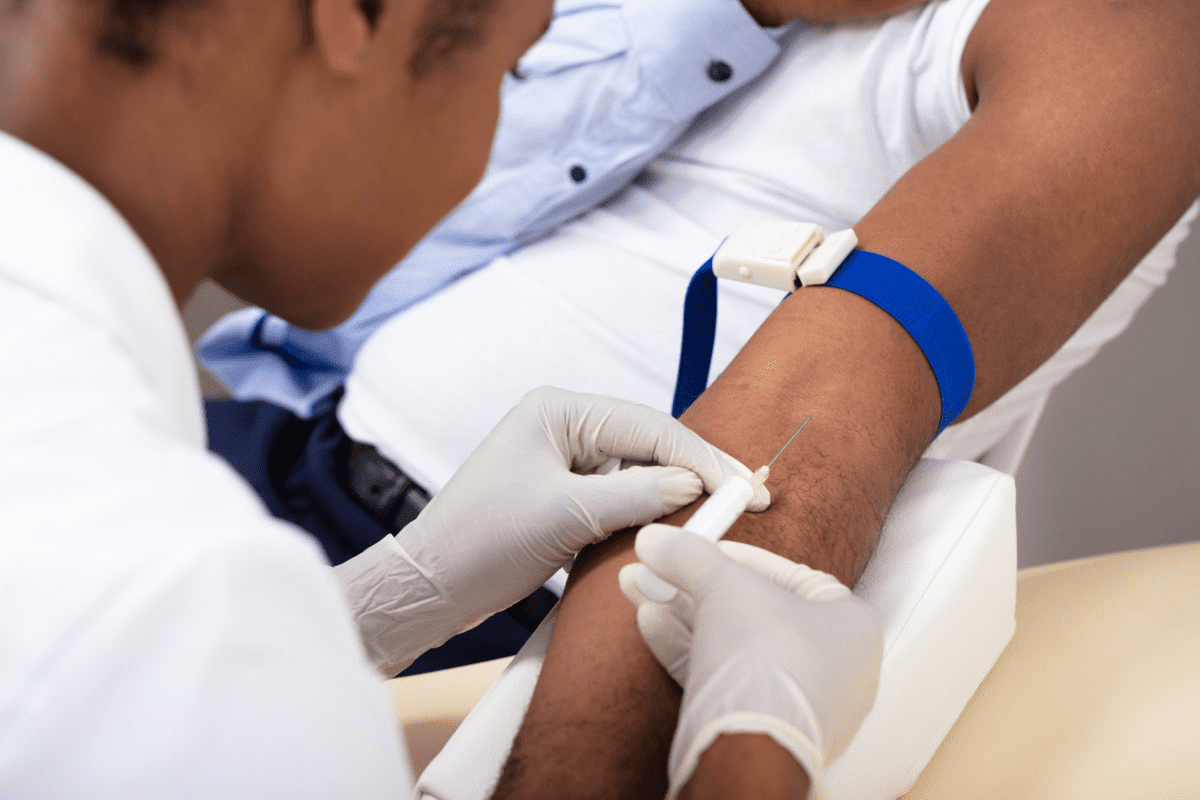
We access the vascular system through the radial or femoral artery. This is done using a sterile method. Local anesthesia is used to reduce discomfort during this step. The choice of access site depends on the patient’s anatomy and the operator’s preference.
Catheter Advancement to the Heart
After securing vascular access, we move the catheter to the heart. This is done under fluoroscopic guidance. It’s a precise step to avoid complications and ensure the catheter is in the right place.
Selective Coronary Artery Cannulation
Next, we engage the catheter with the coronary ostia. Specialized catheters are used for this. Contrast dye is injected to see the coronary arteries. X-ray images are taken to get a clear view of the coronary circulation.
The procedure involves guiding a catheter to the coronary arteries and injecting contrast dye. X-ray images are taken during this. This helps diagnose coronary artery disease accurately.
Interpreting Coronary Angiogram Results
Understanding coronary angiogram results is key to treating coronary artery disease. These results help us decide on the best treatment for patients.
Normal Coronary Anatomy Visualization
A normal coronary angiogram shows no major blockages in the arteries. We look at the images to see if there are any signs of disease.
Identifying and Grading Coronary Stenosis
We focus on finding and measuring coronary stenosis in the angiogram results. This means checking how much the arteries are narrowed or blocked. We use a system to measure this, helping us plan the right treatment.
Quantitative coronary angiography (QCA) gives us exact measurements of stenosis. This is important for deciding if angioplasty or stent placement is needed.
Assessment for Intervention Necessity
The angiogram results help us figure out if a patient needs interventions. We look at how severe the stenosis is and other factors to decide. This is important for creating a treatment plan that fits the patient’s needs.
By carefully looking at the angiogram results, we can find out who needs interventions. This helps improve outcomes and lowers the risk of complications from coronary artery disease.
Post-Procedure Patient Care
At Liv Hospital, we focus on the best care after a coronary angiogram. Our team works together to meet the patient’s physical and emotional needs.
Immediate Monitoring Requirements
We watch the patient’s vital signs closely after the procedure. This includes heart rate, blood pressure, and oxygen levels. It helps us catch any problems early.
- Continuous ECG monitoring to detect any arrhythmias
- Regular checks on the access site for signs of bleeding or hematoma
- Monitoring for symptoms such as chest pain or shortness of breath
Access Site Management and Complications
Managing the access site well is key to avoid complications. We check it often and teach the patient to watch for signs of trouble.
Common complications include:
- Bleeding or hematoma at the access site
- Vascular complications such as pseudoaneurysm or arteriovenous fistula
- Infection at the access site
Discharge Criteria and Instructions
We make sure the patient is ready to go home before we let them leave. They must have stable vital signs and no signs of trouble. We also give them clear instructions for care at home.
Discharge instructions include:
- Information on medications and their possible side effects
- Guidance on activity levels and when to start normal activities again
- Contact information for emergencies
Potential Complications and Risk Management
Coronary angiography is a key tool for diagnosing heart disease. But, like any invasive test, it carries risks. It’s important to know these risks to keep patients safe.
Vascular and Bleeding Complications
Vascular and bleeding issues are common with coronary angiography. These can range from minor bruising to serious problems like pseudoaneurysm formation or retroperitoneal bleeding. Choosing the right patient, using careful access techniques, and proper care after the test can lower these risks.
Monitoring the access site closely is key to managing vascular complications. Having quick plans for any issues that come up is also important. Using ultrasound-guided access can help reduce these risks.
Contrast-Induced Nephropathy Prevention
Contrast-induced nephropathy (CIN) is a big worry for patients with kidney problems. Pre-procedural hydration and using low or iso-osmolar contrast agents can help prevent CIN. Checking a patient’s kidney function before the test and choosing other imaging methods when needed can also help.
| Risk Factor | Prevention Strategy |
|---|---|
| Pre-existing renal impairment | Pre-procedural hydration, use of low/iso-osmolar contrast |
| Diabetes | Careful renal function assessment, consider alternative imaging |
| Dehydration | Pre-procedural hydration |
Cardiac Complications and Emergency Protocols
Cardiac problems during coronary angiography are rare but serious. These can include coronary artery dissection, arrhythmias, or cardiac arrest. It’s vital to have emergency protocols in place, like defibrillators and cardiac life support. Staff should know how to handle these emergencies.
Here’s a table showing some cardiac complications and how to manage them:
| Cardiac Complication | Management Strategy |
|---|---|
| Coronary artery dissection | Immediate stenting or surgical intervention |
| Arrhythmias | Cardiac monitoring, anti-arrhythmic medication |
| Cardiac arrest | Cardiopulmonary resuscitation (CPR), defibrillation |
In summary, coronary angiography is a valuable tool but comes with risks. Knowing these risks and how to manage them is key to patient safety. By being proactive, healthcare providers can reduce complications and improve patient outcomes.
Alternative and Complementary Diagnostic Methods
Cardiac care is advancing, and new diagnostic methods are emerging. The traditional coronary angiogram procedure for heart diagnosis is key, but new options are available. These new methods can sometimes replace or complement the traditional approach.
Non-Invasive Cardiac Imaging Options
Non-invasive cardiac imaging has changed how we diagnose heart disease. Techniques like cardiac magnetic resonance imaging (MRI) and myocardial perfusion imaging let us see the heart and its blood vessels without surgery.
These methods give us important info on the heart’s structure and function. They help spot problems early. They’re great for people at risk of heart disease but without symptoms.
CT Coronary Angiography Comparison
CT coronary angiography is a popular tool now. It uses CT scans to see the coronary arteries. This method is non-invasive and shows detailed images of the heart’s blood vessels.
| Diagnostic Method | Invasiveness | Radiation Exposure | Contrast Use |
|---|---|---|---|
| Coronary Angiogram | Invasive | Moderate | Yes |
| CT Coronary Angiography | Non-Invasive | Moderate to High | Yes |
| Cardiac MRI | Non-Invasive | None | No (or minimal) |
Advanced Invasive Assessment Techniques
Non-invasive methods are valuable, but sometimes invasive techniques are needed. Advanced invasive methods like fractional flow reserve (FFR) and intravascular ultrasound (IVUS) give detailed info on heart disease.
FFR checks the pressure difference across a blockage, helping decide if it’s serious. IVUS gives clear images of the arteries, helping with stent placement and plaque assessment.
These advanced techniques add to what traditional coronary angiography offers. They help us understand heart disease better and make better treatment choices.
Conclusion
The coronary angiogram procedure is key for diagnosing coronary artery disease. It gives vital info for managing and treating the disease. Studies show it’s very important for spotting coronary artery disease, with almost 80 percent of patients showing signs.
Research from the RAND Corporation highlights its value. It’s effective for patients with chronic stable angina and unstable angina. It’s also vital for those with acute myocardial infarction, cardiogenic shock, or evolving myocardial infarction.
This procedure is vital for spotting coronary artery disease and finding the right treatment. Knowing its importance helps patients understand their diagnosis and treatment options. This leads to better heart health.
FAQ
What is a coronary angiogram?
A coronary angiogram is a test that uses X-rays to see the heart’s blood vessels. It helps find blockages or narrowing in the arteries.
Why is a coronary angiogram performed?
This test is used to find heart disease and see how bad the blockages are. It helps doctors decide the best treatment, like angioplasty or surgery.
How is a coronary angiogram done?
To do a coronary angiogram, a thin tube is put through an artery in the leg or arm. It’s guided to the heart’s arteries. Then, a special dye is used to see the arteries on an X-ray.
What are the risks associated with a coronary angiogram?
Risks include bleeding, kidney problems, and heart issues like arrhythmias or heart attacks.
How long does it take to recover from a coronary angiogram?
Recovery time is different for everyone. But most people can go back to normal activities in a few days. They should avoid heavy lifting or hard exercise.
Are there alternative diagnostic methods for coronary artery disease?
Yes, there are other ways to check for heart disease. These include non-invasive tests like CT scans, stress tests, and MRI. There are also more invasive tests.
What is the difference between a coronary angiogram and a CT coronary angiogram?
A coronary angiogram is a test that uses a thin tube to inject dye into the arteries. A CT coronary angiogram uses a CT scan to see the arteries without dye.
Can I undergo a coronary angiogram if I have kidney disease?
If you have kidney disease, there’s a risk of kidney problems from the dye. We check your kidney function first and take steps to protect it.
How is the coronary angiogram procedure performed?
The procedure starts with getting you ready and finding the right spot for the tube. Then, the tube is moved to the heart’s arteries. Next, dye is injected and X-rays are taken.
What are the indications for coronary angiography?
It’s done for symptoms like chest pain or shortness of breath. It’s also for abnormal stress test results or other signs of heart disease.
References
- Coronary angiography. Retrieved from: https://www.mountsinai.org/health-library/tests/coronary-angiography
- Coronary angiography. Retrieved from: https://medlineplus.gov/ency/article/003876.htm
- Coronary angiogram. Retrieved from: https://www.betterhealth.vic.gov.au/health/conditionsandtreatments/coronary-angiogram
- Coronary angiography. Retrieved from: https://www.pennmedicine.org/treatments/coronary-angiography
- CTangiography. Retrieved from: https://www.radiologyinfo.org/en/info/angioct?PdfExport=1


